Related Research Articles
Nikolsky's sign is a clinical dermatological sign, named after Pyotr Nikolsky (1858–1940), a Russian physician who trained and worked in the Russian Empire. The sign is present when slight rubbing of the skin results in exfoliation of the outermost layer. A typical test would be to place the eraser of a pencil on the roof of a lesion and spin the pencil in a rolling motion between the thumb and forefinger. If the lesion is opened, then the Nikolsky's sign is present/positive.

Kindler syndrome is a rare congenital disease of the skin caused by a mutation in the KIND1 gene.
Palisaded neutrophilic and granulomatous dermaititis is usually associated with a well-defined connective tissue disease, lupus erythematosus or rheumatoid arthritis most commonly, and often presents with eroded or ulcerated symmetrically distributed umbilicated papules or nodules on the elbows.
Angioma serpiginosum is characterized by minute, copper-colored to bright red angiomatous puncta that have a tendency to become papular.
Glomeruloid hemangioma is a distinctive vascular tumor first described in 1990 when found to be associated with POEMS syndrome and Castleman disease.

Spindle cell lipoma is an asymptomatic, slow-growing subcutaneous tumor that has a predilection for the posterior back, neck, and shoulders of older men.
Erosive pustular dermatitis of the scalp presents with pustules, erosions, and crusts on the scalp of primarily older Caucasian females, and on biopsy, has a lymphoplasmacytic infiltrate with or without foreign body giant cells and pilosebaceous atrophy.
Tufted folliculitis presents with doll's hair-like bundling of follicular units, and is seen in a wide range of scarring conditions including chronic staphylococcal infection, chronic lupus erythematosus, lichen planopilaris, Graham-Little syndrome, folliculitis decalvans, acne keloidalis nuchae, immunobullous disorders, and dissecting cellulitis.
Atrophodermia vermiculata presents with erythematous follicular papules on the cheeks in childhood and, with time, the lesions develop into pit-like depressions.
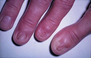
Pterygium inversum unguis or ventral pterygium is characterized by the adherence of the distal portion of the nailbed to the ventral surface of the nail plate. The condition may be present at birth or acquired, and may cause pain with manipulation of small objects, typing, and close manicuring of the nail. secondary due to connective tissue disorders
Pterygium unguis forms as a result of scarring between the proximal nailfold and matrix, with the classic example being lichen planus, though it has been reported to occur as a result of sarcoidosis and Hansen's disease.
Granulosis rubra nasi is a rare familial disease of children, occurring on the nose, cheeks, and chin, characterized by diffuse redness, persistent excessive sweating, and small dark red papules that disappear on diascopic pressure.
Galli–Galli disease is a rare inherited condition that has close resemblance clinically to Dowling-Degos' disease, but is histologically distinct, characterized by skin lesions that are 1- to 2-mm slightly keratotic red to dark brown papules which are focally confluent in a reticulate pattern. The disease is also characterized by slowly progressive and disfiguring reticulate hyperpigmentation of the flexures, clinically and histopathologically diagnostic for Dowling-Degos disease but also associated with suprabasal, nondyskeratotic acantholysis.
Texier's disease is a pseudosclerodermatous reaction that occurs after injection with vitamin K, a subcutaneous sclerosis with or without fasciitis that lasts several years.
Bullous drug reaction most commonly refers to a drug reaction in the erythema multiforme group. These are uncommon reactions to medications, with an incidence of 0.4 to 1.2 per million person-years for toxic epidermal necrolysis and 1.2 to 6.0 per million person-years for Stevens–Johnson syndrome. The primary skin lesions are large erythemas, most often irregularly distributed and of a characteristic purplish-livid color, at times with flaccid blisters.

Linear IgA bullous dermatosis is a rare immune-mediated blistering skin disease frequently associated with medication exposure, especially vancomycin, with men and women being equally affected. It was first described by Tadeusz Chorzelski in 1979 and may be divided into two types:
IgA pemphigus is a subtype of pemphigus with two distinct forms:
Heat urticaria presents within five minutes after the skin has been exposed to heat above 43 °C (109 °F), with the exposed area becoming burned, stinging, and turning red, swollen, and indurated.
Pressure urticaria is a physical urticaria caused by pressure applied to the skin, and is characterized by the development of swelling and pain that usually occurs 3 to 12 hours after local pressure has been applied.
Keratoderma climactericum is a skin condition characterized by hyperkeratosis of the palms and soles beginning at about the time of menopause.
References
- 1 2 Rapini, Ronald P.; Bolognia, Jean L.; Jorizzo, Joseph L. (2007). Dermatology: 2-Volume Set. St. Louis: Mosby. ISBN 1-4160-2999-0.
- ↑ James, William; Berger, Timothy; Elston, Dirk (2005) Andrews' Diseases of the Skin: Clinical Dermatology (10th ed.). Saunders. Page 16. ISBN 0-7216-2921-0.
- ↑ Jonkman, Marcel F.; Horváth, Barbara (2022). "2. Dermatological examination of bullous diseases". In Horváth, Barbara (ed.). Autoimmune Bullous Diseases (2nd ed.). Switzerland: Springer. p. 13. ISBN 978-3-030-91557-5.
- ↑ James, William D.; Elston, Dirk; Treat, James R.; Rosenbach, Misha A.; Neuhaus, Isaac (2020). "2. Cutaneous signs and diagnosis". Andrews' Diseases of the Skin: Clinical Dermatology (13th ed.). Edinburgh: Elsevier. p. 13. ISBN 978-0-323-54753-6.
- ↑ Freiman, Anatoli; Kalia, Sunil; O'Brien, Elizabeth A. (July 2006). "Dermatologic Signs". Journal of Cutaneous Medicine and Surgery. 10 (4): 175–182. doi:10.2310/7750.2006.00042.
| | This dermatology article is a stub. You can help Wikipedia by expanding it. |