Related Research Articles
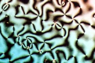
Liquid crystal (LC) is a state of matter whose properties are between those of conventional liquids and those of solid crystals. For example, a liquid crystal can flow like a liquid, but its molecules may be oriented in a common direction as in solid. There are many types of LC phases, which can be distinguished by their optical properties. The contrasting textures arise due to molecules within one area of material ("domain") being oriented in the same direction but different areas having different orientations. An LC material may not always be in an LC state of matter.

Optical rotation, also known as polarization rotation or circular birefringence, is the rotation of the orientation of the plane of polarization about the optical axis of linearly polarized light as it travels through certain materials. Circular birefringence and circular dichroism are the manifestations of optical activity. Optical activity occurs only in chiral materials, those lacking microscopic mirror symmetry. Unlike other sources of birefringence which alter a beam's state of polarization, optical activity can be observed in fluids. This can include gases or solutions of chiral molecules such as sugars, molecules with helical secondary structure such as some proteins, and also chiral liquid crystals. It can also be observed in chiral solids such as certain crystals with a rotation between adjacent crystal planes or metamaterials.
In electrodynamics, circular polarization of an electromagnetic wave is a polarization state in which, at each point, the electromagnetic field of the wave has a constant magnitude and is rotating at a constant rate in a plane perpendicular to the direction of the wave.

Polarization is a property of transverse waves which specifies the geometrical orientation of the oscillations. In a transverse wave, the direction of the oscillation is perpendicular to the direction of motion of the wave. A simple example of a polarized transverse wave is vibrations traveling along a taut string (see image); for example, in a musical instrument like a guitar string. Depending on how the string is plucked, the vibrations can be in a vertical direction, horizontal direction, or at any angle perpendicular to the string. In contrast, in longitudinal waves, such as sound waves in a liquid or gas, the displacement of the particles in the oscillation is always in the direction of propagation, so these waves do not exhibit polarization. Transverse waves that exhibit polarization include electromagnetic waves such as light and radio waves, gravitational waves, and transverse sound waves in solids.
Circular dichroism (CD) is dichroism involving circularly polarized light, i.e., the differential absorption of left- and right-handed light. Left-hand circular (LHC) and right-hand circular (RHC) polarized light represent two possible spin angular momentum states for a photon, and so circular dichroism is also referred to as dichroism for spin angular momentum. This phenomenon was discovered by Jean-Baptiste Biot, Augustin Fresnel, and Aimé Cotton in the first half of the 19th century. Circular dichroism and circular birefringence are manifestations of optical activity. It is exhibited in the absorption bands of optically active chiral molecules. CD spectroscopy has a wide range of applications in many different fields. Most notably, UV CD is used to investigate the secondary structure of proteins. UV/Vis CD is used to investigate charge-transfer transitions. Near-infrared CD is used to investigate geometric and electronic structure by probing metal d→d transitions. Vibrational circular dichroism, which uses light from the infrared energy region, is used for structural studies of small organic molecules, and most recently proteins and DNA.
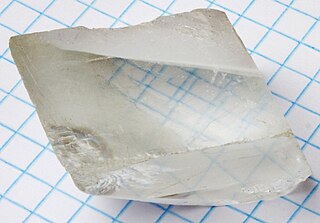
Birefringence is the optical property of a material having a refractive index that depends on the polarization and propagation direction of light. These optically anisotropic materials are described as birefringent or birefractive. The birefringence is often quantified as the maximum difference between refractive indices exhibited by the material. Crystals with non-cubic crystal structures are often birefringent, as are plastics under mechanical stress.
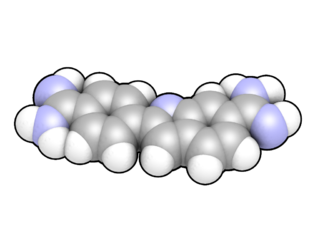
DAPI, or 4′,6-diamidino-2-phenylindole, is a fluorescent stain that binds strongly to adenine–thymine-rich regions in DNA. It is used extensively in fluorescence microscopy. As DAPI can pass through an intact cell membrane, it can be used to stain both live and fixed cells, though it passes through the membrane less efficiently in live cells and therefore provides a marker for membrane viability.

Polarimetry is the measurement and interpretation of the polarization of transverse waves, most notably electromagnetic waves, such as radio or light waves. Typically polarimetry is done on electromagnetic waves that have traveled through or have been reflected, refracted or diffracted by some material in order to characterize that object.

A polarizer or polariser is an optical filter that lets light waves of a specific polarization pass through while blocking light waves of other polarizations. It can filter a beam of light of undefined or mixed polarization into a beam of well-defined polarization, known as polarized light. Polarizers are used in many optical techniques and instruments. Polarizers find applications in photography and LCD technology. In photography, a polarizing filter can be used to filter out reflections.
Raman optical activity (ROA) is a vibrational spectroscopic technique that is reliant on the difference in intensity of Raman scattered right and left circularly polarised light due to molecular chirality.
Fluorescence anisotropy or fluorescence polarization is the phenomenon where the light emitted by a fluorophore has unequal intensities along different axes of polarization. Early pioneers in the field include Aleksander Jablonski, Gregorio Weber, and Andreas Albrecht. The principles of fluorescence polarization and some applications of the method are presented in Lakowicz's book.
Vibrational circular dichroism (VCD) is a spectroscopic technique which detects differences in attenuation of left and right circularly polarized light passing through a sample. It is the extension of circular dichroism spectroscopy into the infrared and near infrared ranges.

Fluorescence is used in the life sciences generally as a non-destructive way of tracking or analysing biological molecules. Some proteins or small molecules in cells are naturally fluorescent, which is called intrinsic fluorescence or autofluorescence. Alternatively, specific or general proteins, nucleic acids, lipids or small molecules can be "labelled" with an extrinsic fluorophore, a fluorescent dye which can be a small molecule, protein or quantum dot. Several techniques exist to exploit additional properties of fluorophores, such as fluorescence resonance energy transfer, where the energy is passed non-radiatively to a particular neighbouring dye, allowing proximity or protein activation to be detected; another is the change in properties, such as intensity, of certain dyes depending on their environment allowing their use in structural studies.

In optical mineralogy, an interference colour chart, also known as the Michel-Levy chart, is a tool first developed by Auguste Michel-Lévy to identify minerals in thin section using a petrographic microscope. With a known thickness of the thin section, minerals have specific and predictable colours in cross-polarized light, and this chart can help identify minerals. The colours are produced by the difference in speed in the fast and slow rays, also known as birefringence.
The following outline is provided as an overview of and topical guide to biophysics:
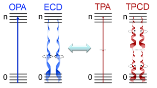
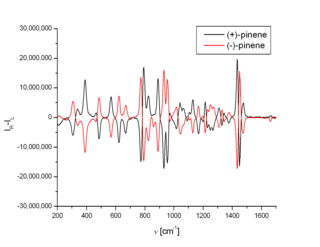
Two-photon circular dichroism (TPCD), the nonlinear counterpart of electronic circular dichroism (ECD), is defined as the differences between the two-photon absorption (TPA) cross-sections obtained using left circular polarized light and right circular polarized light.
Super-resolution dipole orientation mapping (SDOM) is a form of fluorescence polarization microscopy (FPM) that achieved super resolution through polarization demodulation. It was first described by Karl Zhanghao and others in 2016. Fluorescence polarization (FP) is related to the dipole orientation of chromophores, making fluorescence polarization microscopy possible to reveal structures and functions of tagged cellular organelles and biological macromolecules. In addition to fluorescence intensity, wavelength, and lifetime, the fourth dimension of fluorescence—polarization—can also provide intensity modulation without the restriction to specific fluorophores; its investigation in super-resolution microscopy is still in its infancy.

Fluorescence imaging is a type of non-invasive imaging technique that can help visualize biological processes taking place in a living organism. Images can be produced from a variety of methods including: microscopy, imaging probes, and spectroscopy.

Alison Rodger FRSC FRACI FAA CChem is a professor of chemistry at Macquarie University. Her research considers biomacromolecular structures and their characterisation. She is currently developing Raman Linear Difference Spectroscopy and fluorescence detected liner dichroism to understand biomacromolecular structure and interactions with application to the division of bacterial cells.

Anisotropic terahertz microspectroscopy (ATM) is a spectroscopic technique in which molecular vibrations in an anisotropic material are probed with short pulses of terahertz radiation whose electric field is linearly polarized parallel to the surface of the material. The technique has been demonstrated in studies involving single crystal sucrose, fructose, oxalic acid, and molecular protein crystals in which the spatial orientation of molecular vibrations are of interest.
References
- ↑ Bengt Nordén, Alison Rodger and Timothy Dafforn Linear Dichroism and Circular Dichroism. A Textbook on Polarized-Light Spectroscopy. ISBN 978-1-84755-902-9. The Royal Society of Chemistry – London 2010
- ↑ Hannon, M.J., Moreno, V., Prieto, M.J., Molderheim, E., Sletten, E., Meistermann, I., Isaac, C.J., Sanders, K.J., Rodger, A. “Intramolecular DNA coiling mediated by a metallo supramolecular cylinder” Angewandte Chemie, 2001, 40, 879−884
- ↑ Elaine Small, Rachel Marrington, Alison Rodger, David J. Scott, Katherine Sloan, David Roper, Timothy R. Dafforn and Stephen G. Addinall ‘FtsZ Polymer-bundling by the Escherichia coli ZapA Orthologue, YgfE, Involves a Conformational Change in Bound GTP’ 2007, Journal of Molecular Biology, 369: 210-221.
- ↑ Alison Rodger, Rachel Marrington, Michael A. Geeves, Matthew Hicks, Lahari de Alwis, David J. Halsall and Timothy R. Dafforn ‘Looking at long molecules in solution: what happens when they are subjected to Couette flow?’ 2006, Physical Chemistry Chemical Physics, 8: 3161-3171.
- ↑ Heinz Falk, Gunther Vormayr, Leon Margulies, Stephanie Metz and Yehuda Mazur ‘A Linear Dichroism Study of Pyrromethene-, Pyrromethenone- and Bilatriene-abc-Derivates’ 1986, Monatshefte fur Chemie, 117 : 849-858.
- ↑ Hicks, M.R.; Damianoglou, A.; Rodger, A.; Dafforn, T.R.; “Folding and membrane insertion of the pore-forming peptide gramicidin occurs as a concerted process“ Journal of Molecular Biology, 2008, 383, 358-366
- ↑ Gabor Steinbach, Istvan Pomozi, Otto Zsiros, Aniko Pay, Gabor V. Horvat, Gyozo Garab ‘Imaging Fluorescence detected linear dichroism of plant cell walls in laser scanning confocal microscope’ 2008, Cytometry Part A, 73A : 202-208.