Lymphocytic pleocytosis

Last updated
A stain demonstrating an increase in lymphocytes within the cerebrospinal fluid. Concentrations in healthy individuals are usually under 5 white blood cells per mL. CSF pleocytosis with lymphocytic predominance in abnormal CSF smears.png
A stain demonstrating an increase in lymphocytes within the cerebrospinal fluid. Concentrations in healthy individuals are usually under 5 white blood cells per μL.

Lymphocytic pleocytosis is an abnormal increase in the amount of lymphocytes in the cerebrospinal fluid (CSF). It is usually considered to be a sign of infection or inflammation within the nervous system, and is encountered in a number of neurological diseases, such as pseudomigraine, Susac's syndrome, and encephalitis. While lymphocytes make up roughly a quarter of all white blood cells (WBC) in the body, they are generally rare in the CSF. Under normal conditions, there are usually less than 5 white blood cells per μL of CSF. In a pleocytic setting, the number of lymphocytes can jump to more than 1,000 cells per μL. Increases in lymphocyte count are often accompanied by an increase in cerebrospinal protein concentrations in addition to pleocytosis of other types of white blood cells.[ citation needed ]

Contents

Symptoms and signs

Though exact concentrations differ based on the specific disease, mild cases of lymphocytic pleocytosis are considered to begin when lymphocyte counts enter the range of 10-100 cells per mm3. [1] In healthy individuals, only 0-5 white blood cells per μL are normally present in the CSF. [2] In patients with pseudomigraines, studies have shown concentrations ranging from 10 to 760 cells per mm3, with a mean concentration of 199 ± 174 cells per mm3. [3] Increases in white blood cell count to more than 500 cells per mm3 can cause the CSF to appear cloudy when observed during diagnostic tests. The rise in concentration corresponds to an inflammatory immune response typically seen during viral infections. Despite their diversity, diseases featuring lymphocytic pleocytosis share a number of symptoms, most notably headaches and neurological deficits.[ citation needed ]

Causes

Cerebral spinal fluid lymphocytic pleocytosis is generally the result of an immune response to neurovascular inflammation. Many cases point to a viral infection as the root cause of pleocytosis, in which the immune system produces antibodies against neuronal and vascular antigens. This evidence possibly connects it to viral meningitis and Mollaret's disease. [3] Certain non-viral infections, such as Lyme disease have also been considered possible causes. In some diseases, an infection precipitates an autoimmune response, leading to increased lymphocyte levels.[ citation needed ]

Diagnosis

The presence of lymphocytic pleocytosis is generally detected through a lumbar puncture followed by clinical analysis of cerebrospinal fluid. When combined with analysis of the appearance and pressure of the tested CSF, along with measurements for the amount of glucose and proteins present, white blood cell counts can be used to detect or diagnose a number of diseases. Among these are subarachnoid hemorrhage, multiple sclerosis, and the various types of meningitis. While a lumbar puncture may return a WBC count within the normal range of 0-5 cells per μL, this does not rule out the possibility of a disease. [4]

Diseases

Research has found the presence of lymphocytic pleocytosis in the following diseases and documented their respective mechanisms and reactions:

Viral encephalitis

In Encephalitis, the inflammation of the brain leads to a breakdown in neurological function, causing the patient to have symptoms such as fever, confusion, amnesia, personality changes, paralysis, seizures and language dysfunction. A viral infection can directly cause encephalitis or trigger a cascade ending in autoimmunity, with both mechanisms eventually leading to a rise in CSF lymphocyte concentration.[ citation needed ]

For patients with Herpes simplex virus, more than 90% are found to have lymphocytic pleocytosis of varying levels. [5] Intravenous aciclovir, can be used to prevent viral replication, and in the event of persistent lymphocytic pleocytosis, higher doses of aciclovir can also be taken. Studies have shown this treatment combined with valacyclovir to be effective in combating HSV-1 and eventually returning lymphocyte counts to normal. [5]

A viral infection may also result in encephalitis triggered by an autoimmune response. The most ubiquitous form, anti-NMDA receptor encephalitis, is thought to be commonly initiated by herpes infections resulting in an autoimmune response to the NR1 subunit of the NMDA receptor. Lymphocytic pleocytosis is involved in the initial stages of the disease. During this period, lymphocytes can number in the hundreds per mm3, while later on, lymphocyte levels have a tendency to return to equilibrium. It has been proposed that this early spike in lymphocytic concentration is the result of the breaching of the blood–brain barrier during the initial viral infection, giving peripheral antibodies access to the central nervous system and leading to the development of autoimmunity. [6]

Pseudomigraine

Multiple studies have been performed to examine the correlation between pseudomigraines and lymphocytic pleocytosis. A pseudomigraine is characterized by a moderate or severe, typically bilateral throbbing headache accompanied by transient neurological symptoms and lymphocytic pleocytosis. [7] These migraine episodes are recurrent and self-limiting. [8] In these studies, individuals ranging from about 15–40 years of age were examined and the majority of those tested were male. After each migraine episode, the patients were all asymptomatic. When examined via EEG, CT, and MRI forms of imaging, the CT and MRI scans were all normal; however, 30 out of 42 patients had abnormal EEG scans. [3] For 26 of these patients, there was unilateral excessive slowing while 4 of these patients experienced bilateral slowing. [3] In another study, patients displayed an elevated level of lymphocytic pleocytosis with each pseudomigraine episode. [8] [9]

Susac's syndrome

In Susac's syndrome, an autoimmune response damages the blood vessels of the brain, retina and cochlea, leading to a loss of neurological functions. Patients with the syndrome had hearing and vision loss and were found to have higher concentrations of lymphocytes and proteins in their cerebrospinal fluid. Treatment with immunosuppressive drugs like prednisone followed by Azathioprine were found to have significant effects and patients gradually regained lost function, in some cases after only a few weeks. [10]

Treatments

The most appropriate method of returning lymphocyte counts to normal levels is to treat the disease responsible for their increase. For cases in which the root cause is a viral or bacterial infection, drugs that counteract these pathogens have been found to be effective. Since herpes infections often lead to pleocytosis, aciclovir and valacyclovir are commonly prescribed. [11] When pleocytosis is the result of an autoimmune response, immunosuppressive drugs like prednisone can be used.[ citation needed ]

History

Determination of lymphocytic pleocytosis became possible with the advent of the diagnostic lumbar puncture and the technology necessary to analyze the cerebrospinal fluid via microbiological, biochemical, and immunological tests. [8] While the first lumbar punctures were performed in the late 19th century, the tests revealing elevated lymphocyte levels within the CSF were not available until much later. Modern lumbar punctures testing for lymphocyte counts are commonly used to diagnose or rule out certain diseases such as meningitis and determine whether an infection is present within the CSF. [10]

Related Research Articles

<span class="mw-page-title-main">Cerebrospinal fluid</span> Clear, colorless bodily fluid found in the brain and spinal cord

Cerebrospinal fluid (CSF) is a clear, colorless body fluid found within the tissue that surrounds the brain and spinal cord of all vertebrates.

<span class="mw-page-title-main">Encephalitis</span> Inflammation of the brain

Encephalitis is inflammation of the brain. The severity can be variable with symptoms including reduction or alteration in consciousness, headache, fever, confusion, a stiff neck, and vomiting. Complications may include seizures, hallucinations, trouble speaking, memory problems, and problems with hearing.

<span class="mw-page-title-main">Viral meningitis</span> Medical condition

Viral meningitis, also known as aseptic meningitis, is a type of meningitis due to a viral infection. It results in inflammation of the meninges. Symptoms commonly include headache, fever, sensitivity to light and neck stiffness.

<span class="mw-page-title-main">Lumbar puncture</span> Procedure to collect cerebrospinal fluid

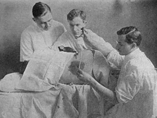
Lumbar puncture (LP), also known as a spinal tap, is a medical procedure in which a needle is inserted into the spinal canal, most commonly to collect cerebrospinal fluid (CSF) for diagnostic testing. The main reason for a lumbar puncture is to help diagnose diseases of the central nervous system, including the brain and spine. Examples of these conditions include meningitis and subarachnoid hemorrhage. It may also be used therapeutically in some conditions. Increased intracranial pressure is a contraindication, due to risk of brain matter being compressed and pushed toward the spine. Sometimes, lumbar puncture cannot be performed safely. It is regarded as a safe procedure, but post-dural-puncture headache is a common side effect if a small atraumatic needle is not used.

Lymphocytic choriomeningitis (LCM) is a rodent-borne viral infectious disease that presents as aseptic meningitis, encephalitis or meningoencephalitis. Its causative agent is lymphocytic choriomeningitis mammarenavirus (LCMV), a member of the family Arenaviridae. The name was coined by Charles Armstrong in 1934.

<span class="mw-page-title-main">Aseptic meningitis</span> Medical condition

Aseptic meningitis is the inflammation of the meninges, a membrane covering the brain and spinal cord, in patients whose cerebral spinal fluid test result is negative with routine bacterial cultures. Aseptic meningitis is caused by viruses, mycobacteria, spirochetes, fungi, medications, and cancer malignancies. The testing for both meningitis and aseptic meningitis is mostly the same. A cerebrospinal fluid sample is taken by lumbar puncture and is tested for leukocyte levels to determine if there is an infection and goes on to further testing to see what the actual cause is. The symptoms are the same for both meningitis and aseptic meningitis but the severity of the symptoms and the treatment can depend on the certain cause.

<span class="mw-page-title-main">Viral encephalitis</span> Medical condition

Viral encephalitis is inflammation of the brain parenchyma, called encephalitis, by a virus. The different forms of viral encephalitis are called viral encephalitides. It is the most common type of encephalitis and often occurs with viral meningitis. Encephalitic viruses first cause infection and replicate outside of the central nervous system (CNS), most reaching the CNS through the circulatory system and a minority from nerve endings toward the CNS. Once in the brain, the virus and the host's inflammatory response disrupt neural function, leading to illness and complications, many of which frequently are neurological in nature, such as impaired motor skills and altered behavior.

<span class="mw-page-title-main">Neurosyphilis</span> Infection of the central nervous system in a patient with syphilis

Neurosyphilis is the infection of the central nervous system in a patient with syphilis. In the era of modern antibiotics, the majority of neurosyphilis cases have been reported in HIV-infected patients. Meningitis is the most common neurological presentation in early syphilis. Tertiary syphilis symptoms are exclusively neurosyphilis, though neurosyphilis may occur at any stage of infection.

<span class="mw-page-title-main">Mollaret's meningitis</span> Medical condition

Mollaret's meningitis is a recurrent or chronic inflammation of the protective membranes covering the brain and spinal cord, known collectively as the meninges. Since Mollaret's meningitis is a recurrent, benign (non-cancerous), aseptic meningitis, it is also referred to as benign recurrent lymphocytic meningitis. It was named for Pierre Mollaret, the French neurologist who first described it in 1944.

The central nervous system (CNS) controls most of the functions of the body and mind. It comprises the brain, spinal cord and the nerve fibers that branch off to all parts of the body. The CNS viral diseases are caused by viruses that attack the CNS. Existing and emerging viral CNS infections are major sources of human morbidity and mortality.

<span class="mw-page-title-main">Meningitis</span> Inflammation of the membranes around the brain and spinal cord

Meningitis is acute or chronic inflammation of the protective membranes covering the brain and spinal cord, collectively called the meninges. The most common symptoms are fever, intense headache, vomiting and neck stiffness and occasionally photophobia.

<span class="mw-page-title-main">Cerebrospinal fluid leak</span> Medical condition

A cerebrospinal fluid leak is a medical condition where the cerebrospinal fluid (CSF) that surrounds the brain and spinal cord leaks out of one or more holes or tears in the dura mater. A CSF leak is classed as either spontaneous (primary), having no known cause, or nonspontaneous (secondary) where it is attributed to an underlying condition. Causes of a primary CSF leak are those of trauma including from an accident or intentional injury, or arising from a medical intervention known as iatrogenic. A basilar skull fracture as a cause can give the sign of CSF leakage from the ear nose or mouth. A lumbar puncture can give the symptom of a post-dural-puncture headache.

<span class="mw-page-title-main">Herpes meningitis</span> Medical condition

Herpes meningitis is inflammation of the meninges, the protective tissues surrounding the spinal cord and brain, due to infection from viruses of the Herpesviridae family - the most common amongst adults is HSV-2. Symptoms are self-limiting over 2 weeks with severe headache, nausea, vomiting, neck-stiffness, and photophobia. Herpes meningitis can cause Mollaret's meningitis, a form of recurrent meningitis. Lumbar puncture with cerebrospinal fluid results demonstrating aseptic meningitis pattern is necessary for diagnosis and polymerase chain reaction is used to detect viral presence. Although symptoms are self-limiting, treatment with antiviral medication may be recommended to prevent progression to Herpes Meningoencephalitis.

<span class="mw-page-title-main">Herpes simplex encephalitis</span> Encephalitis associated with herpes simplex virus

Herpes simplex encephalitis (HSE), or simply herpes encephalitis, is encephalitis due to herpes simplex virus. It is estimated to affect at least 1 in 500,000 individuals per year, and some studies suggest an incidence rate of 5.9 cases per 100,000 live births.

CSF glucose or glycorrhachia is a measurement used to determine the concentration of glucose in cerebrospinal fluid (CSF).

Drug-Induced Aseptic Meningitis (DIAM) is a type of aseptic meningitis related to the use of medications such as nonsteroidal anti-inflammatory drugs (NSAIDs) or biologic drugs such as intravenous immunoglobulin (IVIG). Additionally, this condition generally shows clinical improvement after cessation of the medication, as well as a tendency to relapse with resumption of the medication.

Neurovirology is an interdisciplinary field which represents a melding of clinical neuroscience, virology, immunology, and molecular biology. The main focus of the field is to study viruses capable of infecting the nervous system. In addition to this, the field studies the use of viruses to trace neuroanatomical pathways, for gene therapy, and to eliminate detrimental populations of neural cells.

<span class="mw-page-title-main">Anti-NMDA receptor encephalitis</span> Rare disease which results in brain inflammation

Anti-NMDA receptor encephalitis is a type of brain inflammation caused by antibodies. Early symptoms may include fever, headache, and feeling tired. This is then typically followed by psychosis which presents with false beliefs (delusions) and seeing or hearing things that others do not see or hear (hallucinations). People are also often agitated or confused. Over time, seizures, decreased breathing, and blood pressure and heart rate variability typically occur. In some cases, patients may develop catatonia.

<span class="mw-page-title-main">Neonatal meningitis</span> Medical condition

Neonatal meningitis is a serious medical condition in infants that is rapidly fatal if untreated. Meningitis, an inflammation of the meninges, the protective membranes of the central nervous system, is more common in the neonatal period than any other time in life, and is an important cause of morbidity and mortality globally. Mortality is roughly half in developing countries and ranges from 8%-12.5% in developed countries.

<span class="mw-page-title-main">Chronic meningitis</span> Inflammation of the membranes surrounding the brain and spinal cord lasting longer than 4 weeks

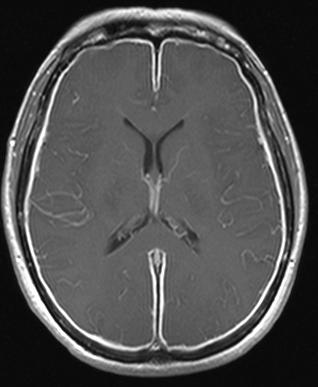
Chronic meningitis is a long-lasting inflammation of the membranes lining the brain and spinal cord. By definition, the duration of signs, symptoms and inflammation in chronic meningitis last longer than 4 weeks. Infectious causes are a leading cause and the infectious organisms responsible for chronic meningitis are different than the organisms that cause acute infectious meningitis. Tuberculosis and the fungi cryptococcus are leading causes worldwide. Chronic meningitis due to infectious causes are more common in those who are immunosuppressed, including those with HIV infection or in children who are malnourished. Chronic meningitis sometimes has a more indolent course than acute meningitis with symptoms developing more insidiously and slowly. Also, some of the infectious agents that cause chronic infectious meningitis such as mycobacterium tuberculosis, many fungal species and viruses are difficult to isolate from the cerebrospinal fluid making diagnosis challenging. No cause is identified during initial evaluation in one third of cases. Magnetic resonance imaging (MRI) of the brain is more sensitive than computed tomography and may show radiological signs that suggest chronic meningitis, however no radiological signs are considered pathognomonic or characteristic. MRI is also normal in many cases further limiting its diagnostic utility.

References

  1. Walker, HK; Hall, WD; Hurst, JW; Jurado, R (1990). Clinical Methods, 3rd edition The History, Physical, and Laboratory Examinations. Boston: Butterworth Publishers. ISBN   978-0-409-90077-4 . Retrieved 21 March 2015.
  2. National Institute of Health. "Cerebral spinal fluid (CSF) collection". www.nlm.nih.gov. National Institute of Health. Retrieved 2015-04-19.
  3. 1 2 3 4 Gómez-Aranda, F; Cañadillas, F; Martí-Massó, JF; Díez-Tejedor, E; Serrano, PJ; Leira, R; Gracia, M; Pascual, J (July 1997). "Pseudomigraine with temporary neurological symptoms and lymphocytic pleocytosis. A report of 50 cases". Brain: A Journal of Neurology. 120 (7): 1105–13. doi:10.1093/brain/120.7.1105. PMID   9236623.
  4. Lumbar Puncture (LP) Interpretation of Cerebrospinal Fluid at eMedicine
  5. 1 2 Skelly, Michael J; Burger, Andrew A; Adekola, Oritsegbubemi (25 September 2012). "Herpes simplex virus-1 encephalitis: a review of current disease management with three case reports". Antiviral Chemistry & Chemotherapy. 23 (1): 13–8. doi:10.3851/IMP2129. PMID   23018202. S2CID   19965194.
  6. Venkatesan, Arun; Benavides, David (February 2015). "Autoimmune Encephalitis and Its Relation to Infection ". Current Neurology and Neuroscience Reports. 15 (3): 3. doi:10.1007/s11910-015-0529-1. PMID   25637289. S2CID   21546062.
  7. Pascual, Julio; Valle, Natalia (June 2003). "Pseudomigraine with lymphocytic pleocytosis". Current Pain and Headache Reports. 7 (3): 224–8. doi:10.1007/s11916-003-0077-2. PMID   12720603. S2CID   27116781.
  8. 1 2 3 Doherty, Carolynne M; Forbes, Raeburn B (2014). "Diagnostic Lumbar Puncture". The Ulster Medical Journal. 83 (2): 93–102. PMC   4113153 . PMID   25075138.
  9. Filina, T; Feja, KN; Tolan, RW (June 2013). "An adolescent with pseudomigraine, transient headache, neurological deficits, and lymphocytic pleocytosis (HaNDL Syndrome): case report and review of the literature". Clinical Pediatrics. 52 (6): 496–502. doi:10.1177/0009922813483358. PMID   23559488. S2CID   35821471.
  10. 1 2 Do, Tau Hung; Fisch, Christiane; Evoy, Francois (March 2004). "Susac syndrome: report of four cases and review of the literature". AJNR. American Journal of Neuroradiology. 25 (3): 382–8. PMC   8158540 . PMID   15037459.
  11. Maguire, Phyllis (February 2008). "How to parse out the possible causes of meningitis". Today's Hospitalist. Retrieved 2020-05-02.