Macro domain

Last updated
Macro
PDB 1zr3 EBI.jpg
Crystal structure of the macro-domain of human core histone variant macroh2a1.1
Identifiers
SymbolMacro
Pfam PF01661
Pfam clan CL0223
InterPro IPR002589
SCOP2 1vhu / SCOPe / SUPFAM
CDD cd02749
Available protein structures:
Pfam   structures / ECOD  
PDB RCSB PDB; PDBe; PDBj
PDBsum structure summary

In molecular biology, the Macro domain (often also written macrodomain) or A1pp domain is a module of about 180 amino acids which can bind ADP-ribose, an NAD metabolite, or related ligands. Binding to ADP-ribose can be either covalent or non-covalent: [1] in certain cases it is believed to bind non-covalently, [2] while in other cases (such as Aprataxin) it appears to bind both non-covalently through a zinc finger motif, and covalently through a separate region of the protein. [3]

Contents

Function

The domain was described originally in association with the ADP-ribose 1-phosphate (Appr-1-P)-processing activity (A1pp) of the yeast YBR022W protein and called A1pp. [4] However, the domain has been renamed Macro as it is the C-terminal domain of mammalian core histone macro-H2A. [5] [6] Macro domain proteins can be found in eukaryotes, in (mostly pathogenic) bacteria, in archaea and in ssRNA viruses, such as coronaviruses, Rubella and Hepatitis E viruses. In vertebrates the domain occurs in e.g. histone macroH2A, predicted poly-ADP-ribose polymerases (PARPs) and B aggressive lymphoma (BAL) protein.

ADP-ribosylation of proteins is an important post-translational modification that occurs in a variety of biological processes, including DNA repair, regulation of transcription, chromatin biology, maintenance of genomic stability, telomere dynamics, [7] cell differentiation and proliferation, [8] necrosis and apoptosis, [9] and long-term memory formation. [10] The Macro domain recognises the ADP-ribose nucleotide and in some cases poly-ADP-ribose, and is thus a high-affinity ADP-ribose-binding module found in a number of otherwise unrelated proteins. [11] ADP-ribosylation of DNA is relatively uncommon and has only been described for a small number of toxins that include pierisin, [12] scabin [13] and DarT. [14] [15] The Macro domain from the antitoxin DarG of the toxin-antitoxin system DarTG, both binds and removes the ADP-ribose modification added to DNA by the toxin DarT. [14] [15] The Macro domain from human, macroH2A1.1, binds an NAD metabolite O-acetyl-ADP-ribose. [16]

ClassSubclassSpeciesActivity
MacroH2A-likeeADP-ribose binding
MacroD-type‘classic’a, b, e, vADP-ribosyl bond hydrolysis
Zn-dependentb, eADP-ribosyl bond hydrolysis
GDAP2-likeeADP-ribose binding
ALC1-likeb, eADP-ribose binding or ADP-ribosyl bond hydrolysis
PARG-likePARG_cateADP-ribosyl bond hydrolysis
mPARG (DUF2263)b, e, vADP-ribosyl bond hydrolysis
Macro2-typee, vADP-ribosyl bond hydrolysis
SUD-M-likevRNA binding
DUF2362eunknown
a, Archaea; b, Bacteria; e, Eukarya; v, Virus


Structure

The 3D structure of the Macro domain describes a mixed alpha/beta fold of a mixed beta sheet sandwiched between four helices with the ligand-binding pocket lies within the fold. [11] Several Macro domain-only domains are shorter than the structure of AF1521 and lack either the first strand or the C-terminal helix 5. Well conserved residues form a hydrophobic cleft and cluster around the AF1521-ADP-ribose binding site. [6] [11] [16] [17]

See also

Related Research Articles

<span class="mw-page-title-main">Nicotinamide adenine dinucleotide</span> Chemical compound which is reduced and oxidized

Nicotinamide adenine dinucleotide (NAD) is a coenzyme central to metabolism. Found in all living cells, NAD is called a dinucleotide because it consists of two nucleotides joined through their phosphate groups. One nucleotide contains an adenine nucleobase and the other nicotinamide. NAD exists in two forms: an oxidized and reduced form, abbreviated as NAD+ and NADH (H for hydrogen), respectively.

<span class="mw-page-title-main">Poly (ADP-ribose) polymerase</span> Family of proteins

Poly (ADP-ribose) polymerase (PARP) is a family of proteins involved in a number of cellular processes such as DNA repair, genomic stability, and programmed cell death.

<span class="mw-page-title-main">Histone-modifying enzymes</span> Type of enzymes

Histone-modifying enzymes are enzymes involved in the modification of histone substrates after protein translation and affect cellular processes including gene expression. To safely store the eukaryotic genome, DNA is wrapped around four core histone proteins, which then join to form nucleosomes. These nucleosomes further fold together into highly condensed chromatin, which renders the organism's genetic material far less accessible to the factors required for gene transcription, DNA replication, recombination and repair. Subsequently, eukaryotic organisms have developed intricate mechanisms to overcome this repressive barrier imposed by the chromatin through histone modification, a type of post-translational modification which typically involves covalently attaching certain groups to histone residues. Once added to the histone, these groups elicit either a loose and open histone conformation, euchromatin, or a tight and closed histone conformation, heterochromatin. Euchromatin marks active transcription and gene expression, as the light packing of histones in this way allows entry for proteins involved in the transcription process. As such, the tightly packed heterochromatin marks the absence of current gene expression.

<span class="mw-page-title-main">ADP-ribosylation</span> Addition of one or more ADP-ribose moieties to a protein.

ADP-ribosylation is the addition of one or more ADP-ribose moieties to a protein. It is a reversible post-translational modification that is involved in many cellular processes, including cell signaling, DNA repair, gene regulation and apoptosis. Improper ADP-ribosylation has been implicated in some forms of cancer. It is also the basis for the toxicity of bacterial compounds such as cholera toxin, diphtheria toxin, and others.

<span class="mw-page-title-main">ETS1</span> Protein-coding gene in the species Homo sapiens

Protein C-ets-1 is a protein that in humans is encoded by the ETS1 gene. The protein encoded by this gene belongs to the ETS family of transcription factors.

<span class="mw-page-title-main">PARP1</span> Mammalian protein found in Homo sapiens

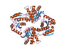
Poly [ADP-ribose] polymerase 1 (PARP-1) also known as NAD+ ADP-ribosyltransferase 1 or poly[ADP-ribose] synthase 1 is an enzyme that in humans is encoded by the PARP1 gene. It is the most abundant of the PARP family of enzymes, accounting for 90% of the NAD+ used by the family. PARP1 is mostly present in cell nucleus, but cytosolic fraction of this protein was also reported.

<span class="mw-page-title-main">ARF1</span> Protein-coding gene in the species Homo sapiens

ADP-ribosylation factor 1 is a protein that in humans is encoded by the ARF1 gene.

<span class="mw-page-title-main">GGA1</span> Protein-coding gene in the species Homo sapiens

ADP-ribosylation factor-binding protein GGA1 is a protein that in humans is encoded by the GGA1 gene.

<span class="mw-page-title-main">Telomeric repeat-binding factor 1</span> Protein-coding gene in humans

Telomeric repeat-binding factor 1 is a protein that in humans is encoded by the TERF1 gene.

<span class="mw-page-title-main">ARF3</span> Protein-coding gene in the species Homo sapiens

ADP-ribosylation factor 3 is a protein that in humans is encoded by the ARF3 gene.

<span class="mw-page-title-main">Tankyrase</span> Enzyme

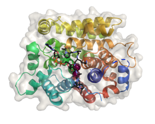
Tankyrase, also known as tankyrase 1, is an enzyme that in humans is encoded by the TNKS gene. It inhibits the binding of TERF1 to telomeric DNA. Tankyrase attracts substantial interest in cancer research through its interaction with AXIN1 and AXIN2, which are negative regulators of pro-oncogenic β-catenin signaling. Importantly, activity in the β-catenin destruction complex can be increased by tankyrase inhibitors and thus such inhibitors are a potential therapeutic option to reduce the growth of β-catenin-dependent cancers.

<span class="mw-page-title-main">HIST1H1A</span>

Histone H1.1 is a protein that in humans is encoded by the HIST1H1A gene.

<span class="mw-page-title-main">PARP4</span> Enzyme

Poly [ADP-ribose] polymerase 4 is an enzyme that in humans is encoded by the PARP4 gene.

<span class="mw-page-title-main">Tankyrase 2</span> Protein-coding gene in the species Homo sapiens

Tankyrase-2 is an enzyme that in humans is encoded by the TNKS2 gene.

<span class="mw-page-title-main">ADP-ribosylhydrolase 3</span> Protein-coding gene in the species Homo sapiens

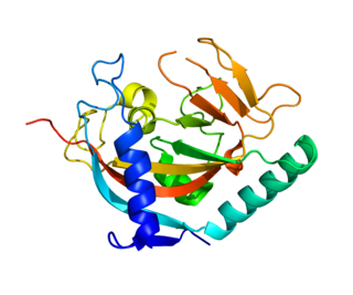
(ADP-ribosyl)hydrolase 3 (ARH3) is an enzyme that in humans is encoded by the ADPRHL2 gene (also called ADPRS). This enzyme reverses the proteins’ post-translational addition of ADP-ribose to serine residues as part of the DNA damage response The enzyme is also known to cleave poly(ADP-ribose) polymers, 1''-O-acetyl-ADP-ribose and alpha-NAD+

<span class="mw-page-title-main">Toxin-antitoxin system</span> Biological process

A toxin-antitoxin system is a set of two or more closely linked genes that together encode both a "toxin" protein and a corresponding "antitoxin". Toxin-antitoxin systems are widely distributed in prokaryotes, and organisms often have them in multiple copies. When these systems are contained on plasmids – transferable genetic elements – they ensure that only the daughter cells that inherit the plasmid survive after cell division. If the plasmid is absent in a daughter cell, the unstable antitoxin is degraded and the stable toxic protein kills the new cell; this is known as 'post-segregational killing' (PSK).

<span class="mw-page-title-main">ADP-ribosylhydrolase</span>

In molecular biology, the (ADP-ribosyl)hydrolase (ARH) family contains enzymes which catalyses the hydrolysis of ADP-ribosyl modifications from proteins, nucleic acids and small molecules.

<span class="mw-page-title-main">PARG</span> Protein-coding gene in the species Homo sapiens

Poly (ADP-ribose) glycohydrolase is an enzyme that in humans is encoded by the PARG gene.

Parthanatos is a form of programmed cell death that is distinct from other cell death processes such as necrosis and apoptosis. While necrosis is caused by acute cell injury resulting in traumatic cell death and apoptosis is a highly controlled process signalled by apoptotic intracellular signals, parthanatos is caused by the accumulation of Poly(ADP ribose) (PAR) and the nuclear translocation of apoptosis-inducing factor (AIF) from mitochondria. Parthanatos is also known as PARP-1 dependent cell death. PARP-1 mediates parthanatos when it is over-activated in response to extreme genomic stress and synthesizes PAR which causes nuclear translocation of AIF. Parthanatos is involved in diseases that afflict hundreds of millions of people worldwide. Well known diseases involving parthanatos include Parkinson's disease, stroke, heart attack, and diabetes. It also has potential use as a treatment for ameliorating disease and various medical conditions such as diabetes and obesity.

References

  1. Hassa PO, Haenni SS, Elser M, Hottiger MO (September 2006). "Nuclear ADP-ribosylation reactions in mammalian cells: where are we today and where are we going?". Microbiol. Mol. Biol. Rev. 70 (3): 789–829. doi:10.1128/MMBR.00040-05. PMC   1594587 . PMID   16959969.
  2. Neuvonen M, Ahola T (January 2009). "Differential activities of cellular and viral macro domain proteins in binding of ADP-ribose metabolites". J. Mol. Biol. 385 (1): 212–25. doi:10.1016/j.jmb.2008.10.045. PMC   7094737 . PMID   18983849.
  3. Ahel I, Ahel D, Matsusaka T, Clark AJ, Pines J, Boulton SJ, West SC (January 2008). "Poly(ADP-ribose)-binding zinc finger motifs in DNA repair/checkpoint proteins". Nature. 451 (7174): 81–5. Bibcode:2008Natur.451...81A. doi:10.1038/nature06420. PMID   18172500. S2CID   4417693.
  4. Martzen MR, McCraith SM, Spinelli SL, Torres FM, Fields S, Grayhack EJ, Phizicky EM (November 1999). "A biochemical genomics approach for identifying genes by the activity of their products". Science. 286 (5442): 1153–5. doi:10.1126/science.286.5442.1153. PMID   10550052.
  5. Aravind L (May 2001). "The WWE domain: a common interaction module in protein ubiquitination and ADP ribosylation". Trends Biochem. Sci. 26 (5): 273–5. doi:10.1016/s0968-0004(01)01787-x. PMID   11343911.
  6. 1 2 Allen MD, Buckle AM, Cordell SC, Löwe J, Bycroft M (July 2003). "The crystal structure of AF1521 a protein from Archaeoglobus fulgidus with homology to the non-histone domain of macroH2A". J. Mol. Biol. 330 (3): 503–11. doi:10.1016/S0022-2836(03)00473-X. PMID   12842467.
  7. Tennen RI, Chua KF (January 2011). "Chromatin regulation and genome maintenance by mammalian SIRT6". Trends in Biochemical Sciences . 36 (1): 39–46. doi:10.1016/j.tibs.2010.07.009. PMC   2991557 . PMID   20729089.
  8. Ji Y, Tulin AV (October 2010). "The roles of PARP1 in gene control and cell differentiation". Current Opinion in Genetics & Development . 20 (5): 512–8. doi:10.1016/j.gde.2010.06.001. PMC   2942995 . PMID   20591646.
  9. Han W, Li X, Fu X (2011). "The macro domain protein family: Structure, functions, and their potential therapeutic implications". Mutation Research . 727 (3): 86–103. doi:10.1016/j.mrrev.2011.03.001. PMC   7110529 . PMID   21421074.
  10. Schreiber V, Dantzer F, Ame JC, de Murcia G (July 2006). "Poly(ADP-ribose): novel functions for an old molecule". Nature Reviews Molecular Cell Biology . 7 (7): 517–28. doi:10.1038/nrm1963. PMID   16829982. S2CID   22030625.
  11. 1 2 3 Karras GI, Kustatscher G, Buhecha HR, Allen MD, Pugieux C, Sait F, Bycroft M, Ladurner AG (June 2005). "The macro domain is an ADP-ribose binding module". EMBO J. 24 (11): 1911–20. doi:10.1038/sj.emboj.7600664. PMC   1142602 . PMID   15902274.
  12. Takamura-Enya, Takeji; Watanabe, Masahiko; Totsuka, Yukari; Kanazawa, Takashi; Matsushima-Hibiya, Yuko; Koyama, Kotaro; Sugimura, Takashi; Wakabayashi, Keiji (2001-10-23). "Mono(ADP-ribosyl)ation of 2′-deoxyguanosine residue in DNA by an apoptosis-inducing protein, pierisin-1, from cabbage butterfly". Proceedings of the National Academy of Sciences. 98 (22): 12414–12419. Bibcode:2001PNAS...9812414T. doi: 10.1073/pnas.221444598 . ISSN   0027-8424. PMC   60068 . PMID   11592983.
  13. Lyons, Bronwyn; Ravulapalli, Ravikiran; Lanoue, Jason; Lugo, Miguel R.; Dutta, Debajyoti; Carlin, Stephanie; Merrill, A. Rod (2016-05-20). "Scabin, a Novel DNA-acting ADP-ribosyltransferase from Streptomyces scabies". The Journal of Biological Chemistry. 291 (21): 11198–11215. doi: 10.1074/jbc.M115.707653 . ISSN   1083-351X. PMC   4900268 . PMID   27002155.
  14. 1 2 Jankevicius, Gytis; Ariza, Antonio; Ahel, Marijan; Ahel, Ivan (2016). "The Toxin-Antitoxin System DarTG Catalyzes Reversible ADP-Ribosylation of DNA". Molecular Cell. 64 (6): 1109–1116. doi:10.1016/j.molcel.2016.11.014. PMC   5179494 . PMID   27939941.
  15. 1 2 Schuller, Marion; Butler, Rachel E.; Ariza, Antonio; Tromans-Coia, Callum; Jankevicius, Gytis; Claridge, Tim D. W.; Kendall, Sharon L.; Goh, Shan; Stewart, Graham R.; Ahel, Ivan (2021-08-18). "Molecular basis for DarT ADP-ribosylation of a DNA base". Nature. 596 (7873): 597–602. doi:10.1038/s41586-021-03825-4. hdl: 2299/25013 . ISSN   1476-4687. PMID   34408320. S2CID   237214909.
  16. 1 2 Kustatscher G, Hothorn M, Pugieux C, Scheffzek K, Ladurner AG (July 2005). "Splicing regulates NAD metabolite binding to histone macroH2A". Nat. Struct. Mol. Biol. 12 (7): 624–5. doi:10.1038/nsmb956. PMID   15965484. S2CID   29456363.
  17. Egloff MP, Malet H, Putics A, Heinonen M, Dutartre H, Frangeul A, Gruez A, Campanacci V, Cambillau C, Ziebuhr J, Ahola T, Canard B (September 2006). "Structural and functional basis for ADP-ribose and poly(ADP-ribose) binding by viral macro domains". J. Virol. 80 (17): 8493–502. doi:10.1128/JVI.00713-06. PMC   1563857 . PMID   16912299.
This article incorporates text from the public domain Pfam and InterPro: IPR002589