
The facial nerve, also known as the seventh cranial nerve, cranial nerve VII, or simply CN VII, is a cranial nerve that emerges from the pons of the brainstem, controls the muscles of facial expression, and functions in the conveyance of taste sensations from the anterior two-thirds of the tongue. The nerve typically travels from the pons through the facial canal in the temporal bone and exits the skull at the stylomastoid foramen. It arises from the brainstem from an area posterior to the cranial nerve VI and anterior to cranial nerve VIII.

In neuroanatomy, the trigeminal nerve (lit. triplet nerve), also known as the fifth cranial nerve, cranial nerve V, or simply CN V, is a cranial nerve responsible for sensation in the face and motor functions such as biting and chewing; it is the most complex of the cranial nerves. Its name (trigeminal, from Latin tri- 'three', and -geminus 'twin') derives from each of the two nerves (one on each side of the pons) having three major branches: the ophthalmic nerve (V1), the maxillary nerve (V2), and the mandibular nerve (V3). The ophthalmic and maxillary nerves are purely sensory, whereas the mandibular nerve supplies motor as well as sensory (or "cutaneous") functions. Adding to the complexity of this nerve is that autonomic nerve fibers as well as special sensory fibers (taste) are contained within it.

The glossopharyngeal nerve, also known as the ninth cranial nerve, cranial nerve IX, or simply CN IX, is a cranial nerve that exits the brainstem from the sides of the upper medulla, just anterior to the vagus nerve. Being a mixed nerve (sensorimotor), it carries afferent sensory and efferent motor information. The motor division of the glossopharyngeal nerve is derived from the basal plate of the embryonic medulla oblongata, whereas the sensory division originates from the cranial neural crest.

In neuroanatomy, the mandibular nerve (V3) is the largest of the three divisions of the trigeminal nerve, the fifth cranial nerve (CN V). Unlike the other divisions of the trigeminal nerve (ophthalmic nerve, maxillary nerve) which contain only afferent fibers, the mandibular nerve contains both afferent and efferent fibers. These nerve fibers innervate structures of the lower jaw and face, such as the tongue, lower lip, and chin. The mandibular nerve also innervates the muscles of mastication.

The mandibular foramen is an opening on the internal surface of the ramus of the mandible. It allows for divisions of the mandibular nerve and blood vessels to pass through.

The inferior alveolar nerve (IAN) (also the inferior dental nerve) is a sensory branch of the mandibular nerve (CN V3) (which is itself the third branch of the trigeminal nerve (CN V)). The nerve provides sensory innervation to the lower/mandibular teeth and their corresponding gingiva as well as a small area of the face (via its mental nerve).

The depressor labii inferioris is a facial muscle. It helps to lower the bottom lip.

The buccal nerve is a sensory nerve of the face arising from the mandibular nerve. It conveys sensory information from the skin of the cheek, and parts of the oral mucosa, periodontium, and gingiva.

The mental foramen is one of two foramina (openings) located on the anterior surface of the mandible. It is part of the mandibular canal. It transmits the terminal branches of the inferior alveolar nerve and the mental vessels.

The zygomatic nerve is a branch of the maxillary nerve. It arises in the pterygopalatine fossa and enters the orbit through the inferior orbital fissure before dividing into its two terminal branches: the zygomaticotemporal nerve and zygomaticofacial nerve.

The inferior alveolar artery is an artery of the head. It is a branch of the maxillary artery. It descends through the infratemporal fossa as part of a neurovascular bundle with the inferior alveolar nerve and vein to the mandibular foramen where it enters and passes anteriorly inside the mandible, supplying the body of mandible and the dental pulp of the lower molar and premolar teeth. Its terminal incisor branch supplies the rest of the lower teeth. Its mental branch exits the mandibula anteriorly through the mental foramen to supply adjacent lip and skin.
The mylohyoid nerve is a mixed nerve of the head. It is a branch of the inferior alveolar nerve. It provides motor innervation the mylohyoid muscle, and the anterior belly of the digastric muscle. It provides sensory innervation to part of the submental area, and sometimes also the mandibular (lower) molar teeth, requiring local anaesthesia for some oral procedures.
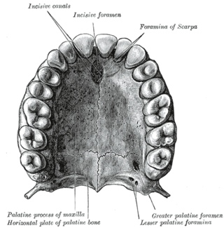
In the human mouth, the incisive foramen is the opening of the incisive canals on the hard palate immediately behind the incisor teeth. It gives passage to blood vessels and nerves. The incisive foramen is situated within the incisive fossa of the maxilla.

The infratemporal fossa is an irregularly shaped cavity that is a part of the skull. It is situated below and medial to the zygomatic arch. It is not fully enclosed by bone in all directions. It contains superficial muscles, including the lower part of the temporalis muscle, the lateral pterygoid muscle, and the medial pterygoid muscle. It also contains important blood vessels such as the middle meningeal artery, the pterygoid plexus, and the retromandibular vein, and nerves such as the mandibular nerve (CN V3) and its branches.

The posterior ethmoidal nerve is a nerve of the head. It is a branch of the nasociliary nerve (itself a branch of the ophthalmic nerve (CN V1)). It provides sensory innervation to the sphenoid sinus and ethmoid sinus, and part of the dura mater in the anterior cranial fossa.

The infraorbital nerve is a branch of the maxillary nerve. It arises in the pterygopalatine fossa. It passes through the inferior orbital fissure to enter the orbit. It travels through the orbit, then enters and traverses the infraorbital canal, exiting the canal at the infraorbital foramen to reach the face. It provides sensory innervation to the skin and mucous membranes around the middle of the face.

The posterior auricular nerve is a nerve of the head. It is a branch of the facial nerve. It communicates with branches from the vagus nerve, the great auricular nerve, and the lesser occipital nerve. Its auricular branch supplies the posterior auricular muscle, the intrinsic muscles of the auricle, and gives sensation to the auricle. Its occipital branch supplies the occipitalis muscle.
The zygomatic branches of the facial nerve (malar branches) are nerves of the face. They run across the zygomatic bone to the lateral angle of the orbit. Here, they supply the orbicularis oculi muscle, and join with filaments from the lacrimal nerve and the zygomaticofacial branch of the maxillary nerve (CN V2).
Inferior alveolar nerve block is a nerve block technique which induces anesthesia (numbness) in the areas of the mouth and face innervated by one of the inferior alveolar nerves which are paired on the left and right side. These areas are the skin and mucous membranes of the lower lip, the skin of the chin, the lower teeth and the labial gingiva of the anterior teeth, all unilaterally to the midline of the side on which the block is administered. However, depending on technique, the long buccal nerve may not be anesthetized by an IANB and therefore an area of buccal gingiva adjacent to the lower posterior teeth will retain normal sensation unless that nerve is anesthetized separately, via a (long) buccal nerve block. The inferior alveolar nerve is a branch of the mandibular nerve, the third division of the trigeminal nerve. This procedure attempts to anaesthetise the inferior alveolar nerve prior to it entering the mandibular foramen on the medial surface of the mandibular ramus.
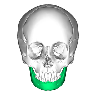
In jawed vertebrates, the mandible, lower jaw, or jawbone is a bone that makes up the lower – and typically more mobile – component of the mouth.

