N. fowleri is typically found in warm bodies of fresh water, such as ponds, lakes, rivers and hot springs. It is found in an amoeboid, temporary flagellate stage or microbial cyst in soil, poorly maintained municipal water supplies, water heaters, near warm-water discharges of industrial plants and in poorly chlorinated or unchlorinated swimming pools. There is no evidence of it living in salt water. As the disease is rare, it is often not considered during diagnosis.[citation needed]
Although infection occurs very rarely,[1] it almost inevitably results in death.[7][8] Of the 128 naegleriasis US cases in the half-century to 2016, only two survived.[9]
Signs and symptoms
Onset of symptoms begins one to twelve days following exposure (with a median of five).[6] Initial symptoms include changes in taste and smell, headache, fever, nausea, vomiting, back pain,[10] and a stiff neck. Secondary symptoms are also meningitis-like including confusion, hallucinations, lack of attention, ataxia, cramp and seizures. After the start of symptoms, the disease progresses rapidly, with death usually occurring anywhere from one to eighteen days later (with a median of five),[11] although it can take longer. In 2013, a man in Taiwan died 25 days after being infected by Naegleria fowleri.[12]
It affects healthy children or young adults who have recently been exposed to bodies of fresh water.[3] Scientists speculate that lower age groups are at a higher risk of contracting the disease because adolescents have a more underdeveloped and porous cribriform plate, through which the amoeba travels to reach the brain.[5]
Cause
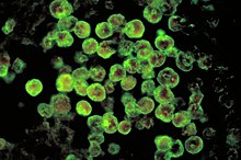
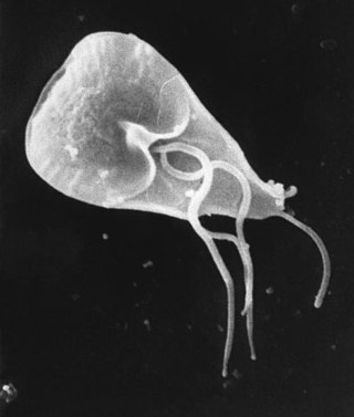
Naegleria fowleri
N. fowleri invades the central nervous system via the nose, specifically through the olfactory mucosa of the nasal tissues. This usually occurs as the result of the introduction of water that has been contaminated with N. fowleri into the nose during activities such as swimming, bathing or nasal irrigation.[13]
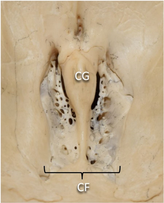
The amoeba follows the olfactory nerve fibers through the cribriform plate of the ethmoid bone into the skull. There, it migrates to the olfactory bulbs and subsequently other regions of the brain, where it feeds on the nerve tissue. The organism then begins to consume cells of the brain, piecemeal through trogocytosis,[14] by means of an amoebostome, a unique actin-rich sucking apparatus extended from its cell surface.[15] It then becomes pathogenic, causing primary amoebic meningoencephalitis (PAM or PAME).[citation needed]
Primary amoebic meningoencephalitis presents symptoms similar to those of bacterial and viral meningitis. Upon abrupt disease onset, a plethora of problems arise. Endogenous cytokines, which release in response to pathogens, affect the hypothalamus' thermoregulatory neurons and cause a rise in body temperature.[16] Additionally, cytokines may act on the vascular organ of the lamina terminalis, leading to the synthesis of prostaglandin (PG) E2 which acts on the hypothalamus, resulting in an increase in body temperature.[17] Also, the release of cytokines and exogenous exotoxins coupled with an increase in intracranial pressure stimulate nociceptors in the meninges[16] creating pain sensations.
The release of cytotoxic molecules in the central nervous system results in extensive tissue damage and necrosis, such as damage to the olfactory nerve through lysis of nerve cells and demyelination.[18] Specifically, the olfactory nerve and bulbs become necrotic and hemorrhagic.[19] Spinal flexion leads to nuchal rigidity, or stiff neck, due to the stretching of the inflamed meninges.[16] The increase in intracranial pressure stimulates the area postrema to create nausea sensations which may lead to brain herniation and damage to the reticular formation.[16] Ultimately, the increase in cerebrospinal fluid from inflammation of the meninges increases intracranial pressure and leads to the destruction of the central nervous system. Although the exact pathophysiology behind the seizures caused by PAM is unknown, scientists speculate that the seizures arise from altered meningeal permeability[16] caused by increased intracranial pressure.
Naegleria fowleri propagates in warm, stagnant bodies of fresh water (typically during the summer months), and enters the central nervous system after insufflation of infected water by attaching itself to the olfactory nerve.[3] It then migrates through the cribriform plate and into the olfactory bulbs of the forebrain,[21] where it multiplies itself greatly by feeding on nerve tissue.
Diagnosis
N. fowleri can be grown in several kinds of liquid axenic media or on non-nutrient agar plates coated with bacteria. Escherichia coli can be used to overlay the non-nutrient agar plate and a drop of cerebrospinal fluid sediment is added to it. Plates are then incubated at 37°C and checked daily for clearing of the agar in thin tracks, which indicate the trophozoites have fed on the bacteria.[22]
Detection in water is performed by centrifuging a water sample with E. coli added, then applying the pellet to a non-nutrient agar plate. After several days, the plate is microscopically inspected and Naegleria cysts are identified by their morphology. Final confirmation of the species' identity can be performed by various molecular or biochemical methods.[23]
Confirmation of Naegleria presence can be done by a so-called flagellation test, where the organism is exposed to a hypotonic environment (distilled water). Naegleria, in contrast to other amoebae, differentiates within two hours into the flagellate state. Pathogenicity can be further confirmed by exposure to high temperature (42°C): Naegleria fowleri is able to grow at this temperature, but the nonpathogenic Naegleria gruberi is not.[citation needed]
Prevention
Michael Beach, a recreational waterborne illness specialist for the Centers for Disease Control and Prevention, stated in remarks to the Associated Press that wearing of nose clips to prevent insufflation of contaminated water would be effective protection against contracting PAM, noting that "You'd have to have water going way up in your nose to begin with".[24]
Advice stated in the press release from Taiwan's Centers for Disease Control recommended people prevent fresh water from entering the nostrils and avoid putting their heads down into fresh water or stirring mud in the water with feet. When starting to suffer from fever, headache, nausea, or vomiting subsequent to any kind of exposure to fresh water, even in the belief that no fresh water has traveled through the nostrils, people with such conditions should be carried to hospital quickly and make sure doctors are well-informed about the history of exposure to fresh water.[25]
Treatment
On the basis of the laboratory evidence and case reports, heroic doses[26] of amphotericin B have been the traditional mainstay of PAM treatment since the first reported survivor in the United States in 1982.[5]
Treatment has often also used combination therapy with multiple other antimicrobials in addition to amphotericin, such as fluconazole, miconazole, rifampicin and azithromycin. They have shown limited success only when administered early in the course of an infection.[27]
While the use of rifampicin has been common, including in all four North American cases of survival, its continued use has been questioned.[5] It only has variable activity in vitro and it has strong effects on the therapeutic levels of other antimicrobials used by inducing cytochrome p450 pathways.[5] Fluconazole is commonly used as it has been shown to have synergistic effects against naegleria when used with amphotericin in vitro.[5]
As of 2015,[update] there was no data on how well miltefosine is able to reach the central nervous system.[5]As of 2015[update] the U.S. CDC offered miltefosine to doctors for the treatment of free-living amoebas including naegleria.[4]
Nevertheless in 2013–2016, three successfully treated cases in the United States utilized the medication miltefosine.[4] In one of the cases, a 12-year-old female, was given miltefosine and targeted temperature management to manage cerebral edema that is secondary to the infection. She survived with no neurological damage. The targeted temperature management commingled with early diagnosis and the miltefosine medication has been attributed with her survival. On the other hand, the other survivor, an 8-year-old male, was diagnosed several days after symptoms appeared and was not treated with targeted temperature management; however, he was administered the miltefosine. He suffered what is likely permanent neurological damage.[4] In 2016, a 16-year-old boy also survived PAM. He was treated with the same protocols of the 12-year-old girl in 2013. He recovered making a near complete neurological recovery; however, he has stated that learning has been more difficult for him since contracting the disease.[4][28]
In 2018, a 10-year-old girl in the Spanish city of Toledo became the first person to have PAM in Spain, and was successfully treated using intravenous and intrathecal amphotericin B.[29]
A 2023 study has showed that the treatment (with usage of benzoxaboroles) of infected mice significantly prolonged survival and showed a 28% cure rate without relapse.[30][31]
Prognosis
This section needs to be updated. Please help update this article to reflect recent events or newly available information.(November 2019)
Since its first description in the 1960s, only seven people worldwide have been reported to have survived PAM out of 450 cases diagnosed, implying a fatality rate of about 98.5%.[3] The survivors include four in the United States, one in Mexico and one in Spain. One of the US survivors had brain damage that is likely permanent, but there are two documented surviving cases in the United States who made a full recovery with no neurological damage; they were both treated with the same protocols.
There is also a fourth survivor in the United States. However, he had a different strain.[4][5]
Epidemiology
The disease is rare and highly lethal: there had only been 381 cases as of 2018.[update][32] Drug treatment research at Aga Khan University in Pakistan has shown that in vitro drug susceptibility tests with some FDA approved drugs used for non-infectious diseases (digoxin and procyclidine were shown to be most effective of the drugs studied) have proved to kill Naegleriafowleri with an amoebicidal rate greater than 95%.[33] The same source has also proposed a device for drug delivery via the transcranial route to the brain.[34]
In the US, the most common states with cases reported of PAM from N. fowleri are the southern states, with Texas and Florida having the highest prevalence. The most commonly affected age group is 5–14-year olds (those who play in water).[35] The number of cases of infection could increase due to climate change, which was posited as the reason for three cases in Minnesota in 2010, 2012, and 2015.[36][37]
As of 2013,[update] the numbers of reported cases were expected to increase simply because of better-informed diagnoses being made both in ongoing cases and in autopsy findings.[38]
History
In 1899, Franz Schardinger first discovered and documented an amoeba he called Amoeba gruberi that could transform into a flagellate.[39] The genus Naegleria was established by Alexis Alexeieff in 1912, who grouped the flagellate amoeba. He coined the term Naegleria after Kurt Nägler, who researched amoebae.[40] It was not until 1965 that doctors Malcolm Fowler and Rodney F. Carter in Adelaide, Australia, reported the first four-human cases of amoebic meningoencephalitis. These cases involved four Australian children, one in 1961 and the rest in 1965, all of whom had succumbed to the illness.[41][42][43] Their work on amebo-flagellates has provided an example of how a protozoan can effectively live both freely in the environment, and in a human host.[44]
In 1966, Butt termed the infection resulting from N. fowleri primary amoebic meningoencephalitis (PAM) to distinguish this central nervous system (CNS) invasion from other secondary invasions made by other amoebae such as Entamoeba histolytica.[44] A retrospective study determined the first documented case of PAM possibly occurred in Britain in 1909.[42] In 1966, four cases were reported in the US. By 1968 the causative organism, previously thought to be a species of Acanthamoeba or Hartmannella, was identified as Naegleria. This same year, occurrence of sixteen cases over a period of three years (1962–1965) was reported in Ústí nad Labem, Czechoslovakia.[45] In 1970, Carter named the species of amoeba N. fowleri, after Malcolm Fowler.[46][47]
Society and culture
Naegleria fowleri is also known as the "brain-eating amoeba". The term has also been applied to Balamuthia mandrillaris, causing some confusion between the two; Balamuthia mandrillaris is unrelated to Naegleria fowleri, and causes a different disease called granulomatous amoebic encephalitis. Unlike naegleriasis, which is usually seen in people with normal immune function, granulomatous amoebic encephalitis is usually seen in people with poor immune function, such as those with HIV/AIDS or leukemia.[48]
Naegleriasis was the topic in Season 2 of the medical mystery drama House, M.D. in the two-part episode titled "Euphoria".[49][50] It is also the topic of the episode "39 Differences" of season 6 of The Good Doctor.[citation needed]
Research
The U.S. National Institutes of Health budgeted $800,000 for research on the disease in 2016.[51]Phenothiazines have been tested in vitro and in animal models of PAM.[52] Improving case detection through increased awareness, reporting, and information about cases might enable earlier detection of infections, provide insight into the human or environmental determinants of infection, and allow improved assessment of treatment effectiveness.[3]
Entamoeba histolytica is an anaerobic parasitic amoebozoan, part of the genus Entamoeba. Predominantly infecting humans and other primates causing amoebiasis, E. histolytica is estimated to infect about 35-50 million people worldwide. E. histolytica infection is estimated to kill more than 55,000 people each year. Previously, it was thought that 10% of the world population was infected, but these figures predate the recognition that at least 90% of these infections were due to a second species, E. dispar. Mammals such as dogs and cats can become infected transiently, but are not thought to contribute significantly to transmission.
Free-living amoebae are a group of protozoa that are important causes of infectious disease in humans and animals.
Naegleria is a genus consisting of 47 described species of protozoa often found in warm aquatic environments as well as soil habitats worldwide. It has three life cycle forms: the amoeboid stage, the cyst stage, and the flagellated stage, and has been routinely studied for its ease in change from amoeboid to flagellated stages. The Naegleria genera became famous when Naegleria fowleri, the causative agent of the usually fatal human and animal disease primary amoebic meningoencephalitis (PAM), was discovered in 1965. Most species in the genus, however, are incapable of causing disease.
Nasal irrigation is a personal hygiene practice in which the nasal cavity is washed to flush out mucus and debris from the nose and sinuses, in order to enhance nasal breathing. Nasal irrigation can also refer to the use of saline nasal spray or nebulizers to moisten the mucous membranes.
In mammalian anatomy, the cribriform plate, horizontal lamina or lamina cribrosa is part of the ethmoid bone. It is received into the ethmoidal notch of the frontal bone and roofs in the nasal cavities. It supports the olfactory bulb, and is perforated by olfactory foramina for the passage of the olfactory nerves to the roof of the nasal cavity to convey smell to the brain. The foramina at the medial part of the groove allow the passage of the nerves to the upper part of the nasal septum while the foramina at the lateral part transmit the nerves to the superior nasal concha.
Meningoencephalitis, also known as herpes meningoencephalitis, is a medical condition that simultaneously resembles both meningitis, which is an infection or inflammation of the meninges, and encephalitis, which is an infection or inflammation of the brain tissue.
Balamuthia mandrillaris is a free-living amoeba that causes the rare but deadly neurological condition granulomatous amoebic encephalitis (GAE). B. mandrillaris is a soil-dwelling amoeba and was first discovered in 1986 in the brain of a mandrill that died in the San Diego Wild Animal Park.
Granulomatous amoebic encephalitis (GAE) is a rare, often fatal, subacute-to-chronic central nervous system disease caused by certain species of free-living amoebae of the genera Acanthamoeba, Balamuthia and Sappinia. The term is most commonly used with Acanthamoeba. In more modern references, the term "balamuthia amoebic encephalitis" (BAE) is commonly used when Balamuthia mandrillaris is the cause.
Miltefosine, sold under the trade name Impavido among others, is a medication mainly used to treat leishmaniasis and free-living amoeba infections such as Naegleria fowleri and Balamuthia mandrillaris. This includes the three forms of leishmaniasis: cutaneous, visceral and mucosal. It may be used with liposomal amphotericin B or paromomycin. It is taken by mouth.
The Florida Department of Health in Orange County is the county health department in Orange County, Florida, formerly known as Orange County Health Department, charged with protecting the health and safety of visitors and residents of that county. The estimated daytime population of Orange County is 1.5 million people. Orange County has an estimated 55 million visitors per year including the major theme parks of Walt Disney World, Universal Orlando, and SeaWorld. The county seat is Orlando, Florida.
The term "brain-eating amoeba" has been used to refer to several microorganisms:
Protozoan infections are parasitic diseases caused by organisms formerly classified in the kingdom Protozoa. These organisms are now classified in the supergroups Excavata, Amoebozoa, Harosa, and Archaeplastida. They are usually contracted by either an insect vector or by contact with an infected substance or surface.
Amoebiasis, or amoebic dysentery, is an infection of the intestines caused by a parasitic amoeba Entamoeba histolytica. Amoebiasis can be present with no, mild, or severe symptoms. Symptoms may include lethargy, loss of weight, colonic ulcerations, abdominal pain, diarrhea, or bloody diarrhea. Complications can include inflammation and ulceration of the colon with tissue death or perforation, which may result in peritonitis. Anemia may develop due to prolonged gastric bleeding.
Sappinia diploidea is a free-living amoeba species.
Amoebic brain abscess is an affliction caused by the anaerobic parasitic protist Entamoeba histolytica. It is extremely rare; the first case being reported in 1849. Brain abscesses resulting from Entamoeba histolytica are difficult to diagnose and very few case reports suggest complete recovery even after the administration of appropriate treatment regimen.
Sappinia is a genus of heterotrophic, lobose amoebae within the family Thecamoebidae. A defining feature of Sappinia, which separates it from its sister genus Thecamoeba, is the presence of two closely apposed nuclei with a central, flattened connection. Sappinia species have two life cycle stages: a trophozoite and a cyst. Up until 2015, only two species had been discovered, Sappinia pedata and Sappinia diploidea. Sequencing of the small subunit rRNA of a particular isolate from a sycamore tree revealed a new species, Sappinia platani.Sappinia species were once thought to be coprozoic, as the first strains were isolated from animal dung. More research has shown that they are typical free-living amoebae, and can be found worldwide in soil, plant litter, and standing decaying plants, as well as freshwater ponds. In 2001, the first and only case of human pathogenesis in Sappinia was confirmed. The patient was a non-immunocompromised 38-year-old male who presented signs of amoebic encephalitis and who patient made a full recovery after treatment with several antimicrobials. The CDC initially classified the causative agent as S. diploidea based on morphological characteristics, but in 2009, Qvarnstrom et al. used molecular data to confirm that the true causative agent was S. pedata.
An amoeba, often called an amoeboid, is a type of cell or unicellular organism with the ability to alter its shape, primarily by extending and retracting pseudopods. Amoebae do not form a single taxonomic group; instead, they are found in every major lineage of eukaryotic organisms. Amoeboid cells occur not only among the protozoa, but also in fungi, algae, and animals.
Naegleria fowleri, also known as the brain-eating amoeba, is a species of the genus Naegleria. It belongs to the phylum Percolozoa and is technically classified as an amoeboflagellate excavate, rather than a true amoeba. This free-living microorganism primarily feeds on bacteria but can become pathogenic in humans, causing an extremely rare, sudden, severe, and usually fatal brain infection known as naegleriasis or primary amoebic meningoencephalitis (PAM).
Amoebic encephalitis or amoebic meningoencephalitis may refer to several potentially fatal diseases that are infections of the central nervous system by free-living amoebae, often referred to in the media as a "brain-eating amoeba" infection:
↑ "Safe Ritual Nasal Rinsing"(PDF). Centers for Disease Control and Prevention. Archived(PDF) from the original on 18 June 2019. Retrieved 28 September 2020.
1 2 3 4 5 Montgomery, Katherine (22 October 2012). "Meningitis". McMaster Pathophysiology Review. Archived from the original on 12 December 2017. Retrieved 29 March 2020.
↑ Cervantes-Sandoval I, Serrano-Luna Jde J, García-Latorre E, Tsutsumi V, Shibayama M (September 2008). "Characterization of brain inflammation during primary amoebic meningoencephalitis". Parasitol. Int. 57 (3): 307–13. doi:10.1016/j.parint.2008.01.006. PMID18374627.
↑ Donald C. Lehman; Mahon, Connie; Manuselis, George (2006). Textbook of Diagnostic Microbiology (3rded.). Philadelphia: Saunders. ISBN978-1-4160-2581-8.[pageneeded]
↑ Bauman, Robert W. (2009). "Microbial Diseases of the Nervous System and Eyes". Microbiology, With Diseases by Body System (2nded.). San Francisco: Pearson Education. p.617.
↑ "Number of Case-Reports of Primary Amebic Meningoencephalitis Caused by Naegleria Fowleri (N=133) by State of Exposure*— United States, 1962–2014".CDC.gov, CDC, www.cdc.gov/parasites/naegleria/pdf/naegleria-state-map-2014.pdf.
↑ Kanwal Farooqi M, Ali S, Ahmed SS (May 2013). "The paradox of primary amoebic meningoencephalitis—a rare disease, but commonly misdiagnosed". J Pak Med Assoc. 63 (5): 667. PMID23758009.
This page is based on this Wikipedia article Text is available under the CC BY-SA 4.0 license; additional terms may apply. Images, videos and audio are available under their respective licenses.