Neurothekeoma

Last updated
Neurothekeoma
Other namesBizarre cutaneous neurofibroma, myxoma of nerve sheath, pacinian neurofibroma
Neurothekeoma2.JPG
Neurothekeoma histology slide
Specialty Oncology   OOjs UI icon edit-ltr-progressive.svg

A Neurothekeoma (NT) is a type of rare benign cutaneous tumor that usually develops on the head and neck. They often occur in the second and early third decades of life and tend to afflict women more frequently than men. [1] First described by Richard L Gallager and Elson B. Helwig, who proposed the term in order to reflect the presumed origin of the lesion from nerve sheath. [2] Microscopically, the lesions described closely resembled the tumor, "nerve sheath myxoma (NSM)", an entity first described by Harkin and Reed. [3] The latter had, through the years, been variously described as bizarre cutaneous neurofibroma, myxoma of nerve sheath, and pacinian neurofibroma. [4]

Contents

Clinically, neurothekeomas present as a solitary nodule of the skin. The most common sites of occurrence are the head and neck and the extremities. The lesions range in size from about 0.5 cm to more than 3 cm. The average patient age is about 25 years, but neurothekeomas may occur at any age. Women are affected about twice often; the male to female ratio is approximately 1:2. [5]

Microscopically, neurothekeoma consists of closely aggregated bundles or fascicles of spindle-shaped cells. The fascicles may or may not have a myxoid background. [6]

Since the time of their first description, it has been reported that neurothekeomas are likely not of nerve sheath origin, as implied by the term. [5] Consequently, neurothekeoma and nerve sheath myxoma are likely not related histogenetically, although they are similar in appearance and in behavior. [5] Based mostly on the quantity of myxoid matrix present, neurothekeomas can have a variety of histologic characteristics, including myxoid, cellular, or mixed-type. The myxoid variety of neurothekeoma has inadvertently included nerve sheath myxoma because to similarity in clinical presentation and histology. However, it appears that the neurothekeoma that Barnhill and Mihm reported in 1990 is a separate and distinct entity from true nerve sheath myxoma. [7] It has been proposed that neurothekeomas are derived from fibrohistiocytic cells rather than the peripheral nerve sheath. [8] [9] [10] [11] Despite having a different histologic appearance, neurothekeomas do not respond with the S100 protein while nerve sheath myxoma do (myxoid, mixed, or cellular).

Difference from nerve sheath myxoma

A rare neuro-ectodermal tumor known as a benign nerve sheath myxoma was originally identified in 1969. [3] In contrast, a cellular variation known as neurothekeoma was characterized in 1980 by Gallager and Helwig. [2] Later on, it was common to use both neurothekeoma and nerve sheath myxoma interchangeably. [12] [13] In 2011, Sheth et al. used microarray analysis to distinguish between neurothekeoma and nerve sheath myxoma based on the genetic expression of the cells. [14] Nerve sheath myxoma is more frequent among young people without gender preference. Between 30 and 40 years of age is when the incidence peaks. [15] The head and neck are rarely affected by the tumor, which is more usually seen in the upper limb. [16] Young women are twice as likely as males to get neurothekeoma, and it usually affects the head and neck rather than the hands. [9] While neurothekeoma can afflict females from infancy to old age with a peak incidence in the twenties, nerve sheath myxoma has a peak incidence in the fourth decade. [17]

Clinical features

Lesions are often a singular subcutaneous dome-shaped papule or nodule measuring less than 2 cm and may be skin-colored, pink to tan, or red to brown. Most lesions are asymptomatic although some may present with pain upon pressure. The lesions tend to grow slowly and are usually superficially located, with rare, deeper involvement of subcutaneous fat or skeletal muscle. The head and neck are the most common locations, but neurothekeomas may also be seen on the shoulder or upper extremities, while involvement of the mucosa is rare. [18] [8] [9] [19] [20] [21] [22]

Treatment

Surgical excision is the treatment of choice. [23] There have been reports of neurothekeomas exhibiting atypical features, raising concern for their potential for aggression even though the majority of these tumors are small (less than 1 cm), have relatively bland histology, little to no cytologic atypia, and only minimal extension into surrounding fat or skeletal muscle. Clinical size more than 1 cm, pleomorphism and enhanced mitotic activity in the cytology, infiltration into skeletal muscle or subcutaneous fat, and vascular invasion are reported as unusual characteristics. Despite the presence of unusual characteristics, documented recurrence rates after total surgical excision are very low. [18] [24] [25]

See also

Related Research Articles

<span class="mw-page-title-main">Liposarcoma</span> Medical condition

Liposarcomas are the most common subtype of soft tissue sarcomas, accounting for at least 20% of all sarcomas in adults. Soft tissue sarcomas are rare neoplasms with over 150 different histological subtypes or forms. Liposarcomas arise from the precursor lipoblasts of the adipocytes in adipose tissues. Adipose tissues are distributed throughout the body, including such sites as the deep and more superficial layers of subcutaneous tissues as well as in less surgically accessible sites like the retroperitoneum and visceral fat inside the abdominal cavity.

<span class="mw-page-title-main">Hemangioendothelioma</span> Medical condition

Hemangioendotheliomas are a family of vascular neoplasms of intermediate malignancy.

<span class="mw-page-title-main">Schwannoma</span> Medical condition

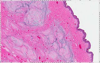
A schwannoma is a usually benign nerve sheath tumor composed of Schwann cells, which normally produce the insulating myelin sheath covering peripheral nerves.

<span class="mw-page-title-main">Granular cell tumor</span> Medical condition

Granular cell tumor is a tumor that can develop on any skin or mucosal surface, but occurs on the tongue 40% of the time.

<span class="mw-page-title-main">Aggressive angiomyxoma</span> Medical condition

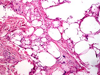
Angiomyxoma is a myxoid tumor involving the blood vessels.

<span class="mw-page-title-main">Hibernoma</span> Medical condition

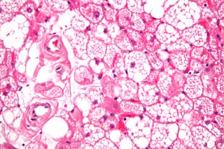
A hibernoma is a benign neoplasm of vestigial brown fat. They were first described under the name ‘pseudolipoma’ by the German physician H. Merkel in 1906 and the term hibernoma was proposed by the French anatomist Louis Gery in 1914 because of its resemblance to brown fat in hibernating animals.

Plexiform fibrohistiocytic tumor is a rare tumor that arises primarily on the upper extremities of children and young adults.

<span class="mw-page-title-main">Atypical fibroxanthoma</span> Medical condition

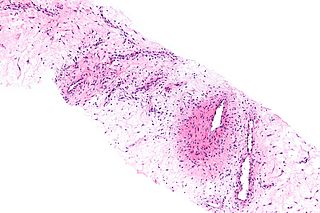
Atypical fibroxanthoma (AFX) of the skin is a low-grade malignancy related to malignant fibrous histiocytoma, which it resembles histologically. Atypical fibroxanthoma manifests as a hard, pink or red papule or nodule that grows over the course of several months and may bleed or ulcerate. They typically occur on the head and neck. Atypical fibroxanthoma is usually asymptomatic.

<span class="mw-page-title-main">Spindle cell lipoma</span> Medical condition

Spindle cell lipoma is an asymptomatic, slow-growing subcutaneous tumor that has a predilection for the posterior back, neck, and shoulders of older men.

<span class="mw-page-title-main">Pleomorphic lipoma</span> Medical condition

Pleomorphic lipomas, like spindle-cell lipomas, occur for the most part on the backs and necks of elderly men, and are characterized by floret giant cells with overlapping nuclei.

<span class="mw-page-title-main">Cutaneous myxoma</span> Medical condition

A cutaneous myxoma, or superficial angiomyxoma, consists of a multilobulated myxoid mass containing stellate or spindled fibroblasts with pools of mucin forming cleft-like spaces. There is often a proliferation of blood vessels and an inflammatory infiltrate. Staining is positive for vimentin, negative for cytokeratin and desmin, and variable for CD34, Factor VIIIa, SMA, MSA and S-100.

A superficial acral fibromyxoma is a type of myxoma and is a rare cutaneous condition characterized by a mesenchymal neoplasm that typically occurs on the digits of middle-aged adults.

<span class="mw-page-title-main">Low-grade fibromyxoid sarcoma</span> Medical condition

Low-grade fibromyxoid sarcoma (LGFMS) is a rare type of low-grade sarcoma first described by H. L. Evans in 1987. LGFMS are soft tissue tumors of the mesenchyme-derived connective tissues; on microscopic examination, they are found to be composed of spindle-shaped cells that resemble fibroblasts. These fibroblastic, spindle-shaped cells are neoplastic cells that in most cases of LGFMS express fusion genes, i.e. genes composed of parts of two different genes that form as a result of mutations. The World Health Organization (2020) classified LGFMS as a specific type of tumor in the category of malignant fibroblastic and myofibroblastic tumors.

Rhabdomyoma is a benign mesenchymal tumor of skeletal muscle, separated into two major categories based on site: Cardiac and extracardiac. They are further separated by histology: fetal, juvenile (intermediate), and adult types. Genital types are recognized, but are often part of either the fetal or juvenile types. The fetal type is thought to recapitulate immature skeletal muscle at about week six to ten of gestational development.

An intraneural perineurioma is a rare benign tumor within the sheath of a single nerve that grows but typically does not recur or metastasize. These lesions are only composed of perineurial cells, cloned from a single cell. They are distinct from schwannoma and neurofibroma.

<span class="mw-page-title-main">Myxoid tumor</span> Connective tissue tumor composed of clear, mucoid substance

A myxoid tumor is a connective tissue tumor with a "myxoid" background, composed of clear, mucoid substance.

Ectomesenchymal chondromyxoid tumor (ECT) is a benign intraoral tumor with presumed origin from undifferentiated (ecto)mesenchymal cells. There are some who think it is a myoepithelial tumor type.

Acral myxoinflammatory fibroblastic sarcoma (AMSF), also termed myxoinflammatory fibroblastic sarcoma (MSF), is a rare, low-grade, soft tissue tumor that the World Health Organization (2020) classified as in the category of rarely metastasizing fibroblastic and myofibroblastic tumors. It is a locally aggressive neoplasm that often recurs at the site of its surgical removal. However, it usually grows slowly and in only 1–2% of cases spreads to distant tissues.

Lipofibromatosis-like neural tumor (LPF-NT) is an extremely rare soft tissue tumor first described by Agaram et al in 2016. As of mid-2021, at least 39 cases of LPF-NT have been reported in the literature. LPF-NT tumors have several features that resemble lipofibromatosis (LPF) tumors, malignant peripheral nerve sheath tumors, spindle cell sarcomas, low-grade neural tumors, peripheral nerve sheath tumors, and other less clearly defined tumors; Prior to the Agaram at al report, LPF-NTs were likely diagnosed as variants or atypical forms of these tumors. The analyses of Agaram at al and subsequent studies uncovered critical differences between LPF-NT and the other tumor forms which suggest that it is a distinct tumor entity differing not only from lipofibromatosis but also the other tumor forms.

Angiofibroma of soft tissue (AFST), also termed angiofibroma, not otherwise specified, is a recently recognized and rare disorder that was classified in the category of benign fibroblastic and myofibroblastic tumors by the World Health Organization in 2020. An AFST tumor is a neoplasm that was first described by A. Mariño-Enríquez and C.D. Fletcher in 2012.

References

  1. Barnhill RL, Mihm MC. Cellular neurothekeoma. A distinctive variant of neurothekeoma mimicking nevomelanocytic tumors. Am J Surg Pathol. 1990 Feb;14(2):113-20
  2. 1 2 Gallager RL, Helwig EB (December 1980). "Neurothekeoma-a benign cutaneous tumor of neural origin". American Journal of Clinical Pathology. 74 (6): 759–764. doi:10.1093/ajcp/74.6.759. PMID   7446487.
  3. 1 2 Harkin, James C.; Richard J. Reed (1969). Tumors of the Peripheral Nervous System. Armed Forces Institute of Pathology Atlas of Tumor Pathology 2Nd series. Washington, D.C.: Armed Forces Institute of Pathology. Retrieved 2024-05-30.
  4. Pulitzer DR, Reed RJ (October 1985). "Nerve-sheath myxoma (perineurial myxoma)". American Journal of Dermatopathology. 7 (5): 407–421. doi:10.1097/00000372-198510000-00003. PMID   4091218. S2CID   12507923.
  5. 1 2 3 Fetsch JF, Laskin WB, Hallman JR, Lupton GP, Miettinen M (July 2007). "Neurothekeoma: an analysis of 178 tumors with detailed immunohistochemical data and long-term patient follow-up information". American Journal of Surgical Pathology. 31 (7): 1103–1114. doi:10.1097/pas.0b013e31802d96af. PMID   17592278. S2CID   13038590.
  6. Reed, Richard J.; Pulitzer, Donald R. (2008), "Chapter 35: Tumors of Neural Tissue", in Elder D; et al. (eds.), Lever's Histopathology of the Skin (Ninth ed.), Philadelphia: Lippincott Raven, ISBN   978-0781773638
  7. Almeida TFA, Verli FD, Dos Santos CRR, Falci SGM, Almeida LY, Almeida LKY, Mesquita ATM, León JE. Multiple Desmoplastic Cellular Neurothekeomas in Child: Report of the First Oral Case and Review of the Literature. Head Neck Pathol. 2018 Mar;12(1):75-81.
  8. 1 2 Fetsch JF, Laskin WB, Hallman JR, Lupton GP, Miettinen M. Neurothekeoma: an analysis of 178 tumors with detailed immunohistochemical data and long-term patient follow-up information. Am J Surg Pathol. 2007 Jul;31(7):1103-14.
  9. 1 2 3 Hornick JL, Fletcher CD. Cellular neurothekeoma: detailed characterization in a series of 133 cases. Am J Surg Pathol. 2007 Mar;31(3):329-40.
  10. Page RN, King R, Mihm MC, Googe PB. Microphthalmia transcription factor and NKI/C3 expression in cellular neurothekeoma. Mod Pathol. 2004 Feb;17(2):230-4.
  11. Yun SJ, Park HS, Lee JB, Kim SJ, Lee SC, Won YH. Myxoid Cellular Neurothekeoma: A New Entity of S100-Negative, CD68-Positive Myxoid Neurothekeoma. Ann Dermatol. 2014 Aug;26(4):510-3.
  12. A.M. Cimino-Mathews, Peripheral nerve sheath tumors, Surgical Pathol. 4 (2011) 761–782
  13. K.M. Benbenisty, A. Andea, J. Metcalf, et al., Atypical cellular neurothekeoma treated with Mohs micrographic surgery, Dermatol. Surg. 32 (2006) 582–587
  14. S. Sheth, X. Li, S. Binder, S.M. Dry, Differential gene expression profiles of neurothekeomas and nerve sheath myxomas by microarray analysis, Mod. Pathol. 24 (2011) 343–354.
  15. I. Ahmed, J. Rawat, S. Singh, A. Sharma, A. Pandey, M. Goel, Neurothekeoma: a sacrococcygeal tumor in a child, J. Pediatr. Surg. 45 (2010) 1037–1039.
  16. S.A. Fanto, E. Fanto, Neurothekeoma of the median nerve: case report, J. Hand Surg. 37A (2012) 1184–1186.
  17. P.W. Allen, Myxoma is not a single entity: a review of the concept of myxoma, Ann. Diagn. Pathol. 4 (2000) 99–123
  18. 1 2 Kao, Erica Y.; Kernig, Mikelle L. (2022). "Neurothekeoma". StatPearls. StatPearls Publishing. PMID   30137810. CC-BY icon.svg Text was copied from this source, which is available under a Creative Commons Attribution 4.0 International License.
  19. Ward JL, Prieto VG, Joseph A, Chevray P, Kronowitz S, Sturgis EM. Neurothekeoma. Otolaryngol Head Neck Surg. 2005 Jan;132(1):86-9.
  20. Rosati LA, Fratamico FC, Eusebi V. Cellular neurothekeoma. Appl Pathol. 1986;4(3):186-91.
  21. Stratton J, Billings SD. Cellular neurothekeoma: analysis of 37 cases emphasizing atypical histologic features. Mod Pathol. 2014 May;27(5):701-10.
  22. Emami N, Zawawi F, Ywakim R, Nahal A, Daniel SJ. Oral cellular neurothekeoma. Case Rep Otolaryngol. 2013;2013:935435.
  23. Boukovalas S, Rogers H, Boroumand N, Cole EL. Cellular Neurothekeoma: A Rare Tumor with a Common Clinical Presentation. Plast Reconstr Surg Glob Open. 2016 Aug;4(8):e1006.
  24. Busam KJ, Mentzel T, Colpaert C, Barnhill RL, Fletcher CD. Atypical or worrisome features in cellular neurothekeoma: a study of 10 cases. Am J Surg Pathol. 1998 Sep;22(9):1067-72.
  25. Wilson AD, Rigby H, Orlando A. Atypical cellular neurothekeoma--a diagnosis to be aware of. J Plast Reconstr Aesthet Surg. 2008;61(2):186-8.