Nocardia

Last updated

Nocardia
Nocardia species close-up detail.jpg
Close-up image of a Nocardia species culture on sheep blood agar
Scientific classification OOjs UI icon edit-ltr.svg
Domain: Bacteria
Phylum: Actinomycetota
Class: Actinomycetia
Order: Mycobacteriales
Family: Nocardiaceae
Genus: Nocardia
Trevisan 1889 (Approved Lists 1980)
Type species
Nocardia asteroides
(Eppinger 1891) Blanchard 1896 (Approved Lists 1980)
Species

See text.

Synonyms [1]
  • MicropolysporaLechevalier et al. 1961 (Approved Lists 1980)
  • AsteroidesPuntoni & Leonardi, 1935
  • Proactinomyces(K.Lehmann & Haag) H.L.Jensen, 1934 [2]
Nocardia as found on a brain biopsy. Nocardia Brain Autopsy.jpg
Nocardia as found on a brain biopsy.

Nocardia is a genus of weakly staining Gram-positive, catalase-positive, rod-shaped bacteria. It forms partially acid-fast beaded branching filaments (acting as fungi, but being truly bacteria). It contains a total of 85 species. Some species are nonpathogenic, while others are responsible for nocardiosis. [3] Nocardia species are found worldwide in soil rich in organic matter. In addition, they are oral microflora found in healthy human gingiva, as well as periodontal pockets. [4] Most Nocardia infections are acquired by inhalation of the bacteria or through traumatic introduction through openings in epithelial barriers.

Contents

Culture and staining

Nocardia colonies have a variable appearance, but most species appear to have aerial hyphae when viewed with a dissecting microscope, particularly when they have been grown on nutritionally limiting media. Nocardia grow slowly on nonselective culture media, and are strict aerobes with the ability to grow in a wide temperature range. Some species are partially acid-fast (meaning a less concentrated solution of sulfuric or hydrochloric acid should be used during the staining procedure) due to the presence of intermediate-length mycolic acids in their cell wall. Majority of strains possess the cord factor (trehalose 6-6' dimycolate), an important virulence factor.

Four different Nocardia species on sheep blood agar demonstrating different colouring Four different Nocardia species.jpg
Four different Nocardia species on sheep blood agar demonstrating different colouring

They are catalase positive and can grow easily on the most commonly used media with colonies becoming evident in 3–5 days. However, prolonged incubation periods (2–3 weeks) are sometimes needed.

Nocardia asteroides Nocardia asteroides.png
Nocardia asteroides

Virulence

The various species of Nocardia are pathogenic bacteria with low virulence; therefore clinically significant disease most frequently occurs as an opportunistic infection in those with a weak immune system, such as small children, the elderly, and the immunocompromised (most typically, HIV). Nocardial virulence factors are the enzymes catalase and superoxide dismutase (which inactivate reactive oxygen species that would otherwise prove toxic to the bacteria), as well as a "cord factor" (which interferes with phagocytosis by macrophages by preventing the fusion of the phagosome with the lysosome).[ citation needed ]

An important virulence factor in many pathogenic strains of Nocardia is trehalose dimycolate, which is also found in the bacterial genus Mycobacterium . This toxin can cause cachexia, a form of muscle wasting. [5]

Clinical disease and microbiological diagnosis

The most commonly encountered species are Nocardia brasiliensis, Nocardia cyriacigeorgica, Nocardia farcinica, and Nocardia nova. Nocardia asteroides is most frequently found species causing noncutaneous invasive disease. Most cases occur as an opportunistic infection in immunocompromised patients. N. brasiliensis is the most common species causing cutaneous nocardiosis. N. caviae is another species of medical interest. The genus is acid-fast to some degree, it stains only weakly Gram positive.

The most common form of human nocardial disease is a slowly progressive pneumonia, the common symptoms of which include cough, dyspnea (shortness of breath), and fever. It is not uncommon for this infection to spread to the pleura or chest wall. Pre-existing pulmonary disease, especially pulmonary alveolar proteinosis, increases the risk of contracting a Nocardia pneumonia. Every organ can be affected if a systemic spread takes place.

Nocardia species are deeply involved in the process of encephalitis as one of its main pathogenic effects. In about 25–33% of people Nocardia infection takes the form of encephalitis and/or brain abscess formation. Nocardia may also cause a variety of cutaneous infections such as actinomycetoma (especially N. brasiliensis), lymphocutaneous disease, cellulitis, and subcutaneous abscesses. [6]

Nocardia isolation from biological specimens can be performed using an agar medium enriched with yeast extract and activated charcoal (BCYE), the same used for Legionella species. Selective media for mycobacteria or fungi can also be inoculated. The most suitable specimens are the sputum, or when clinically necessary, bronchoalveolar lavage or biopsy. Further biochemical tests for species identification are not routinely performed. Serological or cutaneous tests are not available.

Treatment

In most patients with Nocardia infection, the combination of antibiotics is needed. [7] Antibiotic therapy with a sulfonamide, most commonly trimethoprim-sulfamethoxazole, is the treatment of choice. [8] People who take trimethoprim-sulfamethoxazole for other reasons, such as prevention of Pneumocystis jirovecii infection, appear to have fewer Nocardia infections, [9] although this protective effect has been considered unreliable, [10] and some studies have disputed it altogether. [11] Minocycline is usually substituted when a sulfa cannot be given; high-dose imipenem and amikacin have also been used in severe or refractory cases. [8] Linezolid appears to be highly effective against Nocardia, but it is very expensive and may cause severe adverse effects. [12]

Antibiotic therapy is continued for six months (in immunocompetent people) to a year (in immunosuppression), and may need to be continued indefinitely. [8] Proper wound care is also critical.

Genetics

Although Nocardia has interesting and important features such as production of antibiotics and aromatic compound-degrading or -converting enzymes, the genetic study of this organism has been hampered by the lack of genetic tools. However, practical NocardiaE. coli shuttle vectors have been developed recently. [13]

The genera Nocardia and Rhodococcus have been found to be closely related, supported by two conserved signature indels consisting of a one-amino-acid deletion in the alpha subunit of acetyl coenzyme A carboxylase (ACC), and a three-amino-acid insertion in a conserved region of an ATP-binding protein that are specifically shared by species from these two genera. In addition, 14 hypothetical conserved signature proteins have been identified which are unique to the genera Nocardia and Rhodococcus. [14]

Species

Nocardia comprises the following species: [1]

Nomenclature

The genus was named for Edmond Nocard (1850-1903), a French 19th-century veterinarian and biologist. [15]

The genus was circumscribed by Vittore Benedetto Antonio Trevisan di Saint-Léon in Ann. Inst. Pasteur vol.2 (Issue 6) on page 295 in 1888. [2]

Related Research Articles

<i>Salmonella</i> Genus of prokaryotes

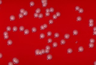
Salmonella is a genus of rod-shaped (bacillus) gram-negative bacteria of the family Enterobacteriaceae. The two known species of Salmonella are Salmonella enterica and Salmonella bongori. S. enterica is the type species and is further divided into six subspecies that include over 2,600 serotypes. Salmonella was named after Daniel Elmer Salmon (1850–1914), an American veterinary surgeon.

<i>Mycoplasma</i> Genus of bacteria

Mycoplasma is a genus of bacteria that, like the other members of the class Mollicutes, lack a cell wall around their cell membranes. Peptidoglycan (murein) is absent. This characteristic makes them naturally resistant to antibiotics that target cell wall synthesis. They can be parasitic or saprotrophic. Several species are pathogenic in humans, including M. pneumoniae, which is an important cause of "walking" pneumonia and other respiratory disorders, and M. genitalium, which is believed to be involved in pelvic inflammatory diseases. Mycoplasma species are among the smallest organisms yet discovered, can survive without oxygen, and come in various shapes. For example, M. genitalium is flask-shaped, while M. pneumoniae is more elongated, many Mycoplasma species are coccoid. Hundreds of Mycoplasma species infect animals.

<i>Pseudomonas</i> Genus of Gram-negative bacteria

Pseudomonas is a genus of Gram-negative bacteria belonging to the family Pseudomonadaceae in the class Gammaproteobacteria. The 313 members of the genus demonstrate a great deal of metabolic diversity and consequently are able to colonize a wide range of niches. Their ease of culture in vitro and availability of an increasing number of Pseudomonas strain genome sequences has made the genus an excellent focus for scientific research; the best studied species include P. aeruginosa in its role as an opportunistic human pathogen, the plant pathogen P. syringae, the soil bacterium P. putida, and the plant growth-promoting P. fluorescens, P. lini, P. migulae, and P. graminis.

<i>Micromonospora</i> Genus of bacteria

Micromonospora is a genus of bacteria of the family Micromonosporaceae. The genus name was first proposed in 1923 by Danish physician Jeppe Ørskov in an attempt to classify what at the time was considered "ray fungi" based on morphology. Members of this genus are found throughout natural soil and sediment environments, as well as in association with roots of plants of various species. The genus is well known for its ability to produce a variety of medically relevant products.

<span class="mw-page-title-main">Micromonosporaceae</span> Family of bacteria

Micromonosporaceae is a family of bacteria of the class Actinomycetia. They are gram-positive, spore-forming soil organisms that form a true mycelium.

Nocardiosis is an infectious disease affecting either the lungs or the whole body. It is due to infection by a bacterium of the genus Nocardia, most commonly Nocardia asteroides or Nocardia brasiliensis.

<i>Actinomyces</i> Genus of bacteria

Actinomyces is a genus of the Actinomycetia class of bacteria. They all are gram-positive and facultatively anaerobic, growing best under anaerobic conditions. Actinomyces species may form endospores, and while individual bacteria are rod-shaped, Actinomyces colonies form fungus-like branched networks of hyphae. The aspect of these colonies initially led to the incorrect assumption that the organism was a fungus and to the name Actinomyces, "ray fungus".

<i>Corynebacterium</i> Genus of bacteria

Corynebacterium is a genus of Gram-positive bacteria and most are aerobic. They are bacilli (rod-shaped), and in some phases of life they are, more specifically, club-shaped, which inspired the genus name.

<i>Aspergillus fumigatus</i> Species of fungus

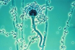
Aspergillus fumigatus is a species of fungus in the genus Aspergillus, and is one of the most common Aspergillus species to cause disease in individuals with an immunodeficiency.

<i>Pseudomonas aeruginosa</i> Species of bacterium

Pseudomonas aeruginosa is a common encapsulated, Gram-negative, aerobic–facultatively anaerobic, rod-shaped bacterium that can cause disease in plants and animals, including humans. A species of considerable medical importance, P. aeruginosa is a multidrug resistant pathogen recognized for its ubiquity, its intrinsically advanced antibiotic resistance mechanisms, and its association with serious illnesses – hospital-acquired infections such as ventilator-associated pneumonia and various sepsis syndromes.

<i>Rhodococcus</i> Genus of bacteria

Rhodococcus is a genus of aerobic, nonsporulating, nonmotile Gram-positive bacteria closely related to Mycobacterium and Corynebacterium. While a few species are pathogenic, most are benign, and have been found to thrive in a broad range of environments, including soil, water, and eukaryotic cells. Some species have large genomes, including the 9.7 megabasepair genome of Rhodococcus sp. RHA1.

The Actinomycetaceae are a family of bacteria in the order Actinomycetales that contains the medically important genus Actinomyces. These organisms are closely related to the mycobacteria, but were originally classified as fungi because they were thought to be transitional forms between bacteria and fungi.

Nocardia brasiliensis is a species of Nocardia. As with most members of Actinomycetota, they contain high guanine and cytosine content. It can cause nocardiosis.

<span class="mw-page-title-main">Pseudonocardiaceae</span> Family of bacteria

The Pseudonocardiaceae are a family of bacteria in the order Actinomycetales and the only member of the suborder Pseudonocardineae.

<span class="mw-page-title-main">Cord factor</span> Chemical compound

Cord factor, or trehalose dimycolate (TDM), is a glycolipid molecule found in the cell wall of Mycobacterium tuberculosis and similar species. It is the primary lipid found on the exterior of M. tuberculosis cells. Cord factor influences the arrangement of M. tuberculosis cells into long and slender formations, giving its name. Cord factor is virulent towards mammalian cells and critical for survival of M. tuberculosis in hosts, but not outside of hosts. Cord factor has been observed to influence immune responses, induce the formation of granulomas, and inhibit tumor growth. The antimycobacterial drug SQ109 is thought to inhibit TDM production levels and in this way disrupts its cell wall assembly.

<i>Bifidobacterium</i> Genus of bacteria

Bifidobacterium is a genus of gram-positive, nonmotile, often branched anaerobic bacteria. They are ubiquitous inhabitants of the gastrointestinal tract though strains have been isolated from the vagina and mouth of mammals, including humans. Bifidobacteria are one of the major genera of bacteria that make up the gastrointestinal tract microbiota in mammals. Some bifidobacteria are used as probiotics.

<i>Actinoplanes</i> Genus of bacteria

Actinoplanes is a genus in the family Micromonosporaceae. They have aerial mycelia and spherical, motile spores. Actinoplanes species produce the pharmaceutically important compounds valienamine, teicoplanin, and ramoplanin.

Nocardia farcinica is a species of bacteria, once thought to be associated with farcy, and a member of the genus Nocardia. This species is very similar in phenotype to Nocardia asteroides, to the degree that some isolates of N. asteroides were later found to be Nocardia farcinica.

Nocardia ignorata is a species of bacteria and a member of the genus Nocardia. Its type strain is IMMIB R-1434T.

Gordonia is a genus of gram-positive, aerobic, catalase-positive bacterium in the Actinomycetota, closely related to the Rhodococcus, Mycobacterium, Skermania, and Nocardia genera. Gordonia bacteria are aerobic,non-motile, and non-sporulating. Gordonia is from the same lineage that includes Mycobacterium tuberculosis. The genus was discovered by Tsukamura in 1971 and named after American bacteriologist Ruth Gordon. Many species are often found in the soil, while other species have been isolated from aquatic environments. Gordonia species are rarely known to cause infections in humans.

References

  1. 1 2 Euzéby JP, Parte AC. "Nocardia". List of Prokaryotic names with Standing in Nomenclature (LPSN). Retrieved June 25, 2022.
  2. 1 2 "Nocardia Trevisan, 1889". www.gbif.org. Retrieved 24 August 2022.
  3. Ryan KJ, Ray CG, eds. (2004). Sherris Medical Microbiology (4th ed.). McGraw Hill. pp. 460–2. ISBN   0-8385-8529-9.
  4. "Nocardia species". GlobalRPH. Retrieved 2023-06-28.
  5. Silva, C. L.; Tincani, I.; Filho, S. L. B.; Faccioli, L. H. (1988-06-01). "Mouse Cachexia Induced by Trehalose Dimycolate from Nocardia asteroides". Microbiology. 134 (6): 1629–1633. doi:10.1099/00221287-134-6-1629. ISSN   1350-0872.
  6. Lovecchio A, Bazzacco G, Di Bella S, Di Meo N, Luzzati R (2022-06-06). "Uncommon lymphocutaneous cellulitis after insect bite: a case report of primary cutaneous nocardiosis and literature review". Le Infezioni in Medicina. 30 (2): 285–292. doi:10.53854/liim-3002-15. PMC   9177186 . PMID   35693062.
  7. Lu SH, Qian ZW, Mou PP, Xie L (November 2020). "Clinical Nocardia species: Identification, clinical characteristics, and antimicrobial susceptibility in Shandong, China". Bosnian Journal of Basic Medical Sciences. 20 (4): 531–538. doi:10.17305/bjbms.2020.4764. PMC   7664795 . PMID   32415818.
  8. 1 2 3 Bartlett JG (October 5, 2007). "Nocardia". Point-of-Care Information Technology ABX Guide. Johns Hopkins University. Archived from the original on December 1, 2009. Retrieved on January 3, 2009. Freely available with registration.
  9. Muñoz P, Muñoz RM, Palomo J, Rodríguez-Creixéms M, Muñoz R, Bouza E (November 1997). "Pneumocystis carinii infection in heart transplant recipients. Efficacy of a weekend prophylaxis schedule". Medicine. 76 (6): 415–422. doi: 10.1097/00005792-199711000-00004 . PMID   9413427.
  10. Peleg AY, Husain S, Qureshi ZA, Silveira FP, Sarumi M, Shutt KA, et al. (May 2007). "Risk factors, clinical characteristics, and outcome of Nocardia infection in organ transplant recipients: a matched case-control study". Clinical Infectious Diseases. 44 (10): 1307–1314. doi: 10.1086/514340 . PMID   17443467.
  11. Khan BA, Duncan M, Reynolds J, Wilkes DS (2008). "Nocardia infection in lung transplant recipients". Clinical Transplantation. 22 (5): 562–566. doi:10.1111/j.1399-0012.2008.00824.x. PMC   3755737 . PMID   18435787.
  12. Jodlowski TZ, Melnychuk I, Conry J (October 2007). "Linezolid for the treatment of Nocardia spp. infections". The Annals of Pharmacotherapy. 41 (10): 1694–1699. doi:10.1345/aph.1K196. PMID   17785610. S2CID   33975237.
  13. Chiba K, Hoshino Y, Ishino K, Kogure T, Mikami Y, Uehara Y, Ishikawa J (February 2007). "Construction of a pair of practical Nocardia-Escherichia coli shuttle vectors". Japanese Journal of Infectious Diseases. 60 (1): 45–47. PMID   17314425. Archived from the original on 2007-08-17.
  14. Gao B, Gupta RS (March 2012). "Phylogenetic framework and molecular signatures for the main clades of the phylum Actinobacteria". Microbiology and Molecular Biology Reviews. 76 (1): 66–112. doi:10.1128/MMBR.05011-11. PMC   3294427 . PMID   22390973.
  15. Burkhardt, Lotte (2022). Eine Enzyklopädie zu eponymischen Pflanzennamen [Encyclopedia of eponymic plant names](pdf) (in German). Berlin: Botanic Garden and Botanical Museum, Freie Universität Berlin. doi:10.3372/epolist2022. ISBN   978-3-946292-41-8 . Retrieved January 27, 2022.

Further reading