Related Research Articles

Lichen planus (LP) is a chronic inflammatory and autoimmune disease that affects the skin, nails, hair, and mucous membranes. It is not an actual lichen, but is named for its appearance. It is characterized by polygonal, flat-topped, violaceous papules and plaques with overlying, reticulated, fine white scale, commonly affecting dorsal hands, flexural wrists and forearms, trunk, anterior lower legs and oral mucosa. The hue may be gray-brown in people with darker skin. Although there is a broad clinical range of LP manifestations, the skin and oral cavity remain as the major sites of involvement. The cause is unknown, but it is thought to be the result of an autoimmune process with an unknown initial trigger. There is no cure, but many different medications and procedures have been used in efforts to control the symptoms.
Papular mucinosis is a rare skin disease. Localized and disseminated cases are called papular mucinosis or lichen myxedematosus while generalized, confluent papular forms with sclerosis are called scleromyxedema. Frequently, all three forms are regarded as papular mucinosis. However, some authors restrict it to only mild cases. Another form, acral persistent papular mucinosis is regarded as a separate entity.
Primary cutaneous amyloidosis is a form of amyloidosis associated with oncostatin M receptor. This type of amyloidosis has been divided into the following types:
Gianotti–Crosti syndrome, also known as infantile papular acrodermatitis, papular acrodermatitis of childhood, and papulovesicular acrolocated syndrome, is a reaction of the skin to a viral infection. Hepatitis B virus and Epstein–Barr virus are the most frequently reported pathogens. Other viruses implicated are hepatitis A virus, hepatitis C virus, cytomegalovirus, coxsackievirus, adenovirus, enterovirus, rotavirus, rubella virus, HIV, and parainfluenza virus.

Acrokeratoelastoidosis of Costa or Acrokeratoelastoidosis is a hereditary form of marginal keratoderma, and can be defined as a palmoplantar keratoderma. It is distinguished by tiny, firm pearly or warty papules on the sides of the hands and, occasionally, the feet. It is less common than the hereditary type of marginal keratoderma, keratoelastoidosis marginalis.
Alopecia mucinosa is a skin disorder that generally presents, but not exclusively, as erythematous plaques or flat patches without hair primarily on the scalp, neck and face. This can also be present on the body as a follicular mucinosis and may represent a systemic disease.
Lichen myxedematosus is a group of cutaneous disorders considered mucinoses. Conditions included in this group are:
Discrete papular lichen myxedematosus is a skin condition caused by fibroblasts producing abnormally large amounts of mucopolysaccharides characterized by the occurrence of waxy, flesh-colored papules.
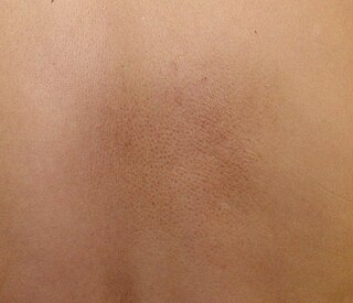
Acral persistent papular mucinosis (APPM) is a rare form of lichen myxedematosus. It is characterized by small papules on the backs of the hands, wrists, and extensor aspects of the distal forearms, with no further clinical or laboratory indications. Lesions tend to persist and may grow in number gradually. Because there are no symptoms, treatment is rarely required.
Self-healing papular mucinosis is a skin condition caused by fibroblasts producing abnormally large amounts of mucopolysaccharides, and may present in adult and juvenile forms. The juvenile variant is also called self-healing juvenile cutaneous mucinosis.
Papular mucinosis of infancy is a skin condition caused by fibroblasts producing abnormally large amounts of mucopolysaccharides, characterized by skin-colored or translucent papules.
Annular elastolytic giant-cell granuloma is a cutaneous condition characterized histologically by a dermal infiltrate of macrophages.
Granuloma multiforme is a cutaneous condition most commonly seen in central Africa, and rarely elsewhere, characterized by skin lesions that are on the upper trunk and arms in sun-exposed areas. It may be confused with tuberculoid leprosy, with which it has clinical similarities. The condition was first noted by Gosset in the 1940s, but it was not until 1964 that Leiker coined the term to describe "a disease resembling leprosy" in his study in Nigeria.
Non-X histiocytoses are a clinically well-defined group of cutaneous syndromes characterized by infiltrates of monocytes/macrophages, as opposed to X-type histiocytoses in which the infiltrates contain Langerhans cells. Conditions included in this group are:
Progressive nodular histiocytosis is a cutaneous condition clinically characterized by the development of two types of skin lesions: superficial papules and deeper larger subcutaneous nodules. Progressive nodular histiocytosis was first reported in 1978 by Taunton et al. It is a subclass of non-Langerhans cell histiocytosis and a subgroup of xanthogranuloma.

Cutaneous lymphoid hyperplasia refers to a groups of benign cutaneous disorders characterized by collections of lymphocytes, macrophages, and dendritic cells in the skin. Conditions included in this groups are:
Weber–Christian disease is a cutaneous condition characterized by recurrent subcutaneous nodules that heal with depression of the overlying skin.
Atypical tuberous myxedema, also known as Jadassohn–Dosseker syndrome, is thought to represent a pure nodular variant of lichen myxedematosus.
Cutaneous lupus mucinosis is a cutaneous condition characterized by lesions that present as asymptomatic skin-colored, at times reddish, 0.5–2 cm papules and nodules.
References
- ↑ Rapini, Ronald P.; Bolognia, Jean L.; Jorizzo, Joseph L. (2007). Dermatology: 2-Volume Set. St. Louis: Mosby. ISBN 978-1-4160-2999-1.
- ↑ Zeng, Rong; Li, Min; Jiang, Yiqun; Liu, Weida (2014). "Nodular Lichen Myxedematosus During Childhood: A Case Report". Pediatric Dermatology. 31 (6). doi:10.1111/pde.12376. ISSN 0736-8046.
- ↑ Rongioletti, Franco (2006). "Lichen Myxedematosus (Papular Mucinosis): New Concepts and Perspectives for an Old Disease". Seminars in Cutaneous Medicine and Surgery. Frontline Medical Communications, Inc. 25 (2): 100–104. doi:10.1016/j.sder.2006.04.001. ISSN 1085-5629.