Osteocyte

Last updated
Osteocyte
Transverse section of bone en.svg
Transverse section of a bone
Bone cells - Osteocytes 1 -- Smart-Servier.png
Illustration showing a single osteocyte
Details
Location Bone
Identifiers
Latin osteocytus
MeSH D010011
TH H2.00.03.7.00003
FMA 66779
Anatomical terms of microanatomy

An osteocyte, an oblate shaped type of bone cell with dendritic processes, is the most commonly found cell in mature bone. It can live as long as the organism itself. [1] The adult human body has about 42 billion of them. [2] Osteocytes do not divide and have an average half life of 25 years. They are derived from osteoprogenitor cells, some of which differentiate into active osteoblasts (which may further differentiate to osteocytes). [1] Osteoblasts/osteocytes develop in mesenchyme.

Contents

In mature bones, osteocytes and their processes reside inside spaces called lacunae (Latin for a pit) and canaliculi, respectively. [1] Osteocytes are simply osteoblasts trapped in the matrix that they secrete. They are networked to each other via long cytoplasmic extensions that occupy tiny canals called canaliculi, which are used for exchange of nutrients and waste through gap junctions.

Although osteocytes have reduced synthetic activity and (like osteoblasts) are not capable of mitotic division, they are actively involved in the routine turnover of bony matrix, through various mechanosensory mechanisms. They destroy bone through a rapid, transient (relative to osteoclasts) mechanism called osteocytic osteolysis. Hydroxyapatite, calcium carbonate and calcium phosphate is deposited around the cell.

Structure

Osteocytes have a stellate shape, approximately 7 micrometers deep and wide by 15 micrometers in length. [3] The cell body varies in size from 5–20 micrometers in diameter and contain 40–60 cell processes per cell, [4] with a cell to cell distance between 20–30 micrometers. [3] A mature osteocyte contains a single nucleus that is located toward the vascular side and has one or two nucleoli and a membrane. [5] The cell also exhibits a reduced size endoplasmic reticulum, Golgi apparatus and mitochondria, and cell processes that radiate largely towards the bone surfaces in circumferential lamellae, or towards a haversian canal and outer cement line typical of osteons in concentric lamellar bone. [5] Osteocytes form an extensive lacunocanalicular network within the mineralized collagen type I matrix, with cell bodies residing within lacunae, and cell/dendritic processes within channels called canaliculi. [6]

An osteocyte in rat bone exposed by resin cast etching An osteocyte in rat bone exposed by resin cast etching.tif
An osteocyte in rat bone exposed by resin cast etching

Development

The fossil record shows that osteocytes were present in bones of jawless fish 400 to 250 million years ago. [7] Osteocyte size has been shown to covary with genome size; and this relationship has been used in paleogenomic research. [8]

During bone formation, an osteoblast is left behind and buried in the bone matrix as an "osteoid osteocyte", which maintains contact with other osteoblasts through extended cellular processes. [9] The process of osteocytogenesis is largely unknown, but the following molecules have been shown to play a crucial role in the production of healthy osteocytes, either in correct numbers or specific distributions: matrix metalloproteinases (MMPs), dentin matrix protein 1 (DMP-1), osteoblast/osteocyte factor 45 (OF45), Klotho, TGF-beta inducible factor (TIEG), lysophosphatidic acid (LPA), E11 antigen, and oxygen. [6] 10–20% of osteoblasts differentiate into osteocytes. [6] Those osteoblasts on the bone surface that are destined for burial as osteocytes slow down matrix production, and are buried by neighboring osteoblasts that continue to produce matrix actively. [10]

HAADF-STEM electron image of a maturing osteocyte (preosteocyte or osteoid osteocyte) at the bone surface, appearing directly above osteoblast-like precursor cells (decalcified matrix). Note the elongated cell processes that are surrounded by the collagen type I matrix and already crossing lamellar boundaries as collagen (and eventually mineral) continues to entomb the cell. Osteocyte.tif
HAADF-STEM electron image of a maturing osteocyte (preosteocyte or osteoid osteocyte) at the bone surface, appearing directly above osteoblast-like precursor cells (decalcified matrix). Note the elongated cell processes that are surrounded by the collagen type I matrix and already crossing lamellar boundaries as collagen (and eventually mineral) continues to entomb the cell.

Palumbo et al. (1990) distinguish three cell types from osteoblast to mature osteocyte: type I preosteocyte (osteoblastic osteocyte), type II preosteocyte (osteoid osteocyte), and type III preosteocyte (partially surrounded by mineral matrix). [10] The embedded "osteoid-osteocyte" must do two functions simultaneously: regulate mineralization and form connective dendritic processes, which requires cleavage of collagen and other matrix molecules. [11] The transformation from motile osteoblast to entrapped osteocyte takes about three days, and during this time, the cell produces a volume of extracellular matrix three times its own cellular volume, which results in 70% volume reduction in the mature osteocyte cell body compared to the original osteoblast volume. [12] The cell undergoes a dramatic transformation from a polygonal shape to a cell that extends dendrites toward the mineralizing front, followed by dendrites that extend to either the vascular space or bone surface. [11] As the osteoblast transitions to an osteocyte, alkaline phosphatase is reduced, and casein kinase II is elevated, as is osteocalcin. [11]

Osteocytes appear to be enriched in proteins that are resistant to hypoxia, which appears to be due to their embedded location and restricted oxygen supply. [13] Oxygen tension may regulate the differentiation of osteoblasts into osteocytes, and osteocyte hypoxia may play a role in disuse-mediated bone resorption. [13]

Function

Although osteocytes are relatively inert cells, they are capable of molecular synthesis and modification, as well as transmission of signals over long distances, in a way similar to the nervous system. [6] They are the most common cell type in bone (31,900 per cubic millimeter in bovine bone to 93,200 per cubic millimeter in rat bone). [6] Most of the receptor activities that play an important role in bone function are present in the mature osteocyte. [6]

Osteocytes are an important regulator of bone mass. [14] [15] Osteocytes contain glutamate transporters that produce nerve growth factors after bone fracture, evidence of a sensing and information transfer system. [6] When osteocytes were experimentally destroyed, the bones showed a significant increase in bone resorption, decreased bone formation, trabecular bone loss, and loss of response to unloading. [6]

Osteocytes are mechanosensor cells that control the activity of osteoblasts and osteoclasts [15] within a basic multicellular unit (BMU), a temporary anatomic structure where bone remodeling occurs. [16] Osteocytes generate an inhibitory signal that is passed through their cell processes to osteoblasts for recruitment to enable bone formation. [17]

Osteocytes are also a key endocrine regulator in the metabolism of minerals such as phosphates. [14] Osteocyte-specific proteins such as sclerostin have been shown to function in mineral metabolism, as well as other molecules such as PHEX, DMP-1, MEPE, and FGF-23, which are highly expressed by osteocytes and regulate phosphate and biomineralization. [11] [15] Osteocyte regulation can be linked to disease. For example, Lynda Bonewald determined that osteocytes make FGF23, which travels through the bloodstream to trigger the release of phosphorus by the kidneys. Without enough phosphorus bones and teeth soften, and muscles become weak, as in X-linked hypophosphatemia. [14] [18] [15] [13]

Sclerostin

Osteocytes synthesize sclerostin, a secreted protein that inhibits bone formation by binding to LRP5/LRP6 coreceptors and blunting Wnt signaling. [15] [7] Sclerostin, the product of the SOST gene, is the first mediator of communication between osteocytes, bone forming osteoblasts and bone resorbing osteoclasts, critical for bone remodeling. [19] Only osteocytes express sclerostin, which acts in a paracrine fashion to inhibit bone formation. [19] Sclerostin is inhibited by parathyroid hormone (PTH) and mechanical loading. [19] Sclerostin antagonizes the activity of BMP (bone morphogenetic protein), a cytokine that induces bone and cartilage formation. [16]

Pathophysiology

Osteonecrosis refers to the classic pattern of cell death and complex osteogenesis and bone resorption processes. Osteocyte necrosis (ON) initiates with hematopoietic and adipocytic cellular necrosis along with interstitial marrow edema. ON happens after about 2 to 3 hours of anoxia; histological signs of osteocytic necrosis do not display until about 24 to 72 hours after hypoxia. ON is first characterized by pyknosis of nuclei, followed by hollow osteocyte lacunae. Capillary revascularization and reactive hyperemia slightly take place at the periphery of the necrosis site, followed by a repair process combining both bone resorption and production that incompletely changes dead with living bone. Nouveau bone overlays onto dead trabeculae along with fragmentary resorption of dead bone. Bone resorption outperforms formation resulting in a net removal of bone, deformed structural integrity of the subchondral trabeculae, joint incongruity, and subchondral fracture. [20]

Clinical significance

Clinically important research of gel based in vitro 3D model for the osteocytic potentiality of human CD34+ stem cells has been described. The results confirm that the human CD34+ stem cells possess unique osteogenic differentiation potential and can be used in the early regeneration of injured bone. [21] Osteocytes die as a consequence of senescence, degeneration/necrosis, apoptosis (programmed cell death), and/or osteoclastic engulfment. [1] The percentage of dead osteocytes in bone increases with age from less than 1% at birth to 75% after age 80. [22] Osteocyte apoptosis is thought to be related to decreased mechanotransduction, which possibly leads to the development of osteoporosis. [23] Apoptotic osteocytes release apoptotic bodies expressing RANKL to recruit osteoclasts. [11]

Mechanical loading increases osteocyte viability in vitro, and contributes to solute transport through the lacuno-canalicular system in bone, which enhances oxygen and nutrient exchange and diffusion to osteocytes. [23] Skeletal unloading has been shown to induce osteocyte hypoxia in vivo, this is when osteocytes undergo apoptosis and recruit osteoclasts to resorb bone. [23] Microdamage in bone occurs as the result of repetitive events of cycling loading, and appears to be associated with osteocyte death by apoptosis, which appear to secrete a signal to target osteoclasts to perform remodeling at a damaged site. [23] Under normal conditions, osteocytes express high amounts of TGF-β and thus repress bone resorption, but when bone grows old, the expression levels of TGF-β decrease, and the expression of osteoclast-stimulatory factors, such as RANKL and M-CSF increases, bone resorption is then enhanced, leading to net bone loss. [23]

Mechanical stimulation of osteocytes results in opening of hemichannels to release PGE2 and ATP, among other biochemical signaling molecules, which play a crucial role in maintaining the balance between bone formation and resorption. [24] Osteocyte cell death can occur in association with pathologic conditions such as osteoporosis and osteoarthritis, which leads to increased skeletal fragility, linked to the loss of ability to sense microdamage and/or signal repair. [11] [25] Oxygen deprivation that occurs as the result of immobilization (bed rest), glucocorticoid treatment, and withdrawal of oxygen have all been shown to promote osteocyte apoptosis. [11] It is now recognized that osteocytes respond in a variety of ways to the presence of implant biomaterials. [26]

See also

Related Research Articles

<span class="mw-page-title-main">Bone</span> Rigid organs that constitute part of the endoskeleton of vertebrates

A bone is a rigid organ that constitutes part of the skeleton in most vertebrate animals. Bones protect the various other organs of the body, produce red and white blood cells, store minerals, provide structure and support for the body, and enable mobility. Bones come in a variety of shapes and sizes and have complex internal and external structures. They are lightweight yet strong and hard and serve multiple functions.

<span class="mw-page-title-main">Bone healing</span> Healing from bone injury

Bone healing, or fracture healing, is a proliferative physiological process in which the body facilitates the repair of a bone fracture.

<span class="mw-page-title-main">Osteoblast</span> Cells secreting extracellular matrix

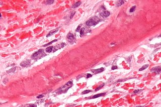
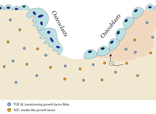
Osteoblasts are cells with a single nucleus that synthesize bone. However, in the process of bone formation, osteoblasts function in groups of connected cells. Individual cells cannot make bone. A group of organized osteoblasts together with the bone made by a unit of cells is usually called the osteon.

<span class="mw-page-title-main">Osteoid</span> Unmineralized, organic portion of the bone matrix

In histology, osteoid is the unmineralized, organic portion of the bone matrix that forms prior to the maturation of bone tissue. Osteoblasts begin the process of forming bone tissue by secreting the osteoid as several specific proteins. When the osteoid becomes mineralized, it and the adjacent bone cells have developed into new bone tissue.

<span class="mw-page-title-main">Osteoclast</span> Cell that breaks down bone tissue

An osteoclast is a type of bone cell that breaks down bone tissue. This function is critical in the maintenance, repair, and remodeling of bones of the vertebral skeleton. The osteoclast disassembles and digests the composite of hydrated protein and mineral at a molecular level by secreting acid and a collagenase, a process known as bone resorption. This process also helps regulate the level of blood calcium.

<span class="mw-page-title-main">Osteoprotegerin</span> Mammalian protein found in Homo sapiens

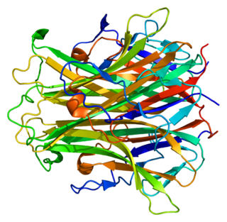
Osteoprotegerin (OPG), also known as osteoclastogenesis inhibitory factor (OCIF) or tumour necrosis factor receptor superfamily member 11B (TNFRSF11B), is a cytokine receptor of the tumour necrosis factor (TNF) receptor superfamily encoded by the TNFRSF11B gene.

<span class="mw-page-title-main">Endochondral ossification</span> Cartilaginous bone development that forms the long bones

Endochondral ossification is one of the two essential processes during fetal development of the mammalian skeletal system by which bone tissue is produced. Unlike intramembranous ossification, the other process by which bone tissue is produced, cartilage is present during endochondral ossification. Endochondral ossification is also an essential process during the rudimentary formation of long bones, the growth of the length of long bones, and the natural healing of bone fractures.

<span class="mw-page-title-main">Intramembranous ossification</span> Mesenchymal bone development that forms the non-long bones

Intramembranous ossification is one of the two essential processes during fetal development of the gnathostome skeletal system by which rudimentary bone tissue is created. Intramembranous ossification is also an essential process during the natural healing of bone fractures and the rudimentary formation of bones of the head.

<span class="mw-page-title-main">Ameloblast</span>

Ameloblasts are cells present only during tooth development that deposit tooth enamel, which is the hard outermost layer of the tooth forming the surface of the crown.

<span class="mw-page-title-main">Dental follicle</span>

The dental follicle, also known as dental sac, is made up of mesenchymal cells and fibres surrounding the enamel organ and dental papilla of a developing tooth. It is a vascular fibrous sac containing the developing tooth and its odontogenic organ. The dental follicle (DF) differentiates into the periodontal ligament. In addition, it may be the precursor of other cells of the periodontium, including osteoblasts, cementoblasts and fibroblasts. They develop into the alveolar bone, the cementum with Sharpey's fibers and the periodontal ligament fibers respectively. Similar to dental papilla, the dental follicle provides nutrition to the enamel organ and dental papilla and also have an extremely rich blood supply.

A cementoblast is a biological cell that forms from the follicular cells around the root of a tooth, and whose biological function is cementogenesis, which is the formation of cementum. The mechanism of differentiation of the cementoblasts is controversial but circumstantial evidence suggests that an epithelium or epithelial component may cause dental sac cells to differentiate into cementoblasts, characterised by an increase in length. Other theories involve Hertwig epithelial root sheath (HERS) being involved.

<span class="mw-page-title-main">Bone resorption</span> Medical condition

Bone resorption is resorption of bone tissue, that is, the process by which osteoclasts break down the tissue in bones and release the minerals, resulting in a transfer of calcium from bone tissue to the blood.

<span class="mw-page-title-main">Sclerostin</span> Protein-coding gene in the species Homo sapiens

Sclerostin is a protein that in humans is encoded by the SOST gene. It is a secreted glycoprotein with a C-terminal cysteine knot-like (CTCK) domain and sequence similarity to the DAN family of bone morphogenetic protein (BMP) antagonists. Sclerostin is produced primarily by the osteocyte but is also expressed in other tissues, and has anti-anabolic effects on bone formation.

<span class="mw-page-title-main">Flat bone</span> Category of bones that function to provide protection or surfaces for muscular attachment

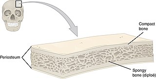
Flat bones are bones whose principal function is either extensive protection or the provision of broad surfaces for muscular attachment. These bones are expanded into broad, flat plates, as in the cranium (skull), the ilium (pelvis), sternum and the rib cage. The flat bones are: the occipital, parietal, frontal, nasal, lacrimal, vomer, hip bone, sternum, ribs, and scapulae.

<span class="mw-page-title-main">RANKL</span> Mammalian protein found in Homo sapiens

Receptor activator of nuclear factor kappa-Β ligand (RANKL), also known as tumor necrosis factor ligand superfamily member 11 (TNFSF11), TNF-related activation-induced cytokine (TRANCE), osteoprotegerin ligand (OPGL), and osteoclast differentiation factor (ODF), is a protein that in humans is encoded by the TNFSF11 gene.

<span class="mw-page-title-main">Lacuna (histology)</span> Small space between lamellae containing osteocytes

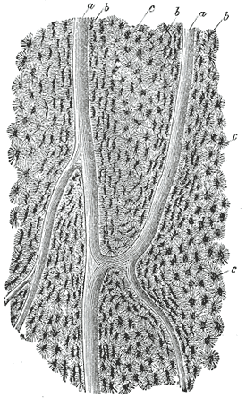
In histology, a lacuna is a small space, containing an osteocyte in bone, or chondrocyte in cartilage.

<span class="mw-page-title-main">Osteonecrosis of the jaw</span> Medical condition

Osteonecrosis of the jaw (ONJ) is a severe bone disease (osteonecrosis) that affects the jaws. Various forms of ONJ have been described since 1861, and a number of causes have been suggested in the literature.

<span class="mw-page-title-main">Bone remodeling</span> Continuous turnover of bone matrix and mineral

In osteology, bone remodeling or bone metabolism is a lifelong process where mature bone tissue is removed from the skeleton and new bone tissue is formed. These processes also control the reshaping or replacement of bone following injuries like fractures but also micro-damage, which occurs during normal activity. Remodeling responds also to functional demands of the mechanical loading.

<span class="mw-page-title-main">Sp7 transcription factor</span> Protein-coding gene in the species Homo sapiens

Transcription factor Sp7, also called osterix (Osx), is a protein that in humans is encoded by the SP7 gene. It is a member of the Sp family of zinc-finger transcription factors It is highly conserved among bone-forming vertebrate species It plays a major role, along with Runx2 and Dlx5 in driving the differentiation of mesenchymal precursor cells into osteoblasts and eventually osteocytes. Sp7 also plays a regulatory role by inhibiting chondrocyte differentiation maintaining the balance between differentiation of mesenchymal precursor cells into ossified bone or cartilage. Mutations of this gene have been associated with multiple dysfunctional bone phenotypes in vertebrates. During development, a mouse embryo model with Sp7 expression knocked out had no formation of bone tissue. Through the use of GWAS studies, the Sp7 locus in humans has been strongly associated with bone mass density. In addition there is significant genetic evidence for its role in diseases such as Osteogenesis imperfecta (OI).

The human skeletal system is a complex organ in constant equilibrium with the rest of the body. In addition to support and structure of the body, bone is the major reservoir for many minerals and compounds essential for maintaining a healthy pH balance. The deterioration of the body with age renders the elderly particularly susceptible to and affected by poor bone health. Illnesses like osteoporosis, characterized by weakening of the bone's structural matrix, increases the risk of hip-fractures and other life-changing secondary symptoms. In 2010, over 258,000 people aged 65 and older were admitted to the hospital for hip fractures. Incidence of hip fractures is expected to rise by 12% in America, with a projected 289,000 admissions in the year 2030. Other sources estimate up to 1.5 million Americans will have an osteoporotic-related fracture each year. The cost of treating these people is also enormous, in 1991 Medicare spent an estimated $2.9 billion for treatment and out-patient care of hip fractures, this number can only be expected to rise.

References

  1. 1 2 3 4 Tate, M. L.; Adamson, J. R.; Tami, A. E.; Bauer, T. W. (2004). "Cells in Focus, The osteocyte". International Journal of Biochemistry & Cell Biology. 36 (1): 1–8. doi:10.1016/S1357-2725(03)00241-3. PMID   14592527.
  2. Buenzli, Pascal R.; Sims, Natalie A. (2015-06-01). "Quantifying the osteocyte network in the human skeleton". Bone. 75: 144–150. doi:10.1016/j.bone.2015.02.016. ISSN   1873-2763. PMID   25708054.
  3. 1 2 Sugawara, Y; Kamioka, H; Honjo, T; Tezuka, K; Takano-Yamamoto, T (2005). "Three dimensional reconstruction of chick calvarial osteocytes and their cell processes using confocal microscopy". Bone. 36 (5): 877–83. doi:10.1016/j.bone.2004.10.008. PMID   15820146.
  4. Tanaka-amioka, K; Kamioka, H; Ris, H; Lim, SS (1998). "Osteocyte shape is dependent on actin filaments and osteocyte processes are unique actin-rich projections". J. Bone Miner. Res. 13 (10): 1555–68. doi: 10.1359/jbmr.1998.13.10.1555 . PMID   9783544. S2CID   5972946.
  5. 1 2 Dudley, HR; Spiro, D (1961). "The fine structure of bone cells". The Journal of Biophysical and Biochemical Cytology. 11 (3): 627–649. doi:10.1083/jcb.11.3.627. PMC   2225143 . PMID   19866598.
  6. 1 2 3 4 5 6 7 8 Noble, SN (2008). "The osteocyte lineage". Archives of Biochemistry and Biophysics. 473 (2): 106–111. doi:10.1016/j.abb.2008.04.009. PMID   18424256.
  7. 1 2 Divieti Pajevic, P (2009). "Regulation of Bone Resorption and Mineral Homeostasis by Osteocytes". IBMS BoneKEy. 6 (2): 63–70. doi:10.1138/20090363.
  8. Organ, C. L.; Shedlock, A. M.; Meade, A.; Pagel, M.; Edwards, S. V. (2007). "Origin of avian genome size and structure in nonavian dinosaurs". Nature. 446 (7132): 180–184. Bibcode:2007Natur.446..180O. doi:10.1038/nature05621. PMID   17344851. S2CID   3031794.
  9. Palumbo, Carla; Palazzini, Silvana; Zaffe, Davide; Marotti, Gastone (1990). "Osteocyte Differentiation in the Tibia of Newborn Rabbit: An Ultrastructural Study of the Formation of Cytoplasmic Processes". Cells Tissues Organs. 137 (4): 350–358. doi:10.1159/000146907. PMID   2368590.
  10. 1 2 Franz-Odendaal, T; Hall, B; Witten, PE (2006). "Buried Alive: How Osteoblasts Become Osteocytes". Developmental Dynamics. 235 (1): 176–190. doi: 10.1002/dvdy.20603 . PMID   16258960. S2CID   42563087.
  11. 1 2 3 4 5 6 7 Bonewald, L (2011). "The Amazing Osteocyte". Journal of Bone and Mineral Research. 26 (2): 229–238. doi:10.1002/jbmr.320. PMC   3179345 . PMID   21254230.
  12. Palumbo, C (1986). "A three dimensional ultrastructural study of osteoid-osteocytes in the tibia of chick embryos". Cell and Tissue Research. 246 (1): 125–131. doi:10.1007/bf00219008. PMID   3779795. S2CID   12594545.
  13. 1 2 3 Dallas, SL; Bonewald, L (2010). "Dynamics of the Transition from Osteoblast to Osteocyte". Ann N Y Acad Sci. 1192 (1): 437–443. Bibcode:2010NYASA1192..437D. doi:10.1111/j.1749-6632.2009.05246.x. PMC   2981593 . PMID   20392270.
  14. 1 2 3 Dance, Amber (23 February 2022). "Fun facts about bones: More than just scaffolding". Knowable Magazine. doi: 10.1146/knowable-022222-1 . Retrieved 8 March 2022.
  15. 1 2 3 4 5 Robling, Alexander G.; Bonewald, Lynda F. (10 February 2020). "The Osteocyte: New Insights". Annual Review of Physiology. 82 (1): 485–506. doi:10.1146/annurev-physiol-021119-034332. hdl:1805/30982. ISSN   0066-4278. PMC   8274561 . PMID   32040934 . Retrieved 8 March 2022.
  16. 1 2 ran Bezooijen Rl, Ran; Papapoulos, SE; Hamdy, NA; ten Dijke, P; Lowik, C (2005). "Control of Bone Formation by Osteocytes". BoneKEy-Osteovision. 2 (12): 33–38.
  17. Marotti, G; Ferretti, M; Muglia, MA; Palumbo, C; Palazzani, SA (1992). "A quantitative evaluation of osteoblast-osteocyte relationships on growing endosteal surface of rabbit tibiae". Bone. 13 (5): 363–368. doi:10.1016/8756-3282(92)90452-3. PMID   1419377.
  18. "Soft and weak bones? Have you heard of X-linked hypophosphatemia (XLH)? Learn more about this disease and its symptoms". XLHLink. Retrieved 9 March 2022.
  19. 1 2 3 Bellido, T (2007). "Osteocyte Apoptosis Induces Bone Resorption and Impairs the Skeletal Response to Weighlessness". BoneKEy-Osteovision. 4 (9): 252–256. doi:10.1138/20070272.
  20. Nahian, Ahmed; AlEssa, Ahmed M. (2021), "Histology, Osteocytes", StatPearls, Treasure Island (FL): StatPearls Publishing, PMID   32644416 , retrieved 2021-06-24
  21. Srikanth, Lokanathan; Sunitha, Manne Mudhu; Kumar, Pasupuleti Santhosh; Chandrasekhar, Chodimella; Vengamma, Bhuma; Sarma, Potukuchi Venkata Gurunadha Krishna (November 2016). "Gel based in vitro 3D model exploring the osteocytic potentiality of human CD34+ stem cells". Molecular Biology Reports. 43 (11): 1233–1242. doi:10.1007/s11033-016-4053-4. ISSN   1573-4978. PMID   27497820. S2CID   13230517.
  22. Tomkinson, A; Reeve, J; Shaw, RW; Noble, BS (1997). "The death of osteocytes via apoptosis accompanies estrogen withdrawal in human bone". The Journal of Clinical Endocrinology and Metabolism. 82 (9): 3128–3135. doi: 10.1210/jcem.82.9.4200 . PMID   9284757.
  23. 1 2 3 4 5 Heino, TJ; Kurata, K; Higaki, H; Vaananen, K (2009). "Evidence of the role of osteocytes in the initiation of targeted remodeling". Technology and Health Care. 17 (1): 49–56. doi:10.3233/THC-2009-0534. PMID   19478405.
  24. Burra, S; Nicolella, DP; Jiang, JX (2011). "Dark horse in osteocyte biology". Communicative and Integrative Biology. 4 (1): 48–50. doi:10.4161/cib.13646. PMC   3073269 . PMID   21509177.
  25. Carpentier, VT; Wong, J; Yeap, Y; Gan, C; Sutton-Smith, P; Badiei, A; Fazzalari, NL; Kuliwaba, JS (2012). "Increased proportion of hypermineralized osteocyte lacunae in osteoporotic and osteoarthritic human trabecular bone: Implications for bone remodeling". Bone. 50 (3): 688–694. doi:10.1016/j.bone.2011.11.021. PMID   22173055.
  26. Shah, F.A.; Thomsen, P.; Palmquist, A. (4 June 2018). "A Review of the Impact of Implant Biomaterials on Osteocytes". Journal of Dental Research. 97 (9): 977–986. doi:10.1177/0022034518778033. PMC   6055115 . PMID   29863948.