Paraneoplastic pemphigus

Last updated
Paraneoplastic Pemphigus
Specialty Dermatology   OOjs UI icon edit-ltr-progressive.svg

Paraneoplastic pemphigus (PNP) is an autoimmune disorder stemming from an underlying tumor. It is hypothesized that antigens associated with the tumor trigger an immune response resulting in blistering of the skin and mucous membranes.

Contents

While patients with malignant and benign tumors are both at risk, malignancy is associated with high mortality rates (near 90%). Current treatment focuses on general wound healing and administering corticosteroids, which has not demonstrated a high success rate. Recent research developments aim to treat the underlying tumor in order to alleviate the symptoms of PNP.

Signs and symptoms

While the presence of lesions is the denominator among patients with PNP, the characteristics of the lesions differ. The five clinical presentations of lesions associated with PNP include:

It is most common that mucous membrane lesions of the oral cavity are presented first. They can involve the oropharynx, nasopharynx, tongue, and vermilion (red portion) of the lips. They are also known to develop in the conjunctiva of the eye, anogenital (perineum) region, and esophagus. Cutaneous lesions tend to follow the onset of mucosal lesions. The blisters often erupt in waves, usually affecting the upper trunk, head, neck, and proximal extremities. Pemphigoid-like lesions are seen more often on the extremities. Lichenoid lesions are more common among children, presenting on the trunk and limbs, ranging from small red scaly papules to extensive violet to brown papules extending to the face and neck. Within the spectrum of lichenoid presentations are wounds that have features of erythema multiforme and graft-vs.-host disease. Scaly lesions on the palms of the hand and soles of the feet have been noted to coincide with the lichenoid lesions. Lesions of varying morphology may present simultaneously and transform from one type to another as the disease progresses. [1]

Mechanism

Underlying cause

PNP is ultimately caused by the presence of a tumor. There is a strong association between the development of PNP and malignancy of the tumor. However, it is not uncommon for the tumor to be benign, as in the case of afflictions such as thymoma and Castleman's disease. Only one patient without a tumor has met the diagnostic criteria for PNP. However, they rapidly reached their demise and it is suggested they may have had an undiagnosed tumor. [1]

Mechanism behind display of major symptoms

The underlying tumor causes circulating and tissue-bound antibodies to direct themselves against antigens in the plakin family, which are involved in the intracellular attachment structures in various levels of the skin/respiratory tract/membranes (keeping skin tissue together throughout the body). The number of target antigens varies on a case by case basis. The variability is likely what accounts for the different presentations of PNP. Through immunoprecipitation, target antigens have been found to include desmoglein-3, desmoglein-1, envoplakin, periplakin, desmoplakin 1, desmoplakin 2, and bullous pemphigoid antigen I.[ citation needed ]

The precise mechanism for how tumors are able to induce autoantibodies toward the plakin proteins is unknown. Suggested theories include tumor production of plakin proteins which initiate an autoimmune response against them, and cross-reactivity of tumor antigens and epidermal antigens. [2] [3]

Once the molecules that hold the various levels of the membranes together are attacked, they are unable to function properly, and the tissue breaks apart. This is manifested as the associated blistering and lesions of PNP.[ citation needed ]

Diagnosis

In order to diagnose paraneoplastic pemphigus, several tests may be performed. Initially, samples are obtained via skin biopsy for routine microscopy and direct immunofluorescence (DIF) testing. The skin sample needs to be obtained from an unaffected area adjacent to a lesion. Testing in more detail follows depending on the results from the DIF. Prompt diagnosis of PNP is crucial due to the high mortality rate of the disease. [4]

Camisa and Helm revised the original criteria from Anhalt et al. [5] into major and minor signs indicating PNP: [6]

Major:

Minor:

Microscopy

Microscopy of the skin sample obtained from the biopsy is used to detect the presence of cleavage within the dermis, epidermal acantholysis (breaking apart of the skin), dyskeratotic keratinocytes and vacuolar changes in the layers of the skin, interfacial dermatitis, and epidermal exocytosis. Presentation of these characteristics suggests PNP. [1]

Direct immunofluorescence testing

The presence of Immunoglobulin G, A, or M in the epidermis is normal. Detection in other locations such as intercellular and areas below the epidermis (subepidermal), as well as along the dermoepidermal junction (area that joins the epidermis and dermis), suggests paraneoplastic pemphigus.[ citation needed ]

Follow-up tests for confirmation

Indirect immunofluorescence (IDIF)

Patients with high concentration of antibodies show intercellular, intraepidermal antibodies as well as along the dermoepidermal junction. Patients with low concentration of antibodies only present with them inside the cells (intercellular).[ citation needed ]

If the results are negative, perform the additional assays regardless. Cases have been confirmed that reported with initial negative DIF and IDIF tests.[ citation needed ]

Assays

Immunoprecipitation, immunoblotting and enzyme-link immunosorbent assay (ELISA)

Poot et al. 2013 determined that immunoprecipitation for antibodies against envoplakin and periplakin or alpha2-macroglobulin-like–1 is the most sensitive test. However, alpha2-macroglobulin-like-1 can also be detected in patients with toxic epidermal necrosis. [2]

Similar diseases with overlapping symptoms

Bullous Pemphigoid, Cicatricial Pemphigoid, Drug Eruptions. Epidermolysis Bullosa, Epidermolysis Bullosa Acquisita, Erythema Multiforme, Lichen Planus, pemphigus vulgaris, Stevens–Johnson syndrome and toxic epidermal necrolysis. [1] [4] [7]

PNP is most commonly mistaken for pemphigus vulgaris, due to the extreme similarities of the lesions that develop. However, the difference lies in the specificity of the autoreactive antibodies in each case. [8]

Treatment

Wound healing

Initial treatment involves addressing any existing infections that may have occurred due to the broken state of the skin. Existing wounds are treated with warm compresses, non-adherent (non-stick) dressing, and topical antibiotic ointment. Immunosuppressive agents are administered in attempt to decrease blistering; this is not often effective. The first medication given aiming to heal the wounds are high dose corticosteroids. This is followed by steroid sparing agents which may reduce steroid intake and therefore lessen the side effects. Skin lesions are more likely to respond to this line of treatment than mucosal lesions. However, a high level of caution is advised in patients with a confirmed malignancy, where immunosuppression is vital and dictates treatment options. If the initial therapy fails to control the symptoms of PNP, and the condition of the patient deteriorates, a more aggressive approach may be necessary. [9]

Medication

Prednisone

Prednisone is an immunosuppressive agent which affects all of the organ systems. Effects on the cellular level include cell activation, replication, differentiation, and mobility. The overall goal is to decrease blistering (inhibition of immediate and delayed hypersensitivity) through decreasing the production of autoantibodies. In order to suppress the production of antibodies, higher doses must be administered. Lesser doses can be prescribed in order to achieve suppression of monocyte function. [10]

Azathioprine

Azathioprine is a steroid-sparing agent used in combination with Prednisone. It functions by inhibiting RNA and DNA synthesis. [11]

Ciclosporin

Ciclosporin is an immunosuppressive agent most often used in organ transplantation that has demonstrated to be effective with skin disorders. It functions by lessening production of autoantibodies and therefore diminishing the development of blisters and erosions. The mechanism of action is by inhibiting the production of T lymphocytes and lymphokines. [12]

Cyclophosphamide

Cyclophosphamide is an immunomodulator used in combination with systemic steroids to remove bone marrow. This is followed by transplanting peripheral blood stem cells. [13]

Prognosis

Quality of life/ Life expectancy

If the lesions are mild, the patient will be subject to a good deal of pain. If the lesions are severe, the overall quality of life is devastating. The impaired skin barrier function commonly leads to localized infection, which sepsis and death may follow. The pain from the oral and pharyngeal ulcers interfere with eating, which can compromise nutritional health.[ citation needed ]

The general prognosis for PNP is poor. It is more hopeful if the tumor is benign, but in the case of malignant tumors, the mortality rate is roughly 90%. The two most commonly associated types of tumors are non-Hodgkin lymphoma and chronic lymphocytic lymphoma; nearly all of these patients die within two years of diagnosis. This is attributed to the effects of the tumor combined with the negative side effects of the medication administered to treat PNP.[ citation needed ]

Roughly 1/3 of the deaths from PNP stem from pulmonary insufficiency which is brought about by the action of PNP on the respiratory mucosa. It manifests as dyspnea and progresses to bronchiolitis obliterans (non-reversible obstructive lung disease) via an unknown mechanism.[ citation needed ]

Risk Factors

As PNP is ultimately caused by the presence of a tumor, it is not contagious. There is no known way to predict who will become afflicted with it. Patients with cancer are therefore a group at risk. Although PNP has been known to affect all age groups, it is more likely to afflict middle-aged to older patients. [14]

Recent research

The Development of ELISA testing for specific diagnosis of PNP was released in 2009. The research focuses on the specific determination of autoantibodies involved in the mechanism of PNP. Specifically, antibodies against envoplakin and periplakin were being investigated. [15] Further use of ELISA testing on these antibodies confirmed the presence of anti-envoplakin and anti-periplakin autoantibodies in patients with PNP. [16]

Further research in 2013 outlined the various types of assays that could be used to determine which antibodies were involved in PNP. Demonstration of certain antibodies in the serum was named as the basis for diagnosis of PNP. This piece labeled PNP as a "multiorgan disease characterized by antibodies against plakins, desmogleins and the α2-macroglobulin-like-1 (A2ML1) protein, in association with an underlying neoplasm". [2]

A study concluded in 2009, summarized in 2010, surrounded the surgical removal of the associated tumor as a means to treat PNP. While 7/22 of the subjects perished due to resulting infection from the body's inability to heal itself after surgery, the other 15 cases survived. This study outlined the importance of early detection and prompt treatment as of utmost important in the treatment of PNP. [17]

In 2011, a case study of a woman with ulcers on the back of her leg reported as being diagnosed with PNP. The underlying tumors are almost exclusively of B-cell lineage. However, T-cells and CD56+ Natural Killer cells have also been postulated to be associated effectors of paraneoplastic pemphigus. This case demonstrated the rare association between Natural Killer cell lymphoma and PNP, suggesting that Natural Killer cells could be involved in the pathogenesis of PNP. The article warned clinicians to be alert to the possibility that paraneoplastic pemphigus in lymphomas not of B-cell lineage. This added to the already complex, not fully understood pathogenesis of PNP. [18]

A study in 2013 outlined the effectiveness of plasma exchange in PNP patients with benign tumors. [19]

The University of Toronto has been working to develop a form of treatment that improves the patient's overall quality of life while remaining economically achievable. They believe they have achieved this through fixed-dose rituximab. It has proven to be effective among auto-immune diseases, but the correct administration process for treating PNP is yet to be defined. The results of the study demonstrated varying levels of remission. [20]

Related Research Articles

<span class="mw-page-title-main">Lichen planus</span> Human chronic inflammatory disease

Lichen planus (LP) is a chronic inflammatory and immune-mediated disease that affects the skin, nails, hair, and mucous membranes. It is not an actual lichen, and is only named that because it looks like one. It is characterized by polygonal, flat-topped, violaceous papules and plaques with overlying, reticulated, fine white scale, commonly affecting dorsal hands, flexural wrists and forearms, trunk, anterior lower legs and oral mucosa. The hue may be gray-brown in people with darker skin. Although there is a broad clinical range of LP manifestations, the skin and oral cavity remain as the major sites of involvement. The cause is unknown, but it is thought to be the result of an autoimmune process with an unknown initial trigger. There is no cure, but many different medications and procedures have been used in efforts to control the symptoms.

<span class="mw-page-title-main">Pemphigus</span> Blistering autoimmune diseases

Pemphigus is a rare group of blistering autoimmune diseases that affect the skin and mucous membranes. The name is derived from the Greek root pemphix, meaning "pustule".

<span class="mw-page-title-main">Bullous pemphigoid</span> Autoimmune disease of skin and connective tissue characterized by large blisters

Bullous pemphigoid is an autoimmune pruritic skin disease which typically occurs in people aged over 60, that may involve the formation of blisters (bullae) in the space between the epidermal and dermal skin layers. It is classified as a type II hypersensitivity reaction, which involves formation of anti-hemidesmosome antibodies, causing a loss of keratinocytes to basement membrane adhesion.

Nikolsky's sign is a clinical dermatological sign, named after Pyotr Nikolsky (1858–1940), a Russian physician who trained and worked in the Russian Empire. The sign is present when slight rubbing of the skin results in exfoliation of the outermost layer. A typical test would be to place the eraser of a pencil on the roof of a lesion and spin the pencil in a rolling motion between the thumb and forefinger. If the lesion is opened, then the Nikolsky's sign is present/positive.

<span class="mw-page-title-main">Pemphigus vulgaris</span> Medical condition

Pemphigus vulgaris is a rare chronic blistering skin disease and the most common form of pemphigus. Pemphigus was derived from the Greek word pemphix, meaning blister. It is classified as a type II hypersensitivity reaction in which antibodies are formed against desmosomes, components of the skin that function to keep certain layers of skin bound to each other. As desmosomes are attacked, the layers of skin separate and the clinical picture resembles a blister. These blisters are due to acantholysis, or breaking apart of intercellular connections through an autoantibody-mediated response. Over time the condition inevitably progresses without treatment: lesions increase in size and distribution throughout the body, behaving physiologically like a severe burn.

Hailey–Hailey disease, or familial benign chronic pemphigus or familial benign pemphigus, was originally described by the Hailey brothers in 1939. It is a genetic disorder that causes blisters to form on the skin.

The desmogleins are a family of desmosomal cadherins consisting of proteins DSG1, DSG2, DSG3, and DSG4. They play a role in the formation of desmosomes that join cells to one another.
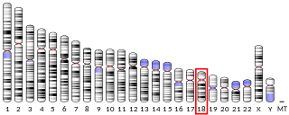
<span class="mw-page-title-main">Desmoglein-3</span> Protein-coding gene in the species Homo sapiens

Desmoglein-3 is a protein that in humans is encoded by the DSG3 gene. In the skin epidermis Desmoglein-3 is expressed in the basal lower layers of the epidermis, and dominates in terms of expression on mucosal surfaces compared to Desmoglein-1.

<span class="mw-page-title-main">Pemphigoid</span> Autoimmune blistering diseases

Pemphigoid is a group of rare autoimmune blistering diseases of the skin, and mucous membranes. As its name indicates, pemphigoid is similar in general appearance to pemphigus, but, unlike pemphigus, pemphigoid does not feature acantholysis, a loss of connections between skin cells.

<span class="mw-page-title-main">Gestational pemphigoid</span> Medical condition

Gestational pemphigoid (GP) is a rare autoimmune variant of the skin disease bullous pemphigoid, and first appears in pregnancy. It presents with tense blisters, small bumps, hives and intense itching, usually starting around the navel before spreading to limbs in mid-pregnancy or shortly after delivery. The head, face and mouth are not usually affected.

A paraneoplastic syndrome is a syndrome that is the consequence of a tumor in the body, specifically due to the production of chemical signaling molecules by tumor cells or by an immune response against the tumor. Unlike a mass effect, it is not due to the local presence of cancer cells.

<span class="mw-page-title-main">Envoplakin</span> Protein-coding gene in the species Homo sapiens

Envoplakin is a protein that in humans is encoded by the EVPL gene.

<span class="mw-page-title-main">Dermatitis herpetiformis</span> Medical condition

Dermatitis herpetiformis (DH) is a chronic autoimmune blistering skin condition, characterised by intensely itchy blisters filled with a watery fluid. DH is a cutaneous manifestation of coeliac disease, although the exact causal mechanism is not known. DH is neither related to nor caused by herpes virus; the name means that it is a skin inflammation having an appearance similar to herpes.

Pemphigus foliaceus is an autoimmune of the skin. Pemphigus foliaceus causes a characteristic inflammatory attack at the subcorneal layer of epidermis, which results in skin lesions that are scaly or crusted erosions with an erythematous (red) base. Mucosal involvement is absent even with widespread disease.

Epidermolysis bullosa acquisita, also known as acquired epidermolysis bullosa, is a longterm autoimmune blistering skin disease. It generally presents with fragile skin that blisters and becomes red with or without trauma. Marked scarring is left with thin skin, milia and nail changes. It typically begins around age 50.

Mucous membrane pemphigoid is a rare chronic autoimmune subepithelial blistering disease characterized by erosive lesions of the mucous membranes and skin. It is one of the pemphigoid diseases that can result in scarring.

<span class="mw-page-title-main">Anti-SSA/Ro autoantibodies</span> Type of anti-nuclear autoantibodies

Anti-SSA autoantibodies are a type of anti-nuclear autoantibodies that are associated with many autoimmune diseases, such as systemic lupus erythematosus (SLE), SS/SLE overlap syndrome, subacute cutaneous lupus erythematosus (SCLE), neonatal lupus and primary biliary cirrhosis. They are often present in Sjögren's syndrome (SS). Additionally, Anti-Ro/SSA can be found in other autoimmune diseases such as systemic sclerosis (SSc), polymyositis/dermatomyositis (PM/DM), rheumatoid arthritis (RA), and mixed connective tissue disease (MCTD), and are also associated with heart arrhythmia.

<span class="mw-page-title-main">Ernst H. Beutner</span> American microbiologist

Ernst H. Beutner was a German-born microbiologist who discovered the role of autoimmunity in pemphigus and pemphigoid using self-designed immunofluorescent methods. For this achievement, he is often regarded as the "Founder of Immunodermatology". He was the author or co-author of over 10 papers, which were each cited over 100 times.

References

Notes
  1. 1 2 3 4 Sehgal, Virendra N.; Srivastava, Govind (2009). "Paraneoplastic pemphigus/paraneoplastic autoimmune multiorgan syndrome". International Journal of Dermatology. 48 (2): 162–9. doi: 10.1111/j.1365-4632.2009.03995.x . PMID   19200194. S2CID   20426868.
  2. 1 2 3 Poot, A.M.; Diercks, G.F.H.; Kramer, D.; Schepens, I.; Klunder, G.; Hashimoto, T.; Borradori, L.; Jonkman, M.F.; Pas, H.H. (2013). "Laboratory diagnosis of paraneoplastic pemphigus". British Journal of Dermatology. 169 (5): 1016–24. doi: 10.1111/bjd.12479 . PMID   23796242. S2CID   207070543.
  3. Proby, Charlotte; Fujii, Yoshiko; Owaribe, Katsushi; Nishikawa, Takeji; Amagai, Masayuki (1999). "Human Autoantibodies against HD1/Plectin in Paraneoplastic Pemphigus". Journal of Investigative Dermatology. 112 (2): 153–6. doi: 10.1046/j.1523-1747.1999.00498.x . PMID   9989789.
  4. 1 2 Paraneoplastic Pemphigus~workup at eMedicine
  5. Anhalt, Grant J.; Kim, SooChan; Stanley, John R.; Korman, Neil J.; Jabs, Douglas A.; Kory, Mark; Izumi, Hiroshi; Ratrie, Harry; Mutasim, Diya; Ariss-Abdo, Lina; Labib, Ramzy S. (1990). "Paraneoplastic Pemphigus". New England Journal of Medicine. 323 (25): 1729–35. doi: 10.1056/NEJM199012203232503 . PMID   2247105.
  6. Camisa, Charles (1993). "Paraneoplastic Pemphigus Is a Distinct Neoplasia-Induced Autoimmune Disease". Archives of Dermatology. 129 (7): 883–6. doi:10.1001/archderm.1993.01680280071014. PMID   8323311.
  7. Frew, John W.; Murrell, Dédée F. (2011). "Paraneoplastic Pemphigus (Paraneoplastic Autoimmune Multiorgan Syndrome): Clinical Presentations and Pathogenesis". Dermatologic Clinics. 29 (3): 419–25, viii. doi:10.1016/j.det.2011.03.018. PMID   21605807.
  8. Maverakis, Emanual; Goodarzi, Heidi; Wehrli, Lisa N.; Ono, Yoko; Garcia, Miki Shirakawa (2012). "The Etiology of Paraneoplastic Autoimmunity". Clinical Reviews in Allergy & Immunology. 42 (2): 135–44. doi:10.1007/s12016-010-8248-5. PMID   21246308. S2CID   33226991.
  9. Paraneoplastic Pemphigus~treatment at eMedicine
  10. "Prednisone Intensol (prednisone) dosing, indications, interactions, adverse effects, and more".
  11. "Azasan, Imuran (azathioprine) dosing, indications, interactions, adverse effects, and more".
  12. "Neoral, Sandimmune (cyclosporine) dosing, indications, interactions, adverse effects and more".
  13. "Cytoxan (cyclophosphamide) dosing, indications, interactions, adverse effects, and more".
  14. "Pemphigus - Symptoms and causes". Mayo Clinic .
  15. Probst, Christian; Schlumberger, Wolfgang; Stöcker, Winfried; Recke, Andreas; Schmidt, Enno; Hashimoto, Takashi; Zhu, Xue Jun; Zillikens, Detlef; Komorowski, Lars (2009). "Development of ELISA for the specific determination of autoantibodies against envoplakin and periplakin in paraneoplastic pemphigus". Clinica Chimica Acta. 410 (1–2): 13–8. doi:10.1016/j.cca.2009.08.022. PMID   19737550.
  16. Huang, Yongchu; Li, Jing; Zhu, Xuejun (2009). "Detection of anti-envoplakin and anti-periplakin autoantibodies by ELISA in patients with paraneoplastic pemphigus". Archives of Dermatological Research. 301 (10): 703–9. doi:10.1007/s00403-008-0901-y. PMID   18820940. S2CID   862396.
  17. Zhang, Jun; Qiao, Qi-lu; Chen, Xi-xue; Liu, Ping; Qiu, Jian-xing; Zhao, Hu; Zhao, Jian-xun; Liu, Yu-cun; Wan, Yuan-lian (2011). "Improved outcomes after complete resection of underlying tumors for patients with paraneoplastic pemphigus: a single-center experience of 22 cases". Journal of Cancer Research and Clinical Oncology. 137 (2): 229–34. doi:10.1007/s00432-010-0874-z. PMID   20390428. S2CID   22375666.
  18. Gill, Harinder; Trendell-Smith, Nigel J.; Loong, Florence; Chan, Johnny Chun-Yin; Kwong, Yok-Lam (2011). "Paraneoplastic pemphigus due to natural-killer/T-cell lymphoma". British Journal of Haematology. 154 (2): 160. doi: 10.1111/j.1365-2141.2011.08645.x . PMID   21517813. S2CID   40794144.
  19. Kitagawa, C; Nakajima, K; Aoyama, Y; Fujioka, A; Nakajima, H; Tarutani, M; Tsuruta, D; Hashimoto, T; Sano, S (2014). "A Typical Case of Paraneoplastic Pemphigus Without Detection of Malignancy: Effectiveness of Plasma Exchange". Acta Dermato Venereologica. 94 (3): 359–61. doi: 10.2340/00015555-1742 . PMID   24162880.
  20. Heelan, Kara; Al-Mohammedi, Faisal; Smith, Myles J.; Knowles, Sandra; Lansang, Perla; Walsh, Scott; Shear, Neil H. (2014). "Durable Remission of Pemphigus With a Fixed-Dose Rituximab Protocol". JAMA Dermatology. 150 (7): 703–8. doi: 10.1001/jamadermatol.2013.6739 . PMID   24500264.