Phosphoglycerate mutase

Last updated
Citrate is structurally similar to the substrate 3-phosphoglycerate. The citrate molecule is shown in green. The suspected catalytically essential histidine residue involved in forming the phosphohistidine complex is directly to the left of the bound citrate molecule. Proximal Residues Bound To Citrate in Active Site of PGM.png
Citrate is structurally similar to the substrate 3-phosphoglycerate. The citrate molecule is shown in green. The suspected catalytically essential histidine residue involved in forming the phosphohistidine complex is directly to the left of the bound citrate molecule.
Phosphoglycerate mutase family
Identifiers
SymbolPGAM
Pfam PF00300
InterPro IPR013078
PROSITE PDOC00158
SCOP2 3pgm / SCOPe / SUPFAM
Available protein structures:
Pfam   structures / ECOD  
PDB RCSB PDB; PDBe; PDBj
PDBsum structure summary
PDB 1e59 A:5-191 1e58 A:5-191 1yjx D:5-193

1yfk A:5-193 1ljd A:5-193 1fzt A:9-170 4pgm B:3-189 3pgm B:3-189 1qhf A:3-189 1bq3 B:3-189 1bq4 B:3-189 1rii A:6-190 1t8p A:5-195 1h2e A:2-151 1h2f A:2-151 1ebb A:2-151 2bif A:251-398 3bif A:251-398 1bif :251-398 1k6m B:253-400 1c80 A:253-400 1c7z A:253-400 1fbt B:253-400 1c81 A:253-400

Contents

2axn A:248-395 1ujc A:2-115 1ujb A:2-115
phosphoglycerate mutase 1 (brain)
Homodimer B-type PGM.png
Identifiers
SymbolPGAM1
Alt. symbolsPGAMA
NCBI gene 5223
HGNC 8888
OMIM 172250
RefSeq NM_002629
UniProt P18669
Other data
EC number 5.4.2.11
Locus Chr. 10 q25.3
Search for
Structures Swiss-model
Domains InterPro
phosphoglycerate mutase 2 (muscle)
Identifiers
SymbolPGAM2
NCBI gene 5224
HGNC 8889
OMIM 261670
RefSeq NM_000290
UniProt P15259
Other data
EC number 5.4.2.11
Locus Chr. 7 p13-p12
Search for
Structures Swiss-model
Domains InterPro
This enzyme is not to be confused with Bisphosphoglycerate mutase which catalyzes the conversion of 1,3-bisphosphoglycerate to 2,3-bisphosphoglycerate.

Phosphoglycerate mutase (PGM) is any enzyme that catalyzes step 8 of glycolysis - the internal transfer of a phosphate group from C-3 to C-2 which results in the conversion of 3-phosphoglycerate (3PG) to 2-phosphoglycerate (2PG) through a 2,3-bisphosphoglycerate intermediate. These enzymes are categorized into the two distinct classes of either cofactor-dependent (dPGM) or cofactor-independent (iPGM). [1] The dPGM enzyme (EC 5.4.2.11) is composed of approximately 250 amino acids and is found in all vertebrates as well as in some invertebrates, fungi, and bacteria. The iPGM (EC 5.4.2.12) class is found in all plants and algae as well as in some invertebrate, fungi, and Gram-positive bacteria. [2] This class of PGM enzyme shares the same superfamily as alkaline phosphatase. [3]

Mechanism

PGM is an isomerase enzyme, effectively transferring a phosphate group (PO43−) from the C-3 carbon of 3-phosphoglycerate to the C-2 carbon forming 2-phosphoglycerate. There are a total of three reactions dPGM can catalyze: a mutase reaction resulting in the conversion of 3PG to 2PG and vice versa, [4] [5] a phosphatase reaction creating phosphoglycerate from 2,3-bisphosphoglycerate, [6] [7] and a synthase reaction producing 2,3-bisphosphoglycerate from 1,3-bisphosphoglycerate similar to the enzyme bisphosphoglycerate mutase[ citation needed ]. Kinetic and structural studies have provided evidence that indicate dPGM and bisphosphoglycerate mutase are paralogous structures. [6] Both enzymes are contained in the superfamily that also contains the phosphatase portion of phosphofructokinase 2 and prostatic acid phosphatase. [8]

The catalyzed mutase reaction involves two separate phosphoryl groups and the ending phosphate on the 2-carbon is not the same phosphate removed from the 3-carbon.
In the cofactor-dependent enzyme's initial state, the active site contains a phosphohistidine complex formed by phosphorylation of a specific histidine residue. [9] When 3-phosphoglycerate enters the active site, the phosphohistidine complex is positioned as to facilitate transfer of phosphate from enzyme to substrate C-2 creating a 2,3-bisphosphoglycerate intermediate.
Dephosphorylation of the enzyme histidine actuates a local allosteric change in enzyme configuration which now aligns the substrates 3-C phosphate group with enzyme active site histidine and facilitates phosphate transfer returning the enzyme to its initial phosphorylated state and releasing product 2-phosphoglycerate. 2,3-bisphosphoglycerate is required a cofactor for dPGM. In contrast, the iPGM class is independent of 2,3-bisphosphoglycerate and catalyzes the intramolecular transfer of the phosphate group on monophosphoglycerates using a phosphoserineintermediate. [10]

Reaction summary

3PG + P-Enzyme → 2,3BPG + Enzyme → 2PG + P-Enzyme

   3-phosphoglycerate         intermediate          2-phosphoglycerate

ΔG°=+1.1kcal/mol

Isozymes

Phosphoglycerate mutase exists primarily as a dimer of two either identical or closely related subunits of about 32kDa. The enzyme is found in organisms as simple as yeast through Homo sapiens and its structure is highly conserved throughout. (Yeast PGM≈74% conserved vs mammal form).

In mammals, the enzyme subunits appear to be either a muscle-derived form (m-type) or other tissue (b-type for brain where the b-isozyme was originally isolated). Existing as a dimer, the enzyme then has 3 isozymes depending on which subunit forms makeup the whole molecule (mm, bb or mb). The mm-type is found mainly in smooth muscle almost exclusively. The mb-isozyme is found in cardiac and skeletal muscle and the bb-type is found in the rest of tissues. [11] While all three isozymes may be found in any tissue, the above distributions are based on prevalence in each.

Interactive pathway map

Click on genes, proteins and metabolites below to link to respective articles. [§ 1]

[[File:
WP534.png go to articlego to articlego to articlego to articlego to articlego to articlego to articlego to articlego to articlego to articlego to articlego to articlego to articlego to articlego to articlego to articlego to articlego to articlego to articlego to articlego to articlego to articlego to articlego to articlego to articlego to articlego to articlego to articlego to articlego to articlego to articlego to articlego to articlego to articlego to articlego to articlego to articlego to articlego to articlego to articlego to articlego to articlego to articlego to articlego to articlego to articlego to articlego to articlego to articlego to articlego to articlego to articlego to articlego to articlego to articlego to articlego to articlego to articlego to articlego to articlego to articlego to articlego to articlego to articlego to articlego to articlego to articlego to articlego to articlego to articlego to articlego to article
[[
]]
[[
]]
[[
]]
[[
]]
[[
]]
[[
]]
[[
]]
[[
]]
[[
]]
[[
]]
[[
]]
[[
]]
[[
]]
[[
]]
[[
]]
[[
]]
[[
]]
[[
]]
[[
]]
[[
]]
[[
]]
[[
]]
[[
]]
[[
]]
[[
]]
[[
]]
[[
]]
[[
]]
[[
]]
[[
]]
[[
]]
[[
]]
[[
]]
[[
]]
[[
]]
[[
]]
[[
]]
[[
]]
[[
]]
[[
]]
[[
]]
[[
]]
[[
]]
[[
]]
[[
]]
[[
]]
[[
]]
[[
]]
[[
]]
[[
]]
[[
]]
[[
]]
[[
]]
[[
]]
[[
]]
[[
]]
[[
]]
[[
]]
[[
]]
[[
]]
[[
]]
[[
]]
[[
]]
[[
]]
[[
]]
[[
]]
[[
]]
[[
]]
[[
]]
[[
]]
[[
]]
[[
]]
[[
]]
[[
]]
[[
]]
WP534.png go to articlego to articlego to articlego to articlego to articlego to articlego to articlego to articlego to articlego to articlego to articlego to articlego to articlego to articlego to articlego to articlego to articlego to articlego to articlego to articlego to articlego to articlego to articlego to articlego to articlego to articlego to articlego to articlego to articlego to articlego to articlego to articlego to articlego to articlego to articlego to articlego to articlego to articlego to articlego to articlego to articlego to articlego to articlego to articlego to articlego to articlego to articlego to articlego to articlego to articlego to articlego to articlego to articlego to articlego to articlego to articlego to articlego to articlego to articlego to articlego to articlego to articlego to articlego to articlego to articlego to articlego to articlego to articlego to articlego to articlego to articlego to article
|alt=Glycolysis and Gluconeogenesis edit]]
Glycolysis and Gluconeogenesis edit
  1. The interactive pathway map can be edited at WikiPathways: "GlycolysisGluconeogenesis_WP534".

Regulation

Phosphoglycerate mutase has a small positive Gibbs free energy and this reaction proceeds easily in both directions. Since it is a reversible reaction, it is not the site of major regulation mechanisms or regulation schemes for the glycolytic pathway.

Anionic molecules such as vanadate, [12] acetate, chloride ion, phosphate, 2-phosphoglycolate, and N-[tris(hydroxymethyl)methyl-2-amino]ethanesulfonate are known inhibitors of the mutase activity of dPGM. Studies have shown dPGM to be sensitive to changes in ionic concentration, where increasing concentrations of salts result in the activation of the enzyme's phosphatase activity while inhibiting its mutase activity. Certain salts, such as KCl, are known to be competitive inhibitors in respect to 2-phosphoglycerate and mutase activity. [13] Both phosphate and 2-phosphoglycolate are competitive inhibitors of mutase activity in respect to the substrates 2-phosphoglycerate and 2,3-bisphosphoglycerate. [14]

Clinical significance

In humans the PGAM2 gene which encodes this enzyme is located on the short arm of chromosome 7.

Deficiency of phosphoglycerate mutase causes glycogen storage disease type X, a rare autosomal recessive genetic disorder with symptoms ranging from mild to moderate; is not thought life-threatening and can be managed with changes in lifestyle.[ citation needed ] This presents as a metabolic myopathy and is one of the many forms of syndromes formerly referred to as muscular dystrophy.[ citation needed ] PGAM1 deficiency affects the liver, while PGAM2 deficiency affects the muscle.

Onset is generally noted as childhood to early adult though some who may be mildly affected by the disorder may not know they have it. Patients with PGAM deficiency are usually asymptomatic, except when they engage in brief, strenuous efforts which may trigger myalgias, cramps, muscle necrosis and myoglobinuria. [15] An unusual pathologic feature of PGAM deficiency is the association with tubular aggregates. The symptoms are an intolerance to physical exertion or activity, cramps and muscle pain. Permanent weakness is rare. The disease is not progressive and has an excellent prognosis.[ citation needed ]

Human proteins containing this domain

BPGM; PFKFB1; PFKFB2; PFKFB3; PFKFB4; PGAM1; PGAM2; PGAM4; PGAM5; STS1; UBASH3A;

Related Research Articles

<span class="mw-page-title-main">Glycolysis</span> Catabolic pathway

Glycolysis is the metabolic pathway that converts glucose into pyruvate, and in most organisms, occurs in the liquid part of cells, the cytosol. The free energy released in this process is used to form the high-energy molecules adenosine triphosphate (ATP) and reduced nicotinamide adenine dinucleotide (NADH). Glycolysis is a sequence of ten reactions catalyzed by enzymes.

<span class="mw-page-title-main">Kinase</span> Enzyme catalyzing transfer of phosphate groups onto specific substrates

In biochemistry, a kinase is an enzyme that catalyzes the transfer of phosphate groups from high-energy, phosphate-donating molecules to specific substrates. This process is known as phosphorylation, where the high-energy ATP molecule donates a phosphate group to the substrate molecule. This transesterification produces a phosphorylated substrate and ADP. Conversely, it is referred to as dephosphorylation when the phosphorylated substrate donates a phosphate group and ADP gains a phosphate group. These two processes, phosphorylation and dephosphorylation, occur four times during glycolysis.

<span class="mw-page-title-main">Phosphorylation</span> Chemical process of introducing a phosphate

In biochemistry, phosphorylation is the attachment of a phosphate group to a molecule or an ion. This process and its inverse, dephosphorylation, are common in biology. Protein phosphorylation often activates many enzymes.

<span class="mw-page-title-main">Phosphofructokinase 1</span> Class of enzymes

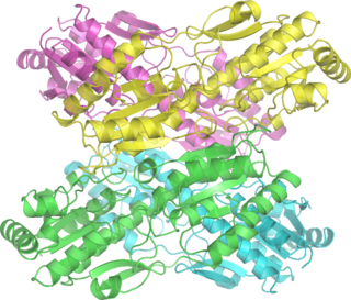
Phosphofructokinase-1 (PFK-1) is one of the most important regulatory enzymes of glycolysis. It is an allosteric enzyme made of 4 subunits and controlled by many activators and inhibitors. PFK-1 catalyzes the important "committed" step of glycolysis, the conversion of fructose 6-phosphate and ATP to fructose 1,6-bisphosphate and ADP. Glycolysis is the foundation for respiration, both anaerobic and aerobic. Because phosphofructokinase (PFK) catalyzes the ATP-dependent phosphorylation to convert fructose-6-phosphate into fructose 1,6-bisphosphate and ADP, it is one of the key regulatory steps of glycolysis. PFK is able to regulate glycolysis through allosteric inhibition, and in this way, the cell can increase or decrease the rate of glycolysis in response to the cell's energy requirements. For example, a high ratio of ATP to ADP will inhibit PFK and glycolysis. The key difference between the regulation of PFK in eukaryotes and prokaryotes is that in eukaryotes PFK is activated by fructose 2,6-bisphosphate. The purpose of fructose 2,6-bisphosphate is to supersede ATP inhibition, thus allowing eukaryotes to have greater sensitivity to regulation by hormones like glucagon and insulin.

<span class="mw-page-title-main">Phosphoglucomutase</span>

Phosphoglucomutase is an enzyme that transfers a phosphate group on an α-D-glucose monomer from the 1 to the 6 position in the forward direction or the 6 to the 1 position in the reverse direction.

Substrate-level phosphorylation is a metabolism reaction that results in the production of ATP or GTP supported by the energy released from another high-energy bond that leads to phosphorylation of ADP or GDP to ATP or GTP (note that the reaction catalyzed by creatine kinase is not considered as "substrate-level phosphorylation"). This process uses some of the released chemical energy, the Gibbs free energy, to transfer a phosphoryl (PO3) group to ADP or GDP. Occurs in glycolysis and in the citric acid cycle.

<span class="mw-page-title-main">Triosephosphate isomerase</span> Enzyme involved in glycolysis

Triose-phosphate isomerase is an enzyme that catalyzes the reversible interconversion of the triose phosphate isomers dihydroxyacetone phosphate and D-glyceraldehyde 3-phosphate.

<span class="mw-page-title-main">Phosphofructokinase 2</span> Class of enzymes

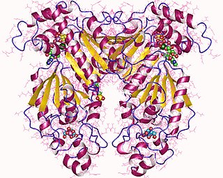
Phosphofructokinase-2 (6-phosphofructo-2-kinase, PFK-2) or fructose bisphosphatase-2 (FBPase-2), is an enzyme indirectly responsible for regulating the rates of glycolysis and gluconeogenesis in cells. It catalyzes formation and degradation of a significant allosteric regulator, fructose-2,6-bisphosphate (Fru-2,6-P2) from substrate fructose-6-phosphate. Fru-2,6-P2 contributes to the rate-determining step of glycolysis as it activates enzyme phosphofructokinase 1 in the glycolysis pathway, and inhibits fructose-1,6-bisphosphatase 1 in gluconeogenesis. Since Fru-2,6-P2 differentially regulates glycolysis and gluconeogenesis, it can act as a key signal to switch between the opposing pathways. Because PFK-2 produces Fru-2,6-P2 in response to hormonal signaling, metabolism can be more sensitively and efficiently controlled to align with the organism's glycolytic needs. This enzyme participates in fructose and mannose metabolism. The enzyme is important in the regulation of hepatic carbohydrate metabolism and is found in greatest quantities in the liver, kidney and heart. In mammals, several genes often encode different isoforms, each of which differs in its tissue distribution and enzymatic activity. The family described here bears a resemblance to the ATP-driven phospho-fructokinases, however, they share little sequence similarity, although a few residues seem key to their interaction with fructose 6-phosphate.

<span class="mw-page-title-main">3-Phosphoglyceric acid</span> Chemical compound

3-Phosphoglyceric acid (3PG, 3-PGA, or PGA) is the conjugate acid of 3-phosphoglycerate or glycerate 3-phosphate (GP or G3P). This glycerate is a biochemically significant metabolic intermediate in both glycolysis and the Calvin-Benson cycle. The anion is often termed as PGA when referring to the Calvin-Benson cycle. In the Calvin-Benson cycle, 3-phosphoglycerate is typically the product of the spontaneous scission of an unstable 6-carbon intermediate formed upon CO2 fixation. Thus, two equivalents of 3-phosphoglycerate are produced for each molecule of CO2 that is fixed. In glycolysis, 3-phosphoglycerate is an intermediate following the dephosphorylation (reduction) of 1,3-bisphosphoglycerate.

A mutase is an enzyme of the isomerase class that catalyzes the movement of a functional group from one position to another within the same molecule. In other words, mutases catalyze intramolecular group transfers. Examples of mutases include bisphosphoglycerate mutase, which appears in red blood cells and phosphoglycerate mutase, which is an enzyme integral to glycolysis. In glycolysis, it changes 3-phosphoglycerate to 2-phosphoglycerate by moving a single phosphate group within a single molecule.

<span class="mw-page-title-main">Enolase</span>

Phosphopyruvate hydratase, usually known as enolase, is a metalloenzyme (EC 4.2.1.11) that catalyses the conversion of 2-phosphoglycerate (2-PG) to phosphoenolpyruvate (PEP), the ninth and penultimate step of glycolysis. The chemical reaction is:

<span class="mw-page-title-main">2,3-Bisphosphoglyceric acid</span> Chemical compound

2,3-Bisphosphoglyceric acid (2,3-BPG), also known as 2,3-diphosphoglyceric acid (2,3-DPG), is a three-carbon isomer of the glycolytic intermediate 1,3-bisphosphoglyceric acid (1,3-BPG).

<span class="mw-page-title-main">Phosphoglycerate kinase</span> Enzyme

Phosphoglycerate kinase is an enzyme that catalyzes the reversible transfer of a phosphate group from 1,3-bisphosphoglycerate (1,3-BPG) to ADP producing 3-phosphoglycerate (3-PG) and ATP :

<span class="mw-page-title-main">Bisphosphoglycerate mutase</span> Enzyme

Bisphosphoglycerate mutase is an enzyme expressed in erythrocytes and placental cells. It is responsible for the catalytic synthesis of 2,3-Bisphosphoglycerate (2,3-BPG) from 1,3-bisphosphoglycerate. BPGM also has a mutase and a phosphatase function, but these are much less active, in contrast to its glycolytic cousin, phosphoglycerate mutase (PGM), which favors these two functions, but can also catalyze the synthesis of 2,3-BPG to a lesser extent.

<span class="mw-page-title-main">Phosphofructokinase</span> Enzyme in glycolysis

Phosphofructokinase (PFK) is a kinase enzyme that phosphorylates fructose 6-phosphate in glycolysis.

<span class="mw-page-title-main">Fructose-bisphosphate aldolase</span>

Fructose-bisphosphate aldolase, often just aldolase, is an enzyme catalyzing a reversible reaction that splits the aldol, fructose 1,6-bisphosphate, into the triose phosphates dihydroxyacetone phosphate (DHAP) and glyceraldehyde 3-phosphate (G3P). Aldolase can also produce DHAP from other (3S,4R)-ketose 1-phosphates such as fructose 1-phosphate and sedoheptulose 1,7-bisphosphate. Gluconeogenesis and the Calvin cycle, which are anabolic pathways, use the reverse reaction. Glycolysis, a catabolic pathway, uses the forward reaction. Aldolase is divided into two classes by mechanism.

Glucose-1,6-bisphosphate synthase is a type of enzyme called a phosphotransferase and is involved in mammalian starch and sucrose metabolism. It catalyzes the transfer of a phosphate group from 1,3-bisphosphoglycerate to glucose-1-phosphate, yielding 3-phosphoglycerate and glucose-1,6-bisphosphate.

<span class="mw-page-title-main">Phosphoglycolate phosphatase</span>

Phosphoglycolate phosphatase(EC 3.1.3.18; systematic name 2-phosphoglycolate phosphohydrolase), also commonly referred to as phosphoglycolate hydrolase, 2-phosphoglycolate phosphatase, P-glycolate phosphatase, and phosphoglycollate phosphatase, is an enzyme responsible for catalyzing the conversion of 2-phosphoglycolate into glycolate and phosphate:

<span class="mw-page-title-main">PGAM2</span> Protein-coding gene in the species Homo sapiens

Phosphoglycerate mutase 2 (PGAM2), also known as muscle-specific phosphoglycerate mutase (PGAM-M), is a phosphoglycerate mutase that, in humans, is encoded by the PGAM2 gene on chromosome 7.

<span class="mw-page-title-main">2-Phosphoglycolate</span> Chemical compound

2-Phosphoglycolate (chemical formula C2H2O6P3-; also known as phosphoglycolate, 2-PG, or PG) is a natural metabolic product of the oxygenase reaction mediated by the enzyme ribulose 1,5-bisphosphate carboxylase (RuBisCo).

References

  1. Johnsen, U; Schönheit, P (September 2007). "Characterization of cofactor-dependent and cofactor-independent phosphoglycerate mutases from Archaea". Extremophiles: Life Under Extreme Conditions. 11 (5): 647–57. doi:10.1007/s00792-007-0094-x. PMID   17576516. S2CID   5836321.
  2. Jedrzejas, MJ (2000). "Structure, function, and evolution of phosphoglycerate mutases: comparison with fructose-2,6-bisphosphatase, acid phosphatase, and alkaline phosphatase". Progress in Biophysics and Molecular Biology. 73 (2–4): 263–87. doi: 10.1016/s0079-6107(00)00007-9 . PMID   10958932.
  3. Galperin, MY; Bairoch, A; Koonin, EV (August 1998). "A superfamily of metalloenzymes unifies phosphopentomutase and cofactor-independent phosphoglycerate mutase with alkaline phosphatases and sulfatases". Protein Science. 7 (8): 1829–35. doi:10.1002/pro.5560070819. PMC   2144072 . PMID   10082381.
  4. Sasaki, R; Utsumi, S; Sugimoto, E; Chiba, H (15 July 1976). "Subunit structure and multifunctional properties of yeast phosphoglyceromutase". European Journal of Biochemistry. 66 (3): 523–33. doi: 10.1111/j.1432-1033.1976.tb10578.x . PMID   182494.
  5. Rose, ZB; Dube, S (25 August 1976). "Rates of phosphorylation and dephosphorylation of phosphoglycerate mutase and bisphosphoglycerate synthase". The Journal of Biological Chemistry. 251 (16): 4817–22. doi: 10.1016/S0021-9258(17)33188-5 . PMID   8447.
  6. 1 2 Rose, ZB; Dube, S (10 December 1978). "Phosphoglycerate mutase. Kinetics and effects of salts on the mutase and bisphosphoglycerate phosphatase activities of the enzyme from chicken breast muscle". The Journal of Biological Chemistry. 253 (23): 8583–92. doi: 10.1016/S0021-9258(17)34332-6 . PMID   213437.
  7. Sasaki, R; Hirose, M; Sugimoto, E; Chiba, H (10 March 1971). "Studies on a role of the 2,3-diphosphoglycerate phosphatase activity in the yeast phosphoglycerate mutase reaction". Biochimica et Biophysica Acta (BBA) - Enzymology. 227 (3): 595–607. doi:10.1016/0005-2744(71)90010-6. PMID   4328052.
  8. Wang, Y; Wei, Z; Liu, L; Cheng, Z; Lin, Y; Ji, F; Gong, W (17 June 2005). "Crystal structure of human B-type phosphoglycerate mutase bound with citrate". Biochemical and Biophysical Research Communications. 331 (4): 1207–15. doi:10.1016/j.bbrc.2005.03.243. PMID   15883004.
  9. Britton, HG; Clarke, JB (March 1969). "The mechanism of the phosphoglycerate mutase reaction". The Biochemical Journal. 112 (1): 10P–11P. doi:10.1042/bj1120010pb. PMC   1187664 . PMID   5774486.
  10. Jedrzejas, MJ; Chander, M; Setlow, P; Krishnasamy, G (28 July 2000). "Mechanism of catalysis of the cofactor-independent phosphoglycerate mutase from Bacillus stearothermophilus. Crystal structure of the complex with 2-phosphoglycerate". The Journal of Biological Chemistry. 275 (30): 23146–53. doi: 10.1074/jbc.m002544200 . PMID   10764795.
  11. Omenn, GS; Cheung, SC (May 1974). "Phosphoglycerate mutase isozyme marker for tissue differentiation in man". American Journal of Human Genetics. 26 (3): 393–9. PMC   1762627 . PMID   4827367.
  12. Song, L; Xu, Z; Yu, X (August 2007). "Molecular cloning and characterization of a phosphoglycerate mutase gene from Clonorchis sinensis". Parasitology Research. 101 (3): 709–14. doi:10.1007/s00436-007-0540-9. PMID   17468884. S2CID   104159.
  13. Grisolia, S; Tecson, J (11 January 1967). "Mercury-induced reversible increase in 2,3-diphosphoglycerate phosphatase and concomitant decrease in mutase activity of animal phosphoglycerate mutases". Biochimica et Biophysica Acta (BBA) - Enzymology. 132 (1): 56–67. doi:10.1016/0005-2744(67)90191-x. PMID   4291574.
  14. Grisolia, S; Cleland, WW (March 1968). "Influence of salt, substrate, and cofactor concentrations on the kinetic and mechanistic behavior of phosphoglycerate mutase". Biochemistry. 7 (3): 1115–21. doi:10.1021/bi00843a032. PMID   5690561.
  15. Salameh J, Goyal N, Choudry R, Camelo-Piragua S, Chong PS (July 2012). "Phosphoglycerate mutase deficiency with tubular aggregates in a patient from panama" (PDF). Muscle Nerve. 47 (1): 138–40. doi:10.1002/mus.23527. hdl: 2027.42/95158 . PMID   23169535. S2CID   34151935.