Poikiloderma vasculare atrophicans

Last updated
Poikiloderma vasculare atrophicans
Other namesParapsoriasis variegata [1] or Parapsoriasis lichenoides [2]
Poikiloderma vasculare atrophicans 2.JPG
Typical skin changes and discoloration described as poikiloderma vasculare atrophicans
Specialty Dermatology   OOjs UI icon edit-ltr-progressive.svg

Poikiloderma vasculare atrophicans (PVA), is a cutaneous condition (skin disease) characterized by hypo- or hyperpigmentation (diminished or heightened skin pigmentation, respectively), telangiectasia and skin atrophy. [3] [4] [5] Other names for the condition include prereticulotic poikiloderma and atrophic parapsoriasis. [6] The condition was first described by pioneer American pediatrician Abraham Jacobi in 1906. [7] PVA causes areas of affected skin to appear speckled red and inflamed, yellowish and/or brown, gray or grayish-black, with scaling and a thinness that may be described as "cigarette paper". [3] On the surface of the skin, these areas may range in size from small patches, to plaques (larger, raised areas), to neoplasms (spreading, tumor-like growths on the skin). [3] [6]

Contents

Mycosis fungoides, a type of skin lymphoma, may be a cause of PVA. The condition may also be caused by, associated with or accompany any of the following conditions or disorders: other skin lymphomas, dermatomyositis, lupus erythematosus, Rothmund–Thomson syndrome, Kindler syndrome, dyskeratosis congenita, and chronic radiodermatitis. [4] Rare causes include arsenic ingestion, and the condition can also be idiopathic. [1] [3] [5]

PVA may be considered a rare variant of cutaneous T-cell lymphoma, a non-Hodgkin's form of lymphoma affecting the skin. [7] It may also be included among a number of similar conditions that are considered as precursors to mycosis fungoides. PVA is believed to be a syndrome closely associated with large-plaque parapsoriasis and its cohort retiform parapsoriasis; including PVA, all three conditions fit within an updated view of the once ambiguous classification scheme known as parapsoriasis. [5]

Presentation

Skinlayers.png
Illu skin02.jpg
The layers of the epidermis (left). Melanocytes (rlght), located in the bottom epidermal layer, produce melanin.

PVA can be characterized by speckled, combined hyper- and hypopigmentation in the plaques or patches of affected skin. [5] Hyperpigmentation is excess coloration, or darkening of the skin, [8] while hypopigmentation is a diminished or pallid coloring to the skin. Pigmentation changes in PVA, apparent in the epidermal (outermost) skin layer, may be attributed to incontinence (leaking out) of melanin from melanocytes into the dermal skin layer below. [5] Inflammation of the skin and cutaneous tissue, common with PVA, [7] also contributes to color changes in the skin, typified by redness. Telangiectasia, the visible "vascular" element of PVA, is the dilation of small blood vessels near the skin surface. [5] Skin atrophy, a wasting-away of the tissue comprising the skin, is a prominent part of PVA and effects the dermal, and particularly the epidermal layer. [5] This, in part, is the result of degenerative liquefaction of the stratum basale (bottom cell-layer) of the epidermis. [5] Atrophy of the skin gives it a thin, dry and wrinkled appearance, which in PVA-affected individuals has been described as "cigarette paper". [7] Hyperkeratosis, a thickening of the stratum corneum (top cell-layer of the epidermis), has also been reported. [5] [9]

Cause

PVA usually has an underlying cause, attributed to existing skin diseases and disorders associated with a cutaneous lymphoma or inflammation. [5] Mycosis fungoides is the common lymphoma believed to cause PVA, although it may be considered a precursor when the lymphoma is occult (hidden) and undiagnosed. [5] Large plaque parapsoriasis is another common causes of PVA. [5] Less common causes include autoimmune-related connective tissue diseases such as lupus, dermatomyositis and scleroderma. [5] Dermatoses and those that are genetically inspired, called genodermatoses, may also be an underlying cause of PVA. Among them, xeroderma pigmentosum and Rothmund–Thomson syndrome (poikiloderma congenita) are thought to be the most prominent. [5] Ingestion of substances containing arsenic, such as arsphenamine, has also been suggested as a least common cause. [5] PVA can also be idiopathic (of unknown cause), as seen in a small number of cases. [5]

Diagnosis

Classification

Poikiloderma vasculare atrophicans, or PVA, indicates that extra or altered skin pigmentation ("poikiloderma") [10] is occurring, associated with heightened visibity of capillaries ("vasculare", referring to telangiectasia) under the skin, related to thinning and wasting away ("atrophicans") of the skin and its tissue. Telangiectasia is an enlargement of capillaries underneath the skin. [10]

PVA also has common names that include parapsoriasis-related terminology (i.e. parapsoriasis variagata, or "variegated" parapsoriasis). [5] Parapsoriasis is a term first used by Brocq in 1902, [11] intended to represent a group comprising a number of uncommon skin disorders, under a once used, now antiquated classification scheme for all inflammatory dermatoses (skin diseases known to be associated with or cause inflammation). [5] Brocq chose the term "parapsoriasis" to illustrate that the dermatoses placed in this group had or would have commonalities with psoriasiasis, including appearance and chronicity (lifelong or indefinite duration). [5] This poorly designated grouping has led to confusion in establishing a nosology (a method of classifying diseases and disorders) that associated or distinguished these disorders, and through the years differing opinions and uses regarding parapsoriasis by both authors and physicians has caused further confusion. [5] In more recent times, after much discussion and growing consensus, parapsoriasis and its terminology has been revisited and re-examined often. Newer thought on parapsoriasis, such as by Sutton (1956) [12] all the way to that by Sehgal, et al. (2007) [13] has cleared much of the confusion and has sparked increased understanding of parapsoriasis and its constituents.

PVA fits within this updated view of parapsoriasis as a syndrome often associated with large plaque parapsoriasis and, or including its variant form, retiform parapsoriasis. [5] Additionally, it may be considered a precursor or variant of the lymphomatous skin disorder mycosis fungoides, which is also associated with large plaque parapsoriasis. [5] Large plaque parapsoriasis consists of inflamed, oddly discolored (such as yellow or blue), web-patterned and scaling plaques on the skin, 10 cm (3.9 in) or larger in diameter. [5] When the condition of the skin encompassed by these plaques worsens and becomes atrophic, it is typically considered retiform parapsoriasis. [5] PVA can occur in either the large plaque or retiform stage, but it can only be considered PVA when its three constituents (poikiloderma, telangiectasia, atrophy) are present. [5] PVA is therefore considered an independent syndrome identified by its constituents, wherever it occurs. [5]

In modern consideration and usage, the solitary term "poikiloderma" has also come to represent all three elements of PVA. [5] When skin diseases and disorders or skin conditions described as dermatoses contain the term poikiloderma in their assessment or diagnosis (such as with Bloom syndrome), this can sometimes be an erroneous usage of the term. [5] Discretion has been advised. [5] Usage of the entire term "poikiloderma vasculare atrophicans" may also be reserved to indicate it as the primary condition affecting the skin in cases where the disorder associated with it is secondary. [5]

See also

Related Research Articles

Skin condition Any medical condition that affects the integumentary system

A skin condition, also known as cutaneous condition, is any medical condition that affects the integumentary system—the organ system that encloses the body and includes skin, nails, and related muscle and glands. The major function of this system is as a barrier against the external environment.

Lichen planus Human chronic inflammatory disease

Lichen planus (LP) is a chronic inflammatory and immune-mediated disease that affects the skin, nails, hair, and mucous membranes. It is not an actual lichen, and is only named that because it looks like one. It is characterized by polygonal, flat-topped, violaceous papules and plaques with overlying, reticulated, fine white scale, commonly affecting dorsal hands, flexural wrists and forearms, trunk, anterior lower legs and oral mucosa. Although there is a broad clinical range of LP manifestations, the skin and oral cavity remain as the major sites of involvement. The cause is unknown, but it is thought to be the result of an autoimmune process with an unknown initial trigger. There is no cure, but many different medications and procedures have been used in efforts to control the symptoms.

Hyperpigmentation Darkening of an area of skin or nails caused by increased melanin

Hyperpigmentation is the darkening of an area of skin or nails caused by increased melanin.

Mycosis fungoides Most common form of cutaneous T-cell lymphoma

Mycosis fungoides, also known as Alibert-Bazin syndrome or granuloma fungoides, is the most common form of cutaneous T-cell lymphoma. It generally affects the skin, but may progress internally over time. Symptoms include rash, tumors, skin lesions, and itchy skin.

Cutaneous T cell lymphoma Medical condition

Cutaneous T cell lymphoma (CTCL) is a class of non-Hodgkin lymphoma, which is a type of cancer of the immune system. Unlike most non-Hodgkin lymphomas, CTCL is caused by a mutation of T cells. The cancerous T cells in the body initially migrate to the skin, causing various lesions to appear. These lesions change shape as the disease progresses, typically beginning as what appears to be a rash which can be very itchy and eventually forming plaques and tumors before spreading to other parts of the body.

PUVA is an ultraviolet light therapy treatment for skin diseases: eczema, psoriasis, graft-versus-host disease, vitiligo, mycosis fungoides, large plaque parapsoriasis, and cutaneous T-cell lymphoma, using the sensitizing effects of the drug psoralen. The psoralen is applied or taken orally to sensitize the skin, then the skin is exposed to UVA.

Chlormethine Chemical compound

Chlormethine, also known as mechlorethamine, mustine, HN2, and embikhin (эмбихин), is a nitrogen mustard sold under the brand name Mustargen among others. It is the prototype of alkylating agents, a group of anticancer chemotherapeutic drugs. It works by binding to DNA, crosslinking two strands and preventing cell duplication. It binds to the N7 nitrogen on the DNA base guanine. As the chemical is a blister agent, its use is strongly restricted within the Chemical Weapons Convention where it is classified as a Schedule 1 substance.

Sézary disease Medical condition

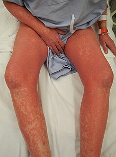
Sézary disease, or Sézary syndrome, is a type of cutaneous T-cell lymphoma that was first described by Albert Sézary. The affected T cells, known as Sézary's cells or Lutzner cells, have pathological quantities of mucopolysaccharides. Sézary disease is sometimes considered a late stage of mycosis fungoides with lymphadenopathy.

Rothmund–Thomson syndrome Rare autosomal recessive skin condition.

Rothmund–Thomson syndrome (RTS) is a rare autosomal recessive skin condition.

Large plaque parapsoriasis are skin lesions that may be included in the modern scheme of cutaneous conditions described as parapsoriasis. These lesions, called plaques, may be irregularly round-shaped to oval and are 10 cm (4 in) or larger in diameter. They can be very thin plaques that are asymptomatic or mildly pruritic. Large-plaque parapsoriasis is a common associate of retiform parapsoriasis, can be accompanied by poikiloderma vasculare atrophicans, and can in rare occasions be a precursor to cutaneous T-cell lymphoma.

Cutaneous lymphoid hyperplasia Medical condition

Cutaneous lymphoid hyperplasia refers to a groups of benign cutaneous disorders characterized by collections of lymphocytes, macrophages, and dendritic cells in the skin. Conditions included in this groups are:

Pagetoid reticulosis is a cutaneous condition, an uncommon lymphoproliferative disorder, sometimes considered a form of mycosis fungoides.

Granulomatous slack skin (GSS) is a rare cutaneous condition, a variant of lymphoma that typically presents in middle-aged adults.

Non-mycosis fungoides CD30− cutaneous large T-cell lymphoma is a cutaneous condition that usually presents as solitary or generalized plaques, nodules, or tumors of short duration.

Pleomorphic T-cell lymphoma is a cutaneous condition characterized by a 5-year survival rate of 62%.

Lutzner cells

Lutzner cells were discovered by Marvin A. Lutzner, Lucien-Marie Pautrier, and Albert Sézary. These cells are described as the smaller forms of Sézary cells, or Sézary-Lutzner cells, and the two variants are recognised as being morphologically different. Aggregates of these cells in mycosis fungoides are known as a Pautrier's microabscesses. They are a form of T-lymphocytes that has been mutated This atypical form of T-lymphocytes contains T-cell receptors on the surface and is found in both the dermis and epidermis layers of the skin. Since Lutzner cells are a mutated form of T-lymphocytes, they develop in bone marrow and are transported to the thymus is order to mature. The production and maturation stages occur before the cell has developed a mutation. Lutzner cells can form cutaneous T-cell lymphoma, which is a form of skin cancer.

Retiform parapsoriasis is a cutaneous condition, considered to be a type of large-plaque parapsoriasis. It is characterized by widespread, ill-defined plaques on the skin, that have a net-like or zebra-striped pattern. Skin atrophy, a wasting away of the cutaneous tissue, usually occurs within the area of these plaques.

Poikiloderma Medical condition

Poikiloderma is a skin condition that consists of areas of hypopigmentation, hyperpigmentation, telangiectasias and atrophy. Poikiloderma of Civatte is most frequently seen on the chest or the neck, characterized by red colored pigment on the skin that is commonly associated with sun damage.

Poikiloderma of Civatte is a cutaneous condition and refers to reticulated red to red-brown skin patches with telangiectasias. It is identifiable as a reddish-brown discoloration on the side of the neck, usually on both sides. It is more common in lighter-skinned individuals, in females rather than in males and more often affects middle-aged to elderly women. This disease is basically a change of the skin due to dilation of the blood vessels in the neck. "Civatte" was the French dermatologist who first identified it in the 1920s.

References

  1. 1 2 Howard MS, Smoller BR (Jun 2000). "Mycosis fungoides: classic disease and variant presentations". Seminars in Cutaneous Medicine and Surgery. 19 (2): 91–99. doi:10.1016/S1085-5629(00)80005-X. PMID   10892710.
  2. Diseases Database (DDB): 10208
  3. 1 2 3 4 Chapman, R. S.; Paul, C. J. (July 1975). "Poikiloderma atrophicans vasculare as a pointer to reticulosis of the skin". Postgraduate Medical Journal. 51 (597): 463–467. doi:10.1136/pgmj.51.597.463. PMC   2496068 . PMID   1103107.
  4. 1 2 Rapini, Ronald P.; Bolognia, Jean L.; Jorizzo, Joseph L. (2007). Dermatology: 2-Volume Set. St. Louis: Mosby. ISBN   978-1-4160-2999-1.
  5. 1 2 3 4 5 6 7 8 9 10 11 12 13 14 15 16 17 18 19 20 21 22 23 24 25 26 27 28 29 30 Lambert WC, Everett MA (Oct 1981). "The nosology of parapsoriasis". J. Am. Acad. Dermatol. 5 (4): 373–395. doi:10.1016/S0190-9622(81)70100-2. PMID   7026622.
  6. 1 2 Bonvalet, D.; Colau-Gohm, K.; Belaïch, S.; Civatte, J.; Degos, R. (Jan 1977). "The difference forms of "parapsoriasis en plaques". A report of 90 cases (author's transl)". Annales de Dermatologie et de Vénéréologie. 104 (1): 18–25. PMID   843023.
  7. 1 2 3 4 Kreuter A, Hoffmamm K, Altmeyer P (Apr 2005). "A case of poikiloderma vasculare atrophicans, a rare variant of cutaneous T-cell lymphoma, responding to extracorporeal photopheresis". Journal of the American Academy of Dermatology. 52 (4): 706–708. doi:10.1016/j.jaad.2004.10.877. PMID   15793532.
  8. "A definition of the term "hyperpigmentation" from Aocd.com" . Retrieved Feb 6, 2010.
  9. "A definition of the term "hyperkeratosis" from Everydayhealth.com" . Retrieved Feb 6, 2010.
  10. 1 2 "A definition of the term "poikiloderma" from Medterms.com" . Retrieved Dec 28, 2009.
  11. Brocq L (1902). "Les parapsoriasis". Ann Dermatol Syphiligr (Paris). 3: 433–468.
  12. Sutton RL (1956). Diseases of the skin. St. Louis, Mo.: The C. V. Mosby Co. pp. 936–941.
  13. Sehgal, V. N.; Srivastava, G.; Aggarwal, A. K. (Nov–Dec 2007). "Parapsoriasis: a complex issue". Skinmed. 6 (6): 280–286. doi:10.1111/j.1540-9740.2007.06490.x. PMID   17975354.