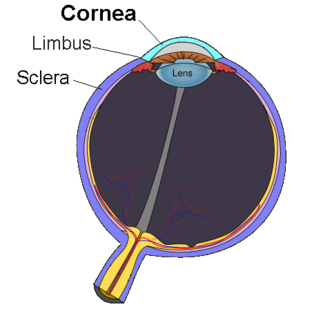
The cornea is the transparent front part of the eye that covers the iris, pupil, and anterior chamber. Along with the anterior chamber and lens, the cornea refracts light, accounting for approximately two-thirds of the eye's total optical power. In humans, the refractive power of the cornea is approximately 43 dioptres. The cornea can be reshaped by surgical procedures such as LASIK.

The corneal endothelium is a single layer of endothelial cells on the inner surface of the cornea. It faces the chamber formed between the cornea and the iris.

Corneal transplantation, also known as corneal grafting, is a surgical procedure where a damaged or diseased cornea is replaced by donated corneal tissue. When the entire cornea is replaced it is known as penetrating keratoplasty and when only part of the cornea is replaced it is known as lamellar keratoplasty. Keratoplasty simply means surgery to the cornea. The graft is taken from a recently deceased individual with no known diseases or other factors that may affect the chance of survival of the donated tissue or the health of the recipient.
Fuchs dystrophy, also referred to as Fuchs endothelial corneal dystrophy (FECD) and Fuchs endothelial dystrophy (FED), is a slowly progressing corneal dystrophy that usually affects both eyes and is slightly more common in women than in men. Although early signs of Fuchs dystrophy are sometimes seen in people in their 30s and 40s, the disease rarely affects vision until people reach their 50s and 60s.
Descemet's membrane is the basement membrane that lies between the corneal proper substance, also called stroma, and the endothelial layer of the cornea. It is composed of different kinds of collagen than the stroma. The endothelial layer is located at the posterior of the cornea. Descemet's membrane, as the basement membrane for the endothelial layer, is secreted by the single layer of squamous epithelial cells that compose the endothelial layer of the cornea.

A corneal ulcer, or ulcerative keratitis, is an inflammatory condition of the cornea involving loss of its outer layer. It is very common in dogs and is sometimes seen in cats. In veterinary medicine, the term corneal ulcer is a generic name for any condition involving the loss of the outer layer of the cornea, and as such is used to describe conditions with both inflammatory and traumatic causes.

Corneal dystrophy is a group of rare hereditary disorders characterised by bilateral abnormal deposition of substances in the transparent front part of the eye called the cornea.

Zinc finger E-box-binding homeobox 1 is a protein that in humans is encoded by the ZEB1 gene.

Collagen alpha-2(VIII) chain is a protein that in humans is encoded by the COL8A2 gene. Mutations of the gene are linked to posterior polymorphous dystrophy type 2.

Collagen alpha-1(VIII) chain is a protein that in humans is encoded by the COL8A1 gene.

Sodium bicarbonate transporter-like protein 11 is a protein that in humans is encoded by the SLC4A11 gene.
Visual system homeobox 1 is a protein that in humans is encoded by the VSX1 gene.
Bullous keratopathy, also known as pseudophakic bullous keratopathy (PBK), is a pathological condition in which small vesicles, or bullae, are formed in the cornea due to endothelial dysfunction.

Meesmann corneal dystrophy (MECD) is a rare hereditary autosomal dominant disease that is characterized as a type of corneal dystrophy and a keratin disease. MECD is characterized by the formation of microcysts in the outermost layer of the cornea, known as the anterior corneal epithelium. The anterior corneal epithelium also becomes fragile. This usually affects both eyes rather than a single eye and worsens over time. There are two phenotypes, Meesmann corneal dystrophy 1 (MECD1) and Meesmann corneal dystrophy 2 (MECD2), which affect the genes KRT3 and KRT12, respectively. A heterozygous mutation in either of these genes will lead to a single phenotype. Many with Meesmann corneal dystrophy are asymptomatic or experience mild symptoms.

Anterior segment mesenchymal dysgenesis, or simply anterior segment dysgenesis (ASD), is a failure of the normal development of the tissues of the anterior segment of the eye. It leads to anomalies in the structure of the mature anterior segment, associated with an increased risk of glaucoma and corneal opacity.

Congenital hereditary corneal dystrophy (CHED) is a form of corneal endothelial dystrophy that presents at birth.
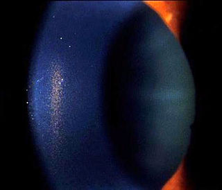
Krukenberg's spindle is the name given to the pattern formed on the inner surface of the cornea by pigmented iris cells that are shed during the mechanical rubbing of posterior pigment layer of the iris with the zonules that are deposited as a result of the currents of the aqueous humor. The sign was described in 1899 by Friedrich Ernst Krukenberg (1871-1946), who was a German pathologist specialising in ophthalmology.
Pre Descemet's endothelial keratoplasty (PDEK) is a kind of endothelial keratoplasty, where the pre descemet's layer (PDL) along with descemet's membrane (DM) and endothelium is transplanted. Conventionally in a corneal transplantation, doctors use a whole cornea or parts of the five layers of the cornea to perform correction surgeries. In May 2013, Dr Harminder Dua discovered a sixth layer between the stroma and the descemet membrane which was named after him as the Dua's layer. In the PDEK technique, doctors take the innermost two layers of the cornea, along with the Dua's layer and graft it in the patient's eye.
Descemet membrane endothelial keratoplasty (DMEK) is a method of corneal transplantation. The DMEK technique involves the removal of a very thin sheet of tissue from the posterior side of a person's cornea, replacing it with the two innermost layers of corneal tissue from a donor's eyeball. The two corneal layers which are exchanged are the Descemet's membrane and the corneal endothelium. The person's corneal tissue is gently excised and replaced with the donor tissue via small 'clear corneal incisions' (small corneal incisions just anterior to the corneal limbus. The donor tissue is tamponaded against the person's exposed posterior corneal stroma by injecting a small air bubble into the anterior chamber. To ensure the air tamponade is effective, it is necessary for people to strictly posture so that they are looking up at the ceiling during the recovery period and until the air bubble has fully resorbed.

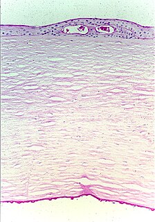
The human cornea is a transparent membrane which allows light to pass through it. The word corneal opacification literally means loss of normal transparency of cornea. The term corneal opacity is used particularly for the loss of transparency of cornea due to scarring. Transparency of the cornea is dependent on the uniform diameter and the regular spacing and arrangement of the collagen fibrils within the stroma. Alterations in the spacing of collagen fibrils in a variety of conditions including corneal edema, scars, and macular corneal dystrophy is clinically manifested as corneal opacity. The term "corneal blindness" is commonly used to describe blindness due to corneal opacity.
