Related Research Articles
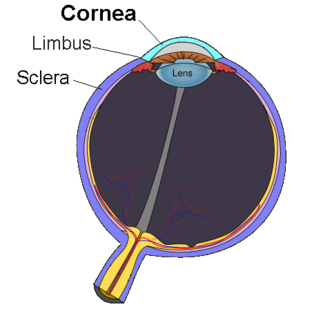
The cornea is the transparent front part of the eye that covers the iris, pupil, and anterior chamber. Along with the anterior chamber and lens, the cornea refracts light, accounting for approximately two-thirds of the eye's total optical power. In humans, the refractive power of the cornea is approximately 43 dioptres. The cornea can be reshaped by surgical procedures such as LASIK.
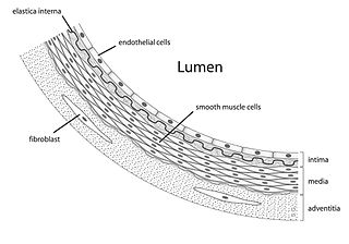
The endothelium is a single layer of squamous endothelial cells that line the interior surface of blood vessels, and lymphatic vessels. The endothelium forms an interface between circulating blood or lymph in the lumen and the rest of the vessel wall. Endothelial cells form the barrier between vessels and tissue and control the flow of substances and fluid into and out of a tissue.

The corneal endothelium is a single layer of endothelial cells on the inner surface of the cornea. It faces the chamber formed between the cornea and the iris.

Corneal transplantation, also known as corneal grafting, is a surgical procedure where a damaged or diseased cornea is replaced by donated corneal tissue. When the entire cornea is replaced it is known as penetrating keratoplasty and when only part of the cornea is replaced it is known as lamellar keratoplasty. Keratoplasty simply means surgery to the cornea. The graft is taken from a recently deceased individual with no known diseases or other factors that may affect the chance of survival of the donated tissue or the health of the recipient.
Fuchs dystrophy, also referred to as Fuchs endothelial corneal dystrophy (FECD) and Fuchs endothelial dystrophy (FED), is a slowly progressing corneal dystrophy that usually affects both eyes and is slightly more common in women than in men. Although early signs of Fuchs dystrophy are sometimes seen in people in their 30s and 40s, the disease rarely affects vision until people reach their 50s and 60s.
Recurrent corneal erosion is a disorder of the eyes characterized by the failure of the cornea's outermost layer of epithelial cells to attach to the underlying basement membrane. The condition is excruciatingly painful because the loss of these cells results in the exposure of sensitive corneal nerves. This condition can often leave patients with temporary blindness due to extreme light sensitivity (photophobia).
Descemet's membrane is the basement membrane that lies between the corneal proper substance, also called stroma, and the endothelial layer of the cornea. It is composed of different kinds of collagen than the stroma. The endothelial layer is located at the posterior of the cornea. Descemet's membrane, as the basement membrane for the endothelial layer, is secreted by the single layer of squamous epithelial cells that compose the endothelial layer of the cornea.

Corneal dystrophy is a group of rare hereditary disorders characterised by bilateral abnormal deposition of substances in the transparent front part of the eye called the cornea.

Carbohydrate sulfotransferase 6 is an enzyme that in humans is encoded by the CHST6 gene.

Collagen alpha-2(VIII) chain is a protein that in humans is encoded by the COL8A2 gene. Mutations of the gene are linked to posterior polymorphous dystrophy type 2.

Collagen alpha-1(VIII) chain is a protein that in humans is encoded by the COL8A1 gene.

Sodium bicarbonate transporter-like protein 11 is a protein that in humans is encoded by the SLC4A11 gene.
Visual system homeobox 1 is a protein that in humans is encoded by the VSX1 gene.

Meesmann corneal dystrophy (MECD) is a rare hereditary autosomal dominant disease that is characterized as a type of corneal dystrophy and a keratin disease. MECD is characterized by the formation of microcysts in the outermost layer of the cornea, known as the anterior corneal epithelium. The anterior corneal epithelium also becomes fragile. This usually affects both eyes rather than a single eye and worsens over time. There are two phenotypes, Meesmann corneal dystrophy 1 (MECD1) and Meesmann corneal dystrophy 2 (MECD2), which affect the genes KRT3 and KRT12, respectively. A heterozygous mutation in either of these genes will lead to a single phenotype. Many with Meesmann corneal dystrophy are asymptomatic or experience mild symptoms.

Congenital stromal corneal dystrophy (CSCD) is an extremely rare, autosomal dominant form of corneal dystrophy. Only 4 families have been reported to have the disease by 2009. The main features of the disease are numerous opaque flaky or feathery areas of clouding in the stroma that multiply with age and eventually preclude visibility of the endothelium. Strabismus or primary open angle glaucoma was noted in some of the patients. Thickness of the cornea stays the same, Descemet's membrane and endothelium are relatively unaffected, but the fibrils of collagen that constitute stromal lamellae are reduced in diameter and lamellae themselves are packed significantly more tightly.
Epithelial basement membrane dystrophy (EBMD) is a disorder of the eye that can cause pain and dryness.

Congenital hereditary corneal dystrophy (CHED) is a form of corneal endothelial dystrophy that presents at birth.
Hereditary benign intraepithelial dyskeratosis is a rare autosomal dominant disease of the conjunctiva and the oral mucosa caused by a duplication of chromosome 4q35. In the mouth it appears similar to white sponge nevus, with painless, diffuse, folded and spongy white plaques. In the eye it appears as gelatinous plaques on bulbar perilimbal conjunctiva.
Keratoendotheliitis fugax hereditaria is an autosomal dominantly inherited disease of the cornea, caused by a point mutation in cryopyrin that in humans is encoded by the NLRP3 gene located on the long arm of chromosome 1.

The human cornea is a transparent membrane which allows light to pass through it. The word corneal opacification literally means loss of normal transparency of cornea. The term corneal opacity is used particularly for the loss of transparency of cornea due to scarring. Transparency of the cornea is dependent on the uniform diameter and the regular spacing and arrangement of the collagen fibrils within the stroma. Alterations in the spacing of collagen fibrils in a variety of conditions including corneal edema, scars, and macular corneal dystrophy is clinically manifested as corneal opacity. The term "corneal blindness" is commonly used to describe blindness due to corneal opacity.
References
- ↑ Schmid E, Lisch W, Philipp W, Lechner S, Göttinger W, Schlötzer-Schrehardt U, Müller T, Utermann G, Janecke AR (March 2006). "A new, X-linked endothelial corneal dystrophy". Am. J. Ophthalmol. 141 (3): 478–487. doi:10.1016/j.ajo.2005.10.020. PMID 16490493.
- ↑ Aldave AJ, Han J, Frausto RF (Aug 2013). "Genetics of the corneal endothelial dystrophies: an evidence-based review". Clinical Genetics. 84 (2): 109–19. doi:10.1111/cge.12191. PMC 3885339 . PMID 23662738.
- ↑ Frausto RF, Wang C, Aldave AJ (6 Nov 2014). "Transcriptome analysis of the human corneal endothelium". Investigative Ophthalmology & Visual Science. 55 (12): 7821–30. doi:10.1167/iovs.14-15021. PMC 4258927 . PMID 25377225.