Protein aggregation

Last updated
Misfolded proteins can form protein aggregates or amyloid fibrils, get degraded, or refold back to its native structure. Protein Aggregation.jpg
Misfolded proteins can form protein aggregates or amyloid fibrils, get degraded, or refold back to its native structure.

In molecular biology, protein aggregation is a phenomenon in which intrinsically-disordered or mis-folded proteins aggregate (i.e., accumulate and clump together) either intra- or extracellularly. [1] [2] Protein aggregates have been implicated in a wide variety of diseases known as amyloidoses, including ALS, Alzheimer's, Parkinson's and prion disease. [3] [4]

Contents

After synthesis, proteins typically fold into a particular three-dimensional conformation that is the most thermodynamically favorable: their native state. [5] This folding process is driven by the hydrophobic effect: a tendency for hydrophobic (water-fearing) portions of the protein to shield themselves from the hydrophilic (water-loving) environment of the cell by burying into the interior of the protein. Thus, the exterior of a protein is typically hydrophilic, whereas the interior is typically hydrophobic.

Protein structures are stabilized by non-covalent interactions and disulfide bonds between two cysteine residues. The non-covalent interactions include ionic interactions and weak van der Waals interactions. Ionic interactions form between an anion and a cation and form salt bridges that help stabilize the protein. Van der Waals interactions include nonpolar interactions (i.e. London dispersion force) and polar interactions (i.e. hydrogen bonds, dipole-dipole bond). These play an important role in a protein's secondary structure, such as forming an alpha helix or a beta sheet, and tertiary structure. Interactions between amino acid residues in a specific protein are very important in that protein's final structure.

When there are changes in the non-covalent interactions, as may happen with a change in the amino acid sequence, the protein is susceptible to misfolding or unfolding. In these cases, if the cell does not assist the protein in re-folding, or degrade the unfolded protein, the unfolded/misfolded protein may aggregate, in which the exposed hydrophobic portions of the protein may interact with the exposed hydrophobic patches of other proteins. [6] [7] There are three main types of protein aggregates that may form: amorphous aggregates, oligomers, and amyloid fibrils. [8]

Causes

Protein aggregation can occur due to a variety of causes. There are four classes that these causes can be categorized into, which are detailed below.

Mutations

Mutations that occur in the DNA sequence may or may not affect the amino acid sequence of the protein. When the sequence is affected, a different amino acid may change the interactions between the side chains that affect the folding of the protein. This can lead to exposed hydrophobic regions of the protein that aggregate with the same misfolded/unfolded protein or a different protein. [9]

In addition to mutations in the affected proteins themselves, protein aggregation could also be caused indirectly through mutations in proteins in regulatory pathways such as the refolding pathway (molecular chaperones) or the ubiquitin-proteasome pathway (ubiquitin ligases). [10] Chaperones help with protein refolding by providing a safe environment for the protein to fold. Ubiquitin ligases target proteins for degradation through ubiquitin modification. [11]

Problems with protein synthesis

Protein aggregation can be caused by problems that occur during transcription or translation. During transcription, DNA is copied into mRNA, forming a strand of pre-mRNA that undergoes RNA processing to form mRNA. [12] During translation, ribosomes and tRNA help translate the mRNA sequence into an amino acid sequence. [12] If problems arise during either step, making an incorrect mRNA strand and/or an incorrect amino acid sequence, this can cause the protein to misfold, leading to protein aggregation.[ citation needed ]

Environmental stresses

Environmental stresses such as extreme temperatures and pH or oxidative stress can also lead to protein aggregation. [13] One such disease is cryoglobulinemia.

Extreme temperatures can weaken and destabilize the non-covalent interactions between the amino acid residues. pHs outside of the protein's pH range can change the protonation state of the amino acids, which can increase or decrease the non-covalent interactions. This can also lead to less stable interactions and result in protein unfolding.

Oxidative stress can be caused by radicals such as reactive oxygen species (ROS). These unstable radicals can attack the amino acid residues, leading to oxidation of side chains (e.g. aromatic side chains, methionine side chains) and/or cleavage of the polypeptide bonds. [14] This can affect the non-covalent interactions that hold the protein together correctly, which can cause protein destabilization, and may cause the protein to unfold. [13]

Aging

Cells have mechanisms that can refold or degrade protein aggregates. However, as cells age, these control mechanisms are weakened and the cell is less able to resolve the aggregates. [13]

The hypothesis that protein aggregation is a causative process in aging is testable now since some models of delayed aging are in hand. If the development of protein aggregates was an aging independent process, slowing down aging will show no effect on the rate of proteotoxicity over time. However, if aging is associated with decline in the activity of protective mechanisms against proteotoxicity, the slow aging models would show reduced aggregation and proteotoxicity. To address this problem several toxicity assays have been done in C. elegans. These studies indicated that reducing the activity of insulin/IGF signaling (IIS), a prominent aging regulatory pathway protects from neurodegeneration-linked toxic protein aggregation. The validity of this approach has been tested and confirmed in mammals as reducing the activity of the IGF-1 signaling pathway protected Alzheimer's model mice from the behavioral and biochemical impairments associated with the disease. [15]

Aggregate localization

Several studies have shown that cellular responses to protein aggregation are well-regulated and organized. Protein aggregates localize to specific areas in the cell, and research has been done on these localizations in prokaryotes (E.coli) and eukaryotes (yeast, mammalian cells). [16] From the macroscopic point of view, positron emission tomography tracers are used for certain misfolded proitein. [17] Recently, a team of researchers led by Dr. Alessandro Crimi has proposed a machine learning method to predict future deposition in the brain. [18]


Bacteria

The aggregates in bacteria asymmetrically end up at one of the poles of the cell, the "older pole." After the cell divides, the daughter cells with the older pole gets the protein aggregate and grows more slowly than daughter cells without the aggregate. This provides a natural selection mechanism for reducing protein aggregates in the bacterial population. [19]

Yeast

A scheme of a yeast cell harboring JUNQ and IPOD inclusions.png

Most of the protein aggregates in yeast cells get refolded by molecular chaperones. However, some aggregates, such as the oxidatively damaged proteins or the proteins marked for degradation, cannot be refolded. Rather, there are two compartments that they can end up in. Protein aggregates can be localized at the Juxtanuclear quality-control compartment (JUNQ), which is near the nuclear membrane, or at the Insoluble Protein deposit (IPOD), near the vacuole in yeast cells. [13] Protein aggregates localize at JUNQ when they are ubiquitinated and targeted for degradation. The aggregated and insoluble proteins localize at IPOD as a more permanent deposition. There is evidence that the proteins here may be removed by autophagy. [20] These two pathways work together in that the proteins tend to come to the IPOD when the proteasome pathway is being overworked. [20]

Mammalian cells

In mammalian cells, these protein aggregates are termed "aggresomes" and they are formed when the cell is diseased. This is because aggregates tend to form when there are heterologous proteins present in the cell, which can arise when the cell is mutated. Different mutates of the same protein may form aggresomes of different morphologies, ranging from diffuse dispersion of soluble species to large puncta, which in turn bear different pathogenicity. [21] The E3 ubiquitin ligase is able to recognize misfolded proteins and ubiquinate them. HDAC6 can then bind to the ubiquitin and the motor protein dynein to bring the marked aggregates to the microtubule organizing center (MTOC). There, they pack together into a sphere that surrounds the MTOC. They bring over chaperones and proteasomes and activate autophagy. [22]

Elimination

There are two main protein quality control systems in the cell that are responsible for eliminating protein aggregates. Misfolded proteins can get refolded by the bi-chaperone system or degraded by the ubiquitin proteasome system or autophagy. [23]

Refolding

The bi-chaperone system utilizes the Hsp70 (DnaK-DnaJ-GrpE in E. coli and Ssa1-Ydj1/Sis1-Sse1/Fe1 in yeast) and Hsp100 (ClpB in E. coli and Hsp104 in yeast) chaperones for protein disaggregation and refolding. [24]

Hsp70 interacts with the protein aggregates and recruits Hsp100. Hsp70 stabilizes an activated Hsp100. Hsp100 proteins have aromatic pore loops that are used for threading activity to disentangle single polypeptides. This threading activity can be initiated at the N-terminus, C-terminus or in the middle of the polypeptide. The polypeptide gets translocated through Hsp100 in a series of steps, utilizing an ATP at each step. [24] The polypeptide unfolds and is then allowed to refold either by itself or with the help of heat shock proteins. [25]

Degradation

Misfolded proteins can be eliminated through the ubiquitin-proteasome system (UPS). This consists of an E1-E2-E3 pathway that ubiquinates proteins to mark them for degradation. In eukaryotes, the proteins get degraded by the 26S proteasome. In mammalian cells, the E3 ligase, carboxy-terminal Hsp70 interacting protein (CHIP), targets Hsp70-bound proteins. In yeast, the E3 ligases Doa10 and Hrd1 have similar functions on endoplasmic reticulum proteins. [26] On the molecular level, degradation rate of aggregates vary from protein to protein due to their different internal environments, and thus different accessibility for protease molecules. [27]

Ubiquitylation.svg

Misfolded proteins can also be eliminated through autophagy, in which the protein aggregates are delivered to the lysosome. [26]

Macro-micro-autophagy.gif

Toxicity

Although it has been thought that the mature protein aggregates themselves are toxic, evidence suggests that it is in fact immature protein aggregates that are most toxic. [28] [29] The hydrophobic patches of these aggregates can interact with other components of the cell and damage them. The hypotheses are that the toxicity of protein aggregates is related to mechanisms of the sequestration of cellular components, the generation of reactive oxygen species and the binding to specific receptors in the membrane or through the disruption of membranes. [30] A quantitative assay has been used to determine that higher molecular weight species are responsible for the membrane permeation. [31] It is known that protein aggregates in vitro can destabilize artificial phospholipid bilayers, leading to permeabilization of the membrane.[ citation needed ]

In biomanufacturing

Protein aggregation is also a common phenomenon in the biopharmaceutical manufacturing process, which may pose risks to patients via generating adverse immune responses. [32]

See also

Related Research Articles

<span class="mw-page-title-main">Proteasome</span> Protein complexes which degrade unnecessary or damaged proteins by proteolysis

Proteasomes are protein complexes which degrade unneeded or damaged proteins by proteolysis, a chemical reaction that breaks peptide bonds. Enzymes that help such reactions are called proteases.

<span class="mw-page-title-main">Protein folding</span> Change of a linear protein chain to a 3D structure

Protein folding is the physical process by which a protein, after synthesis by a ribosome as a linear chain of amino acids, changes from an unstable random coil into a more ordered three-dimensional structure. This structure permits the protein to become biologically functional.

<span class="mw-page-title-main">Chaperone (protein)</span> Proteins assisting in protein folding

In molecular biology, molecular chaperones are proteins that assist the conformational folding or unfolding of large proteins or macromolecular protein complexes. There are a number of classes of molecular chaperones, all of which function to assist large proteins in proper protein folding during or after synthesis, and after partial denaturation. Chaperones are also involved in the translocation of proteins for proteolysis.

Heat shock proteins (HSPs) are a family of proteins produced by cells in response to exposure to stressful conditions. They were first described in relation to heat shock, but are now known to also be expressed during other stresses including exposure to cold, UV light and during wound healing or tissue remodeling. Many members of this group perform chaperone functions by stabilizing new proteins to ensure correct folding or by helping to refold proteins that were damaged by the cell stress. This increase in expression is transcriptionally regulated. The dramatic upregulation of the heat shock proteins is a key part of the heat shock response and is induced primarily by heat shock factor (HSF). HSPs are found in virtually all living organisms, from bacteria to humans.

<span class="mw-page-title-main">Hsp70</span> Family of heat shock proteins

The 70 kilodalton heat shock proteins are a family of conserved ubiquitously expressed heat shock proteins. Proteins with similar structure exist in virtually all living organisms. Intracellularly localized Hsp70s are an important part of the cell's machinery for protein folding, performing chaperoning functions, and helping to protect cells from the adverse effects of physiological stresses. Additionally, membrane-bound Hsp70s have been identified as a potential target for cancer therapies and their extracellularly localized counterparts have been identified as having both membrane-bound and membrane-free structures.

<span class="mw-page-title-main">Chaperonin</span> InterPro Family

HSP60, also known as chaperonins (Cpn), is a family of heat shock proteins originally sorted by their 60kDa molecular mass. They prevent misfolding of proteins during stressful situations such as high heat, by assisting protein folding. HSP60 belong to a large class of molecules that assist protein folding, called molecular chaperones.

<span class="mw-page-title-main">Heat shock response</span> Type of cellular stress response

The heat shock response (HSR) is a cell stress response that increases the number of molecular chaperones to combat the negative effects on proteins caused by stressors such as increased temperatures, oxidative stress, and heavy metals. In a normal cell, proteostasis must be maintained because proteins are the main functional units of the cell. Many proteins take on a defined configuration in a process known as protein folding in order to perform their biological functions. If these structures are altered, critical processes could be affected, leading to cell damage or death. The heat shock response can be employed under stress to induce the expression of heat shock proteins (HSP), many of which are molecular chaperones, that help prevent or reverse protein misfolding and provide an environment for proper folding.

<span class="mw-page-title-main">Endoplasmic-reticulum-associated protein degradation</span>

Endoplasmic-reticulum-associated protein degradation (ERAD) designates a cellular pathway which targets misfolded proteins of the endoplasmic reticulum for ubiquitination and subsequent degradation by a protein-degrading complex, called the proteasome.

In eukaryotic cells, an aggresome refers to an aggregation of misfolded proteins in the cell, formed when the protein degradation system of the cell is overwhelmed. Aggresome formation is a highly regulated process that possibly serves to organize misfolded proteins into a single location.

The unfolded protein response (UPR) is a cellular stress response related to the endoplasmic reticulum (ER) stress. It has been found to be conserved between mammalian species, as well as yeast and worm organisms.

<span class="mw-page-title-main">HSPA1A</span> Protein-coding gene in the species Homo sapiens

Heat shock 70 kDa protein 1, also termed Hsp72, is a protein that in humans is encoded by the HSPA1A gene. As a member of the heat shock protein 70 family and a chaperone protein, it facilitates the proper folding of newly translated and misfolded proteins, as well as stabilize or degrade mutant proteins. In addition, Hsp72 also facilitates DNA repair. Its functions contribute to biological processes including signal transduction, apoptosis, protein homeostasis, and cell growth and differentiation. It has been associated with an extensive number of cancers, neurodegenerative diseases, cell senescence and aging, and inflammatory diseases such as Diabetes mellitus type 2 and rheumatoid arthritis.

<span class="mw-page-title-main">PSMD4</span> Enzyme found in humans

26S proteasome non-ATPase regulatory subunit 4, also as known as 26S Proteasome Regulatory Subunit Rpn10, is an enzyme that in humans is encoded by the PSMD4 gene. This protein is one of the 19 essential subunits that contributes to the complete assembly of 19S proteasome complex.

<span class="mw-page-title-main">PSMC1</span> Enzyme found in humans

26S protease regulatory subunit 4, also known as 26S proteasome AAA-ATPase subunit Rpt2, is an enzyme that in humans is encoded by the PSMC1 gene. This protein is one of the 19 essential subunits of a complete assembled 19S proteasome complex. Six 26S proteasome AAA-ATPase subunits together with four non-ATPase subunits form the base sub complex of 19S regulatory particle for proteasome complex.

<span class="mw-page-title-main">PSMD7</span> Enzyme found in humans

26S proteasome non-ATPase regulatory subunit 7, also known as 26S proteasome non-ATPase subunit Rpn8, is an enzyme that in humans is encoded by the PSMD7 gene.

<span class="mw-page-title-main">PSMD1</span> Protein found in humans

26S proteasome non-ATPase regulatory subunit 1, also as known as 26S Proteasome Regulatory Subunit Rpn2, is a protein that in humans is encoded by the PSMD1 gene. This protein is one of the 19 essential subunits that contributes to the complete assembly of 19S proteasome complex.

<span class="mw-page-title-main">PSMD14</span> Protein-coding gene in the species Homo sapiens

26S proteasome non-ATPase regulatory subunit 14, also known as 26S proteasome non-ATPase subunit Rpn11, is an enzyme that in humans is encoded by the PSMD14 gene. This protein is one of the 19 essential subunits of the complete assembled 19S proteasome complex. Nine subunits Rpn3, Rpn5, Rpn6, Rpn7, Rpn8, Rpn9, Rpn11, SEM1, and Rpn12 form the lid sub complex of the 19S regulatory particle of the proteasome complex.

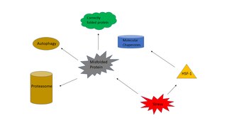
Proteostasis is the dynamic regulation of a balanced, functional proteome. The proteostasis network includes competing and integrated biological pathways within cells that control the biogenesis, folding, trafficking, and degradation of proteins present within and outside the cell. Loss of proteostasis is central to understanding the cause of diseases associated with excessive protein misfolding and degradation leading to loss-of-function phenotypes, as well as aggregation-associated degenerative disorders. Therapeutic restoration of proteostasis may treat or resolve these pathologies.

<span class="mw-page-title-main">JUNQ and IPOD</span> Types of cytosolic protein inclusion bodies

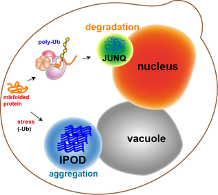
JUNQ and IPOD are types of cytosolic protein inclusion bodies in eukaryotes.

Chemical chaperones are a class of small molecules that function to enhance the folding and/or stability of proteins. Chemical chaperones are a broad and diverse group of molecules, and they can influence protein stability and polypeptide organization through a variety of mechanisms. Chemical chaperones are used for a range of applications, from production of recombinant proteins to treatment of protein misfolding in vivo.

Chaperones, also called molecular chaperones, are proteins that assist other proteins in assuming their three-dimensional fold, which is necessary for protein function. However, the fold of a protein is sensitive to environmental conditions, such as temperature and pH, and thus chaperones are needed to keep proteins in their functional fold across various environmental conditions. Chaperones are an integral part of a cell's protein quality control network by assisting in protein folding and are ubiquitous across diverse biological taxa. Since protein folding, and therefore protein function, is susceptible to environmental conditions, chaperones could represent an important cellular aspect of biodiversity and environmental tolerance by organisms living in hazardous conditions. Chaperones also affect the evolution of proteins in general, as many proteins fundamentally require chaperones to fold or are naturally prone to misfolding, and therefore mitigates protein aggregation.

References

  1. Aguzzi A, O'Connor T (March 2010). "Protein aggregation diseases: pathogenicity and therapeutic perspectives". Nature Reviews. Drug Discovery. 9 (3): 237–248. doi:10.1038/nrd3050. PMID   20190788. S2CID   5756683.
  2. Stefani M, Dobson CM (November 2003). "Protein aggregation and aggregate toxicity: new insights into protein folding, misfolding diseases and biological evolution". Journal of Molecular Medicine. 81 (11): 678–699. doi:10.1007/s00109-003-0464-5. PMID   12942175. S2CID   23544974.
  3. De Felice FG, Vieira MN, Meirelles MN, Morozova-Roche LA, Dobson CM, Ferreira ST (July 2004). "Formation of amyloid aggregates from human lysozyme and its disease-associated variants using hydrostatic pressure". FASEB Journal. 18 (10): 1099–1101. doi:10.1096/fj.03-1072fje. PMID   15155566. S2CID   13647147.
  4. Tanzi RE, Bertram L (February 2005). "Twenty years of the Alzheimer's disease amyloid hypothesis: a genetic perspective". Cell. 120 (4): 545–555. doi: 10.1016/j.cell.2005.02.008 . PMID   15734686. S2CID   206559875.
  5. Brüning A, Jückstock J (2015-01-01). "Misfolded proteins: from little villains to little helpers in the fight against cancer". Frontiers in Oncology. 5: 47. doi: 10.3389/fonc.2015.00047 . PMC   4338749 . PMID   25759792.
  6. Gething MJ, Sambrook J (January 1992). "Protein folding in the cell". Nature. 355 (6355): 33–45. Bibcode:1992Natur.355...33G. doi:10.1038/355033a0. PMID   1731198. S2CID   4330003.
  7. Roberts CJ (December 2007). "Non-native protein aggregation kinetics". Biotechnology and Bioengineering. 98 (5): 927–938. doi: 10.1002/bit.21627 . PMID   17705294. S2CID   21787377.
  8. Cox DL, Nelson MM (2013). Lehninger Principles of Biochemistry. New York: W.H. Freeman. p. 143. ISBN   978-1-4292-3414-6.
  9. Palanikumar L, Karpauskaite L, Al-Sayegh M, Chehade I, Alam M, Hassan S, et al. (June 2021). "Protein mimetic amyloid inhibitor potently abrogates cancer-associated mutant p53 aggregation and restores tumor suppressor function". Nature Communications. 12 (1): 3962. Bibcode:2021NatCo..12.3962P. doi:10.1038/s41467-021-23985-1. PMC   8233319 . PMID   34172723.
  10. Berke SJ, Paulson HL (June 2003). "Protein aggregation and the ubiquitin proteasome pathway: gaining the UPPer hand on neurodegeneration". Current Opinion in Genetics & Development. 13 (3): 253–261. doi:10.1016/S0959-437X(03)00053-4. PMID   12787787.
  11. Grillari J, Grillari-Voglauer R, Jansen-Dürr P (2010). Urbani F, Magariños R, Puertas S, Carretero M, Rodriguez V, Liñares R, et al. (eds.). Post-translational modification of cellular proteins by ubiquitin and ubiquitin-like molecules: role in cellular senescence and aging. Springer Science+Business Media. pp. 172–196. ISBN   978-1-4419-7002-2.
  12. 1 2 Weaver RF (2012). Molecular Biology. New York: McGraw-Hill. pp. 122–156, 523–600. ISBN   978-0-07-352532-7.
  13. 1 2 3 4 Tyedmers J, Mogk A, Bukau B (November 2010). "Cellular strategies for controlling protein aggregation". Nature Reviews. Molecular Cell Biology. 11 (11): 777–788. doi:10.1038/nrm2993. PMID   20944667. S2CID   22449895.
  14. Stadtman ER, Levine RL (December 2003). "Free radical-mediated oxidation of free amino acids and amino acid residues in proteins". Amino Acids. 25 (3–4): 207–218. doi:10.1007/s00726-003-0011-2. PMID   14661084. S2CID   26844881.
  15. Morley JF, Brignull HR, Weyers JJ, Morimoto RI (August 2002). "The threshold for polyglutamine-expansion protein aggregation and cellular toxicity is dynamic and influenced by aging in Caenorhabditis elegans". Proceedings of the National Academy of Sciences of the United States of America. 99 (16): 10417–10422. Bibcode:2002PNAS...9910417M. doi: 10.1073/pnas.152161099 . PMC   124929 . PMID   12122205.
  16. Coquel AS, Jacob JP, Primet M, Demarez A, Dimiccoli M, Julou T, et al. (April 2013). "Localization of protein aggregation in Escherichia coli is governed by diffusion and nucleoid macromolecular crowding effect". PLOS Computational Biology. 9 (4): e1003038. arXiv: 1303.1904 . Bibcode:2013PLSCB...9E3038C. doi: 10.1371/journal.pcbi.1003038 . PMC   3636022 . PMID   23633942.
  17. Scott JJ (January 1987). "The reinnervation of cat muscle spindles by skeletofusimotor axons". Brain Research. 401 (1): 152–154. doi:10.1016/0006-8993(87)91175-9. PMID   2949798. S2CID   42447184.
  18. Gherardini L, Zajdel A, Pini L, Crimi A (October 2023). "Prediction of misfolded proteins spreading in Alzheimer's disease using machine learning and spreading models". Cerebral Cortex. doi: 10.1093/cercor/bhad380 . PMC   10724880 . PMID   37833822.
  19. Bednarska NG, Schymkowitz J, Rousseau F, Van Eldere J (September 2013). "Protein aggregation in bacteria: the thin boundary between functionality and toxicity". Microbiology. 159 (Pt 9): 1795–1806. doi: 10.1099/mic.0.069575-0 . PMID   23894132.
  20. 1 2 Takalo M, Salminen A, Soininen H, Hiltunen M, Haapasalo A (2013-03-08). "Protein aggregation and degradation mechanisms in neurodegenerative diseases". American Journal of Neurodegenerative Disease. 2 (1): 1–14. PMC   3601466 . PMID   23516262.
  21. Wan W, Zeng L, Jin W, Chen X, Shen D, Huang Y, et al. (December 2021). "A Solvatochromic Fluorescent Probe Reveals Polarity Heterogeneity upon Protein Aggregation in Cells". Angewandte Chemie. 60 (49): 25865–25871. doi:10.1002/anie.202107943. PMID   34562048. S2CID   237626399.
  22. Garcia-Mata R, Gao YS, Sztul E (June 2002). "Hassles with taking out the garbage: aggravating aggresomes". Traffic. 3 (6): 388–396. doi: 10.1034/j.1600-0854.2002.30602.x . PMID   12010457. S2CID   305786.
  23. Gregersen N, Bolund L, Bross P (October 2005). "Protein misfolding, aggregation, and degradation in disease". Molecular Biotechnology. 31 (2): 141–150. doi:10.1385/MB:31:2:141. PMID   16170215. S2CID   36403914.
  24. 1 2 Mogk A, Kummer E, Bukau B (2015-01-01). "Cooperation of Hsp70 and Hsp100 chaperone machines in protein disaggregation". Frontiers in Molecular Biosciences. 2: 22. doi: 10.3389/fmolb.2015.00022 . PMC   4436881 . PMID   26042222.
  25. Liberek K, Lewandowska A, Zietkiewicz S (January 2008). "Chaperones in control of protein disaggregation". The EMBO Journal. 27 (2): 328–335. doi:10.1038/sj.emboj.7601970. PMC   2234349 . PMID   18216875.
  26. 1 2 Chen B, Retzlaff M, Roos T, Frydman J (August 2011). "Cellular strategies of protein quality control". Cold Spring Harbor Perspectives in Biology. 3 (8): a004374. doi:10.1101/cshperspect.a004374. PMC   3140689 . PMID   21746797.
  27. Wan W, Zeng L, Jin W, Chen X, Shen D, Huang Y, et al. (December 2021). "A Solvatochromic Fluorescent Probe Reveals Polarity Heterogeneity upon Protein Aggregation in Cells". Angewandte Chemie. 60 (49): 25865–25871. doi:10.1002/anie.202107943. PMID   34562048. S2CID   237626399.
  28. Zhu YJ, Lin H, Lal R (June 2000). "Fresh and nonfibrillar amyloid beta protein(1-40) induces rapid cellular degeneration in aged human fibroblasts: evidence for AbetaP-channel-mediated cellular toxicity". FASEB Journal. 14 (9): 1244–1254. doi:10.1096/fasebj.14.9.1244. PMID   10834946. S2CID   42263619.
  29. Nilsberth C, Westlind-Danielsson A, Eckman CB, Condron MM, Axelman K, Forsell C, et al. (September 2001). "The 'Arctic' APP mutation (E693G) causes Alzheimer's disease by enhanced Abeta protofibril formation". Nature Neuroscience. 4 (9): 887–893. doi:10.1038/nn0901-887. PMID   11528419. S2CID   13516479.
  30. Soto C (January 2003). "Unfolding the role of protein misfolding in neurodegenerative diseases". Nature Reviews. Neuroscience. 4 (1): 49–60. doi:10.1038/nrn1007. PMID   12511861. S2CID   205499427.
  31. Flagmeier P, De S, Wirthensohn DC, Lee SF, Vincke C, Muyldermans S, et al. (June 2017). "Ultrasensitive Measurement of Ca2+ Influx into Lipid Vesicles Induced by Protein Aggregates". Angewandte Chemie. 56 (27): 7750–7754. doi:10.1002/anie.201700966. PMC   5615231 . PMID   28474754.
  32. Vázquez-Rey M, Lang DA (July 2011). "Aggregates in monoclonal antibody manufacturing processes". Biotechnology and Bioengineering. 108 (7): 1494–1508. doi:10.1002/bit.23155. PMID   21480193. S2CID   33285577.