Protist shell

Last updated

Comparative coccolithophore sizes.png
Size comparison between the relatively large coccolithophore Scyphosphaera apsteinii and the relatively small but ubiquitous coccolithophore Emiliania huxleyi [1]

Many protists have protective shells or tests, [2] usually made from silica (glass) or calcium carbonate (chalk). Protists are a diverse group of eukaryote organisms that are not plants, animals, or fungi. They are typically microscopic unicellular organisms that live in water or moist environments.

Contents

Protists shells are often tough, mineralised forms that resist degradation, and can survive the death of the protist as a microfossil. Although protists are typically very small, they are ubiquitous. Their numbers are such that their shells play a huge part in the formation of ocean sediments and in the global cycling of elements and nutrients.

The role of protist shells depends on the type of protist. Protists such as diatoms and radiolaria have intricate, glass-like shells made of silica that are hard and protective, and serve as a barrier to prevent water loss. The shells have small pores that allow for gas exchange and nutrient uptake. Coccolithophores and foraminifera also have hard protective shells, but the shells are made of calcium carbonate. These shells can help with buoyancy, allowing the organisms to float in the water column and move around more easily.

In addition to protection and support, protist shells also serve scientists as a means of identification. By examining the characteristics of the shells, different species of protists can be identified and their ecology and evolution can be studied.

Protists

Cellular life likely originated as single-celled prokaryotes (including modern bacteria and archaea) and later evolved into more complex eukaryotes. Eukaryotes include organisms such as plants, animals, fungi and "protists". Protists are usually single-celled and microscopic. They can be heterotrophic, meaning they obtain nutrients by consuming other organisms, or autotrophic, meaning they produce their own food through photosynthesis or chemosynthesis, or mixotrophic, meaning they produce their own food through a mixture of those methods.

The term protist came into use historically to refer to a group of biologically similar organisms; however, modern research has shown it to be a paraphyletic group that does not contain all descendants of a common ancestor. As such it does not constitute a clade and is not currently in formal scientific use. Nonetheless, the term continues to be used informally to refer to those eukaryotes that cannot be classified as plants, fungi or animals.

Most protists are too small to be seen with the naked eye. They are highly diverse organisms currently organised into 18 phyla, but are not easy to classify. [3] [4] Studies have shown high protist diversity exists in oceans, deep sea-vents and river sediments, suggesting large numbers of eukaryotic microbial communities have yet to be discovered. [5] [6] As eukaryotes, protists possess within their cell at least one nucleus, as well as organelles such as mitochondria and Golgi bodies. Many protists are asexual but can reproduce rapidly through mitosis or by fragmentation; others (including foraminifera) may reproduce either sexually or asexually. [7]

In contrast to the cells of bacteria and archaea, the cells of protists and other eukaryotes are highly organised. Plants, animals and fungi are usually multi-celled and are typically macroscopic. Most protists are single-celled and microscopic, but there are exceptions, and some marine protists are neither single-celled nor microscopic, such as seaweed.

Silicon-based shells

A diatom, enclosed in a silica cell wall 20110123 185042 Diatom.jpg
A diatom, enclosed in a silica cell wall

Although silicon is readily available in the form of silicates, very few organisms use it directly. Diatoms, radiolaria, and siliceous sponges use biogenic silica as a structural material for their skeletons. In more advanced plants, the silica phytoliths (opal phytoliths) are rigid microscopic bodies occurring in the cell; some plants, including rice, need silica for their growth. [8] [9] [10] Silica has been shown to improve plant cell wall strength and structural integrity in some plants. [11]

Diatoms

Diatoms form a (disputed) phylum containing about 100,000 recognised species of mainly unicellular algae. Diatoms generate about 20 per cent of the oxygen produced on the planet each year, [12] take in over 6.7 billion metric tons of silicon each year from the waters in which they live, [13] and contribute nearly half of the organic material found in the oceans.

Diatoms are enclosed in protective silica (glass) shells called frustules. The beautifully engineered and intricate structure of many of these frustules is such that they are often referred to as "jewels of the sea". [14] Each frustule is made from two interlocking parts covered with tiny holes through which the diatom exchanges nutrients and wastes. [15] The frustules of dead diatoms drift to the ocean floor where, over millions of years, they can build up as much as half a mile deep. [16]

Diatoms uses silicon in the biogenic silica (BSiO2) form, [17] which is taken up by the silicon transport protein to be predominantly used in constructing these protective cell wall structures. [18] Silicon enters the ocean in a dissolved form such as silicic acid or silicate. [19] Since diatoms are one of the main users of these forms of silicon, they contribute greatly to the concentration of silicon throughout the ocean. Silicon forms a nutrient-like profile in the ocean due to the diatom productivity in shallow depths, which means there is less concentration of silicon in the upper ocean and more concentration of silicon in the deep ocean. [19]

Diatom productivity in the upper ocean contribute to the amount of silicon exported to the lower ocean. [20] When diatom cells are lysed in the upper ocean, their nutrients like, iron, zinc, and silicon, are brought to the lower ocean through a process called marine snow. Marine snow involves the downward transfer of particulate organic matter by vertical mixing of dissolved organic matter. [21] Availability of silicon appears crucial for diatom productivity, and as long as silicic acid is available for diatoms to utilize, the diatoms contribute other important nutrient concentrations in the deep ocean. [22]

In coastal zones, diatoms serve as the major phytoplanktonic organisms and greatly contribute to biogenic silica production. In the open ocean, however, diatoms have a reduced role in global annual silica production. Diatoms in North Atlantic and North Pacific subtropical gyres contribute only about 6% of global annual marine silica production, while the Southern Ocean produces about one-third of the global marine biogenic silica. [23] The Southern Ocean is referred to as having a "biogeochemical divide", since only minuscule amounts of silicon is transported out of this region. [24]

Diatom shapes
Haeckel Diatomea.jpg
Haeckel Diatomea 4.jpg
Drawings by Haeckel 1904
Diatoms
Diatoms have a silica shell (frustule) with radial (centric) or bilateral (pennate) symmetry
Different diatom frustule shapes and sizes Different shapes of diatoms.png
Different diatom frustule shapes and sizes
Structure of a centric diatom frustule Structure of diatom frustules.png
Structure of a centric diatom frustule
Diatoms
Marine diatoms SEM2.jpg
Diatoms, major components of marine plankton, have silica skeletons called frustules. "The microscopic structures of diatoms help them manipulate light, leading to hopes they could be used in new technologies for light detection, computing or robotics. [27]
SEM images of pores in diatom frustules.webp
SEM images of pores in diatom frustules [28]

Diatom frustules have been accumulating for over 100 million years, leaving rich deposits of nano and microstructured silicon oxide in the form of diatomaceous earth around the globe. The evolutionary causes for the generation of nano and microstructured silica by photosynthetic algae are not yet clear. However, in 2018 it was shown that absorption of ultraviolet light by nanostructured silica protects the DNA in the algal cells, and this may be an evolutionary cause for the formation of the glass cages. [28] [29]

Triparma laevis and shell.jpg
Triparma laevis and a drawing of its silicate shell, scale bar = 1 μm.
Triparma laevis exploded shell.jpg
Exploded drawing of the shell, D = dorsal plate, G = girdle plate, S = shield plate and V = ventral plate.
Triparma laevis belongs to the Bolidophyceae, a sister taxon to the diatoms. [30] [31]
External videos
Nuvola apps kaboodle.svg Diatoms: Tiny factories you can see from space
Nuvola apps kaboodle.svg How diatoms build their beautiful shellsJourney to the Microcosmos

Radiolarians

Radiolarian shapes
Haeckel Phaeodaria 1.jpg
Haeckel Stephoidea edit.jpg
Drawings by Haeckel 1904

Radiolarians are unicellular predatory protists encased in elaborate globular shells usually made of silica and pierced with holes. Their name comes from the Latin for "radius". They catch prey by extending parts of their body through the holes. As with the silica frustules of diatoms, radiolarian shells can sink to the ocean floor when radiolarians die and become preserved as part of the ocean sediment. These remains, as microfossils, provide valuable information about past oceanic conditions. [32]

An animation of the diversity of radiolarian shells Animation of radiolarian diversity.gif
An animation of the diversity of radiolarian shells
Fossil radiolarian
Micro-CT model of radiolarian, Triplococcus acanthicus.png
X-ray microtomography of Triplococcus acanthicus. This is a microfossil from the Middle Ordovician with four nested spheres. The innermost sphere is highlighted red. Each segment is shown at the same scale. [34]
Turing and radiolarian morphology
Spherical radiolarian.jpg
Shell of a spherical radiolarian
Radiolarians - Actinomma sol (33732012006).jpg
Shell micrographs
Computer simulations of Turing patterns on a sphere closely replicate some radiolarian shell patterns [35]
External videos
Nuvola apps kaboodle.svg Radiolarian geometry
Nuvola apps kaboodle.svg Ernst Haeckel's radiolarian engravings

Calcium-based shells

Coccolithophores

Coccolithophores are minute unicellular photosynthetic protists with two flagella for locomotion. Most of them are protected by a shell called a coccosphere. Coccospheres are covered with ornate circular plates or scales called coccoliths. The coccoliths are made from calcium carbonate. The term coccolithophore derives from the Greek for a seed carrying stone, referring to their small size and the coccolith stones they carry. Under the right conditions they bloom, like other phytoplankton, and can turn the ocean milky white. [36]

Coccolithophores
9Calcidiscus leptoporus, diploid, SEM, showing coccoliths.tif
Have plates called coccoliths
Discoaster surculus 01.jpg
Extinct fossil
Coccolithophores build calcite skeletons important to the marine carbon cycle [37]

There are benefits for protists that carry protective shells. The diagram on the left below shows some benefits coccolithophore get from carrying coccoliths. In the diagram, (A) represents accelerated photosynthesis including carbon concentrating mechanisms (CCM) and enhanced light uptake via scattering of scarce photons for deep-dwelling species. (B) represents protection from photodamage including sunshade protection from ultraviolet light (UV) and photosynthetic active radiation (PAR) and energy dissipation under high-light conditions. (C) represents armour protection includes protection against viral/bacterial infections and grazing by selective and nonselective grazers. [38]

Benefits of having shells
Benefits of calcification in coccolithophores.jpg
Benefits in coccolithophore calcification [38] – see text above
Costs of having shells
Calcification and energetic costs of a coccolithophore cell.jpg
Energetic costs in coccolithophore calcification [38]

There are also costs for protists that carry protective shells. The diagram on the right above shows some of the energetic costs coccolithophore incur from carrying coccoliths. In the diagram, the energetic costs are reported in percentage of total photosynthetic budget. (A) represents transport processes include the transport into the cell from the surrounding seawater of primary calcification substrates Ca2+ and HCO3− (black arrows) and the removal of the end product H+ from the cell (gray arrow). The transport of Ca2+ through the cytoplasm to the coccolith vesicle (CV) is the dominant cost associated with calcification. (B) represents metabolic processes include the synthesis of coccolith-associated polysaccharides (CAPs – gray rectangles) by the Golgi complex (white rectangles) that regulate the nucleation and geometry of CaCO3 crystals. The completed coccolith (gray plate) is a complex structure of intricately arranged CAPs and CaCO3 crystals. (C) Mechanical and structural processes account for the secretion of the completed coccoliths that are transported from their original position adjacent to the nucleus to the cell periphery, where they are transferred to the surface of the cell. [38]

Foraminiferans

Foraminiferan shapes
Haeckel Thalamphora.jpg
Haeckel Thalamophora 12.jpg
Drawings by Haeckel 1904

Like radiolarians, foraminiferans (forams for short) are single-celled predatory protists, also protected with shells that have holes in them. Their name comes from the Latin for "hole bearers". Their shells, often called tests, may be single-chambered or multi-chambered; multi-chambered forams add more chambers as they grow. The most famous of these are made of calcite, but tests may also be made of aragonite, agglutinated sediment particles, chiton, or (rarely) of silica. Most forams are benthic, but about 40 living species are planktic. [39] They are widely researched with well established fossil records which allow scientists to infer a lot about past environments and climates. [32] Some foraminifera lack tests altogether. [40]

Foraminiferans
Foram-globigerina hg.jpg
Empty Foraminiferan test, showing multiple chambers and pores
G bulloides Brady 1884.jpg
...and in life, showing pseudopodia streaming from pores
Foraminiferans are important unicellular zooplankton protists, often with calcite tests
External videos
Nuvola apps kaboodle.svg foraminiferans
Nuvola apps kaboodle.svg Foraminiferal networks and growth
Benthic foraminifera Favulina hexagona, together with nanofossils enclosed inside the shell hexagons Favulina hexagona.png
Benthic foraminifera Favulina hexagona, together with nanofossils enclosed inside the shell hexagons

Other shells

Testate amoeba Collection Penard MHNG Specimen 531-1-2 Pamphagus granulatus.tif
Testate amoeba

The cell body of many choanoflagellates is surrounded by a distinguishing extracellular matrix or periplast. These cell coverings vary greatly in structure and composition and are used by taxonomists for classification purposes. Many choanoflagellates build complex basket-shaped "houses", called lorica, from several silica strips cemented together. [43] The functional significance of the periplast is unknown, but in sessile organisms, it is thought to aid attachment to the substrate. In planktonic organisms, there is speculation that the periplast increases drag, thereby counteracting the force generated by the flagellum and increasing feeding efficiency. [43] [44]

External videos
Nuvola apps kaboodle.svg Testate amoebas: blobby, modest shell dwellers                         Journey to the Microcosmos

Microfossils and sediments

Diatomaceous earth is a soft, siliceous, sedimentary rock made up of microfossils in the form of the frustules (shells) of single cell diatoms. This sample consists of a mixture of centric (radially symmetric) and pennate (bilaterally symmetric) diatoms. Click 3 times to fully enlarge. Diatomaceous Earth BrightField.jpg
Diatomaceous earth is a soft, siliceous, sedimentary rock made up of microfossils in the form of the frustules (shells) of single cell diatoms. This sample consists of a mixture of centric (radially symmetric) and pennate (bilaterally symmetric) diatoms. Click 3 times to fully enlarge.

The shells or skeletons of many protists survive over geological time scales as microfossils. Microfossils are fossils that are generally between 0.001mm and 1 mm in size, [45] the study of which requires the use of light or electron microscopy. Fossils which can be studied by the naked eye or low-powered magnification, such as a hand lens, are referred to as macrofossils.

Microfossils are a common feature of the geological record, from the Precambrian to the Holocene. They are most common in marine sediments, but also occur in brackish water, fresh water and terrestrial sedimentary deposits. While every kingdom of life is represented in the microfossil record, the most abundant forms are protist skeletons or cysts from the Chrysophyta, Pyrrhophyta, Sarcodina, acritarchs and chitinozoans, together with pollen and spores from the vascular plants.

In 2017, fossilized microorganisms, or microfossils, were discovered in hydrothermal vent precipitates in the Nuvvuagittuq Belt that may be as old as 4.28 billion years old, the oldest record of life on Earth, suggesting "an almost instantaneous emergence of life" (in a geological time-scale sense), after ocean formation 4.41 billion years ago, and not long after the formation of the Earth 4.54 billion years ago. [46] [47] [48] [49] Nonetheless, life may have started even earlier, at nearly 4.5 billion years ago, as claimed by some researchers. [50] [51]

See also

Related Research Articles

<span class="mw-page-title-main">Diatom</span> Class of microalgae, found in the oceans, waterways and soils of the world

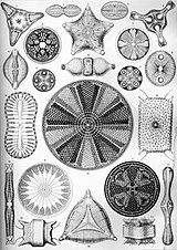
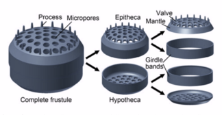
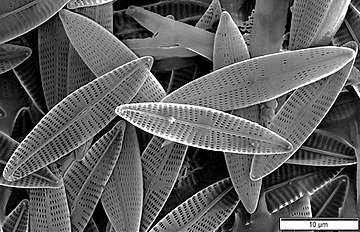
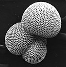
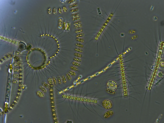
A diatom is any member of a large group comprising several genera of algae, specifically microalgae, found in the oceans, waterways and soils of the world. Living diatoms make up a significant portion of the Earth's biomass: they generate about 20 to 50 percent of the oxygen produced on the planet each year, take in over 6.7 billion tonnes of silicon each year from the waters in which they live, and constitute nearly half of the organic material found in the oceans. The shells of dead diatoms can reach as much as a half-mile deep on the ocean floor, and the entire Amazon basin is fertilized annually by 27 million tons of diatom shell dust transported by transatlantic winds from the African Sahara, much of it from the Bodélé Depression, which was once made up of a system of fresh-water lakes.

<span class="mw-page-title-main">Coccolithophore</span> Unicellular algae responsible for the formation of chalk

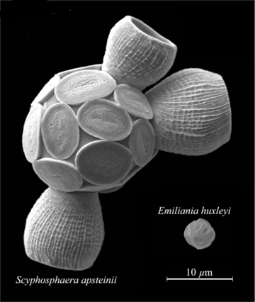
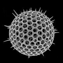
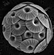
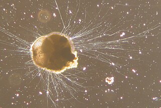
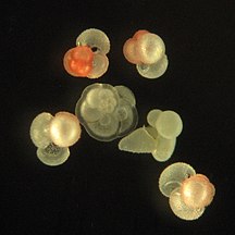
Coccolithophores, or coccolithophorids, are single-celled organisms which are part of the phytoplankton, the autotrophic (self-feeding) component of the plankton community. They form a group of about 200 species, and belong either to the kingdom Protista, according to Robert Whittaker's five-kingdom system, or clade Hacrobia, according to a newer biological classification system. Within the Hacrobia, the coccolithophores are in the phylum or division Haptophyta, class Prymnesiophyceae. Coccolithophores are almost exclusively marine, are photosynthetic, and exist in large numbers throughout the sunlight zone of the ocean.

<span class="mw-page-title-main">Zooplankton</span> Heterotrophic protistan or metazoan members of the plankton ecosystem

Zooplankton are the animal component of the planktonic community, having to consume other organisms to thrive. Plankton are aquatic organisms that are unable to swim effectively against currents. Consequently, they drift or are carried along by currents in the ocean, or by currents in seas, lakes or rivers.

<span class="mw-page-title-main">Radiolaria</span> Phylum of single-celled organisms

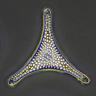
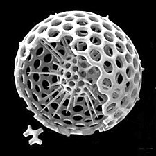
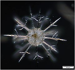
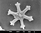
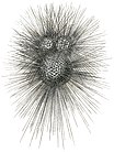
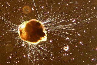
The Radiolaria, also called Radiozoa, are protozoa of diameter 0.1–0.2 mm that produce intricate mineral skeletons, typically with a central capsule dividing the cell into the inner and outer portions of endoplasm and ectoplasm. The elaborate mineral skeleton is usually made of silica. They are found as zooplankton throughout the global ocean. As zooplankton, radiolarians are primarily heterotrophic, but many have photosynthetic endosymbionts and are, therefore, considered mixotrophs. The skeletal remains of some types of radiolarians make up a large part of the cover of the ocean floor as siliceous ooze. Due to their rapid change as species and intricate skeletons, radiolarians represent an important diagnostic fossil found from the Cambrian onwards.

<span class="mw-page-title-main">Coccolith</span> Calcium carbonate scales covering some phytoplankton species

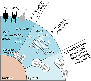
Coccoliths are individual plates or scales of calcium carbonate formed by coccolithophores and cover the cell surface arranged in the form of a spherical shell, called a coccosphere.

<span class="mw-page-title-main">Micropaleontology</span> Branch of paleontology that studies microfossils

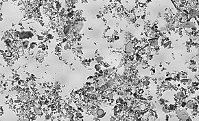
Micropaleontology is the branch of paleontology (palaeontology) that studies microfossils, or fossils that require the use of a microscope to see the organism, its morphology and its characteristic details.

<span class="mw-page-title-main">Foraminifera</span> Phylum of amoeboid protists

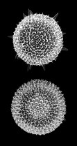
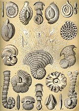
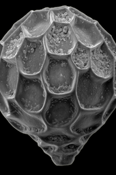
Foraminifera are single-celled organisms, members of a phylum or class of Cercozoan protists characterized by streaming granular ectoplasm for catching food and other uses; and commonly an external shell of diverse forms and materials. Tests of chitin are believed to be the most primitive type. Most foraminifera are marine, the majority of which live on or within the seafloor sediment, while a smaller number float in the water column at various depths, which belong to the suborder Globigerinina. Fewer are known from freshwater or brackish conditions, and some very few (nonaquatic) soil species have been identified through molecular analysis of small subunit ribosomal DNA.

<span class="mw-page-title-main">Microfossil</span> Fossil that requires the use of a microscope to see it

A microfossil is a fossil that is generally between 0.001 mm and 1 mm in size, the visual study of which requires the use of light or electron microscopy. A fossil which can be studied with the naked eye or low-powered magnification, such as a hand lens, is referred to as a macrofossil.

<span class="mw-page-title-main">Biomineralization</span> Process by which living organisms produce minerals

Biomineralization, also written biomineralisation, is the process by which living organisms produce minerals, often resulting in hardened or stiffened mineralized tissues. It is an extremely widespread phenomenon: all six taxonomic kingdoms contain members that are able to form minerals, and over 60 different minerals have been identified in organisms. Examples include silicates in algae and diatoms, carbonates in invertebrates, and calcium phosphates and carbonates in vertebrates. These minerals often form structural features such as sea shells and the bone in mammals and birds.

<span class="mw-page-title-main">Biogenic silica</span> Type of biogenic mineral

Biogenic silica (bSi), also referred to as opal, biogenic opal, or amorphous opaline silica, forms one of the most widespread biogenic minerals. For example, microscopic particles of silica called phytoliths can be found in grasses and other plants.

<span class="mw-page-title-main">Test (biology)</span> Hard shell of some spherical marine animals

In biology, a test is the hard shell of some spherical marine animals and protists, notably sea urchins and microorganisms such as testate foraminiferans, radiolarians, and testate amoebae. The term is also applied to the covering of scale insects. The related Latin term testa is used for the hard seed coat of plant seeds.

<span class="mw-page-title-main">Marine sediment</span>

Marine sediment, or ocean sediment, or seafloor sediment, are deposits of insoluble particles that have accumulated on the seafloor. These particles either have their origins in soil and rocks and have been transported from the land to the sea, mainly by rivers but also by dust carried by wind and by the flow of glaciers into the sea, or they are biogenic deposits from marine organisms or from chemical precipitation in seawater, as well as from underwater volcanoes and meteorite debris.

<span class="mw-page-title-main">Siliceous ooze</span> Biogenic pelagic sediment located on the deep ocean floor

Siliceous ooze is a type of biogenic pelagic sediment located on the deep ocean floor. Siliceous oozes are the least common of the deep sea sediments, and make up approximately 15% of the ocean floor. Oozes are defined as sediments which contain at least 30% skeletal remains of pelagic microorganisms. Siliceous oozes are largely composed of the silica based skeletons of microscopic marine organisms such as diatoms and radiolarians. Other components of siliceous oozes near continental margins may include terrestrially derived silica particles and sponge spicules. Siliceous oozes are composed of skeletons made from opal silica SiO2·nH2O, as opposed to calcareous oozes, which are made from skeletons of calcium carbonate (CaCO3·nH2O) organisms (i.e. coccolithophores). Silica (Si) is a bioessential element and is efficiently recycled in the marine environment through the silica cycle. Distance from land masses, water depth and ocean fertility are all factors that affect the opal silica content in seawater and the presence of siliceous oozes.

<span class="mw-page-title-main">Marine microorganisms</span> Any life form too small for the naked human eye to see that lives in a marine environment

Marine microorganisms are defined by their habitat as microorganisms living in a marine environment, that is, in the saltwater of a sea or ocean or the brackish water of a coastal estuary. A microorganism is any microscopic living organism or virus, that is too small to see with the unaided human eye without magnification. Microorganisms are very diverse. They can be single-celled or multicellular and include bacteria, archaea, viruses and most protozoa, as well as some fungi, algae, and animals, such as rotifers and copepods. Many macroscopic animals and plants have microscopic juvenile stages. Some microbiologists also classify viruses as microorganisms, but others consider these as non-living.

<span class="mw-page-title-main">Nassellaria</span> Order of single-celled organisms

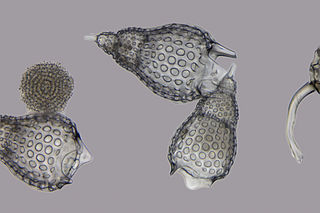
Nassellaria is an order of Rhizaria belonging to the class Radiolaria. The organisms of this order are characterized by a skeleton cross link with a cone or ring.

<span class="mw-page-title-main">Marine biogenic calcification</span> Shell formation mechanism

Marine biogenic calcification is the production of calcium carbonate by organisms in the global ocean.

<span class="mw-page-title-main">Marine protists</span> Protists that live in saltwater or brackish water

Marine protists are defined by their habitat as protists that live in marine environments, that is, in the saltwater of seas or oceans or the brackish water of coastal estuaries. Life originated as marine single-celled prokaryotes and later evolved into more complex eukaryotes. Eukaryotes are the more developed life forms known as plants, animals, fungi and protists. Protists are the eukaryotes that cannot be classified as plants, fungi or animals. They are mostly single-celled and microscopic. The term protist came into use historically as a term of convenience for eukaryotes that cannot be strictly classified as plants, animals or fungi. They are not a part of modern cladistics because they are paraphyletic.

<span class="mw-page-title-main">Foraminifera test</span> Shell of a particular type of protist

Foraminiferal tests are the tests of Foraminifera.

<span class="mw-page-title-main">Protists in the fossil record</span>

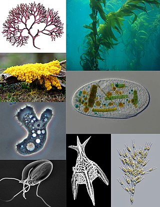
A protist is any eukaryotic organism that is not an animal, plant, or fungus. While it is likely that protists share a common ancestor, the last eukaryotic common ancestor, the exclusion of other eukaryotes means that protists do not form a natural group, or clade. Therefore, some protists may be more closely related to animals, plants, or fungi than they are to other protists. However, like algae, invertebrates and protozoans, the grouping is used for convenience.

Global paleoclimate indicators are the proxies sensitive to global paleoclimatic environment changes. They are mostly derived from marine sediments. Paleoclimate indicators derived from terrestrial sediments, on the other hand, are commonly influenced by local tectonic movements and paleogeographic variations. Factors governing the Earth's climate system include plate tectonics, which controls the configuration of continents, the interplay between the atmosphere and the ocean, and the Earth's orbital characteristics. Global paleoclimate indicators are established based on the information extracted from the analyses of geologic materials, including biological, geochemical and mineralogical data preserved in marine sediments. Indicators are generally grouped into three categories; paleontological, geochemical and lithological.

References

  1. Gafar, N. A., Eyre, B. D. and Schulz, K. G. (2019) "A comparison of species specific sensitivities to changing light and carbonate chemistry in calcifying marine phytoplankton". Scientific Reports, 9(1): 1–12. doi:10.1038/s41598-019-38661-0. CC-BY icon.svg Material was copied from this source, which is available under a Creative Commons Attribution 4.0 International License.
  2. "Groups of Protists | Boundless Biology". courses.lumenlearning.com. Retrieved 16 February 2021.
  3. Cavalier-Smith T (December 1993). "Kingdom protozoa and its 18 phyla". Microbiological Reviews. 57 (4): 953–94. doi:10.1128/mmbr.57.4.953-994.1993. PMC   372943 . PMID   8302218.
  4. Corliss JO (1992). "Should there be a separate code of nomenclature for the protists?". BioSystems. 28 (1–3): 1–14. doi:10.1016/0303-2647(92)90003-H. PMID   1292654.
  5. Slapeta J, Moreira D, López-García P (2005). "The extent of protist diversity: insights from molecular ecology of freshwater eukaryotes". Proceedings of the Royal Society B: Biological Sciences. 272 (1576): 2073–81. doi:10.1098/rspb.2005.3195. PMC   1559898 . PMID   16191619.
  6. Moreira D, López-García P (2002). "The molecular ecology of microbial eukaryotes unveils a hidden world" (PDF). Trends in Microbiology. 10 (1): 31–8. doi:10.1016/S0966-842X(01)02257-0. PMID   11755083.
  7. Foraminifera: notes for a short course organized by M.A. Buzas and B.K. Sen Gupta: prepared for the short course on foraminifera sponsored by the Paleontological Society, held at New Orleans, Louisiana, October 17, 1982. Thomas W. Broadhead, Paleontological Society. [Knoxville, Tenn.]: University of Tennessee, Dept. of Geological Sciences. 1982. ISBN   0-910249-05-9. OCLC   9276403.{{cite book}}: CS1 maint: others (link)
  8. Rahman, Atta-ur- (2008). "Silicon". Studies in Natural Products Chemistry. Vol. 35. p. 856. ISBN   978-0-444-53181-0.
  9. Exley, C. (1998). "Silicon in life:A bioinorganic solution to bioorganic essentiality". Journal of Inorganic Biochemistry. 69 (3): 139–144. doi:10.1016/S0162-0134(97)10010-1.
  10. Epstein, Emanuel (1999). "SILICON". Annual Review of Plant Physiology and Plant Molecular Biology. 50: 641–664. doi:10.1146/annurev.arplant.50.1.641. PMID   15012222.
  11. Kim, Sang Gyu; Kim, Ki Woo; Park, Eun Woo; Choi, Doil (2002). "Silicon-Induced Cell Wall Fortification of Rice Leaves: A Possible Cellular Mechanism of Enhanced Host Resistance to Blast". Phytopathology. 92 (10): 1095–103. doi: 10.1094/PHYTO.2002.92.10.1095 . PMID   18944220.
  12. The Air You're Breathing? A Diatom Made That
  13. Treguer, P.; Nelson, D. M.; Van Bennekom, A. J.; Demaster, D. J.; Leynaert, A.; Queguiner, B. (1995). "The Silica Balance in the World Ocean: A Reestimate". Science. 268 (5209): 375–9. Bibcode:1995Sci...268..375T. doi:10.1126/science.268.5209.375. PMID   17746543. S2CID   5672525.
  14. Ireland, T., "Engineering with algae". Biologist, 63(5): 10.
  15. Wassilieff, Maggy (2006) "Plankton - Plant plankton", Te Ara - the Encyclopedia of New Zealand. Accessed: 2 November 2019.
  16. "King's College London - Lake Megachad". www.kcl.ac.uk. Retrieved 5 May 2018.
  17. Bidle, Kay D.; Manganelli, Maura; Azam, Farooq (6 December 2002). "Regulation of Oceanic Silicon and Carbon Preservation by Temperature Control on Bacteria". Science. 298 (5600): 1980–1984. Bibcode:2002Sci...298.1980B. doi:10.1126/science.1076076. ISSN   0036-8075. PMID   12471255. S2CID   216994.
  18. Durkin, Colleen A.; Koester, Julie A.; Bender, Sara J.; Armbrust, E. Virginia (2016). "The evolution of silicon transporters in diatoms". Journal of Phycology. 52 (5): 716–731. doi:10.1111/jpy.12441. ISSN   1529-8817. PMC   5129515 . PMID   27335204.
  19. 1 2 Dugdale, R. C.; Wilkerson, F. P. (30 December 2001). "Sources and fates of silicon in the ocean: the role of diatoms in the climate and glacial cycles". Scientia Marina. 65 (S2): 141–152. doi: 10.3989/scimar.2001.65s2141 . ISSN   1886-8134.
  20. Baines, Stephen B.; Twining, Benjamin S.; Brzezinski, Mark A.; Krause, Jeffrey W.; Vogt, Stefan; Assael, Dylan; McDaniel, Hannah (December 2012). "Significant silicon accumulation by marine picocyanobacteria". Nature Geoscience. 5 (12): 886–891. Bibcode:2012NatGe...5..886B. doi:10.1038/ngeo1641. ISSN   1752-0908.
  21. Turner, Jefferson T. (January 2015). "Zooplankton fecal pellets, marine snow, phytodetritus and the ocean's biological pump". Progress in Oceanography. 130: 205–248. Bibcode:2015PrOce.130..205T. doi:10.1016/j.pocean.2014.08.005. ISSN   0079-6611.
  22. Yool, Andrew; Tyrrell, Toby (2003). "Role of diatoms in regulating the ocean's silicon cycle". Global Biogeochemical Cycles. 17 (4): n/a. Bibcode:2003GBioC..17.1103Y. doi: 10.1029/2002GB002018 . ISSN   1944-9224. S2CID   16849373.
  23. Tréguer, Paul J.; De La Rocha, Christina L. (3 January 2013). "The World Ocean Silica Cycle". Annual Review of Marine Science. 5 (1): 477–501. doi:10.1146/annurev-marine-121211-172346. PMID   22809182.
  24. Marinov, I.; Gnanadesikan, A.; Toggweiler, J. R.; Sarmiento, J. L. (June 2006). "The Southern Ocean biogeochemical divide". Nature. 441 (7096): 964–967. Bibcode:2006Natur.441..964M. doi:10.1038/nature04883. PMID   16791191. S2CID   4428683.
  25. Arsenieff, L.; Simon, N.; Rigaut-Jalabert, F.; Le Gall, F.; Chaffron, S.; Corre, E.; Com, E.; Bigeard, E.; Baudoux, A.C. (2018). "First Viruses Infecting the Marine Diatom Guinardia delicatula". Frontiers in Microbiology. 9: 3235. doi: 10.3389/fmicb.2018.03235 . PMC   6334475 . PMID   30687251.
  26. Zhang, D.; Wang, Y.; Cai, J.; Pan, J.; Jiang, X.; Jiang, Y. (2012). "Bio-manufacturing technology based on diatom micro- and nanostructure". Chinese Science Bulletin. 57 (30): 3836–3849. Bibcode:2012ChSBu..57.3836Z. doi: 10.1007/s11434-012-5410-x .
  27. Biodegradable glitter and pollution-eating microalgae: the new materials inspired by nature Horizon, 28 May 2020.
  28. 1 2 Aguirre, L. E., Ouyang, L., Elfwing, A., Hedblom, M., Wulff, A. and Inganäs, O. (2018) "Diatom frustules protect DNA from ultraviolet light". Scientific reports, 8(1): 1–6. doi:10.1038/s41598-018-21810-2. CC-BY icon.svg Material was copied from this source, which is available under a Creative Commons Attribution 4.0 International License.
  29. De Tommasi, E., Congestri, R., Dardano, P., De Luca, A.C., Managò, S., Rea, I. and De Stefano, M. (2018) "UV-shielding and wavelength conversion by centric diatom nanopatterned frustules". Scientific Reports, 8(1): 1–14. doi : 10.1038/s41598-018-34651-w. CC-BY icon.svg Material was copied from this source, which is available under a Creative Commons Attribution 4.0 International License.
  30. Booth, B.C. and Marchant, H.J. (1987) "Parmales, a new order of marine chrysophytes, with descriptions of three new genera and seven new species". Journal of Phycology, 23: 245–260. doi:10.1111/j.1529-8817.1987.tb04132.x.
  31. Kuwata, A., Yamada, K., Ichinomiya, M., Yoshikawa, S., Tragin, M., Vaulot, D. and Lopes dos Santos, A. (2018) "Bolidophyceae, a sister picoplanktonic group of diatoms – a review". Frontiers in Marine Science, 5: 370. doi:10.3389/fmars.2018.00370. CC-BY icon.svg Material was copied from this source, which is available under a Creative Commons Attribution 4.0 International License.
  32. 1 2 Wassilieff, Maggy (2006) "Plankton - Animal plankton", Te Ara - the Encyclopedia of New Zealand. Accessed: 2 November 2019.
  33. Kachovich, Sarah (2018) "Minds over Methods: Linking microfossils to tectonics" Blog of the Tectonics and Structural Geology Division of the European Geosciences Union.
  34. Kachovich, S., Sheng, J. and Aitchison, J. C., 2019. Adding a new dimension to investigations of early radiolarian evolution. Scientific reports, 9(1), pp. 1–10. doi:10.1038/s41598-019-42771-0. CC-BY icon.svg Material was copied from this source, which is available under a Creative Commons Attribution 4.0 International License.
  35. Varea, C.; Aragon, J. L.; Barrio, R. A. (1999). "Turing patterns on a sphere". Physical Review E. 60 (4): 4588–4592. Bibcode:1999PhRvE..60.4588V. doi:10.1103/PhysRevE.60.4588. PMID   11970318.
  36. Wassilieff, Maggy (2006) "A coccolithophore", Te Ara - the Encyclopedia of New Zealand. Accessed: 2 November 2019.
  37. Rost, B. and Riebesell, U. (2004) "Coccolithophores and the biological pump: responses to environmental changes". In: Coccolithophores: From Molecular Processes to Global Impact, pages 99–125, Springer. ISBN   9783662062784.
  38. 1 2 3 4 Monteiro, F.M., Bach, L.T., Brownlee, C., Bown, P., Rickaby, R.E., Poulton, A.J., Tyrrell, T., Beaufort, L., Dutkiewicz, S., Gibbs, S. and Gutowska, M.A. (2016) "Why marine phytoplankton calcify". Science Advances, 2(7): e1501822. doi : 10.1126/sciadv.1501822. CC-BY icon.svg Material was copied from this source, which is available under a Creative Commons Attribution 4.0 International License.
  39. Hemleben, C.; Anderson, O.R.; Spindler, M. (1989). Modern Planktonic Foraminifera. Springer-Verlag. ISBN   978-3-540-96815-3.
  40. Pawlowski, Jan; Bolivar, Ignacio; Fahrni, Jose F.; Vargas, Colomban De; Bowser, Samuel S. (November 1999). "Molecular Evidence That Reticulomyxa Filosa Is A Freshwater Naked Foraminifer". The Journal of Eukaryotic Microbiology. 46 (6): 612–617. doi:10.1111/j.1550-7408.1999.tb05137.x. ISSN   1066-5234. PMID   10568034. S2CID   36497475.
  41. Favulina hexagona European Geosciences Union, 9 November 2020.
  42. Foraminifera: History of Study, University College London. Retrieved: 18 November 2019.
  43. 1 2 Leadbeater BS, Thomsen H (2000). "Order Choanoflagellida". An Illustrated Guide to the Protozoa, Second Edition. Lawrence: Society of Protozoologists. 451: 14–38.
  44. Leadbeater BS, Kelly M (2001). "Evolution of animals choanoflagellates and sponges". Water and Atmosphere Online. 9 (2): 9–11.
  45. Drewes, Charlie. "Discovering Devonian Microfossils". Iowa State University. Retrieved 4 March 2017.
  46. Dodd, Matthew S.; Papineau, Dominic; Grenne, Tor; slack, John F.; Rittner, Martin; Pirajno, Franco; O'Neil, Jonathan; Little, Crispin T. S. (2 March 2017). "Evidence for early life in Earth's oldest hydrothermal vent precipitates" (PDF). Nature. 543 (7643): 60–64. Bibcode:2017Natur.543...60D. doi: 10.1038/nature21377 . PMID   28252057.
  47. Zimmer, Carl (1 March 2017). "Scientists Say Canadian Bacteria Fossils May Be Earth's Oldest". The New York Times . Retrieved 2 March 2017.
  48. Ghosh, Pallab (1 March 2017). "Earliest evidence of life on Earth 'found". BBC News . Retrieved 2 March 2017.
  49. Dunham, Will (1 March 2017). "Canadian bacteria-like fossils called oldest evidence of life". Reuters . Retrieved 1 March 2017.
  50. Staff (20 August 2018). "A timescale for the origin and evolution of all of life on Earth". Phys.org . Retrieved 20 August 2018.
  51. Betts, Holly C.; Putick, Mark N.; Clark, James W.; Williams, Tom A.; Donoghue, Philip C.J.; Pisani, Davide (20 August 2018). "Integrated genomic and fossil evidence illuminates life's early evolution and eukaryote origin". Nature . 2 (10): 1556–1562. doi:10.1038/s41559-018-0644-x. PMC   6152910 . PMID   30127539.

Further references