
A radiologic sign is an objective indication of some medical fact (that is, a medical sign) that is detected by a physician during radiologic examination with medical imaging [1] (for example, via an X-ray, CT scan, MRI scan, or sonographic scan).
A radiologic sign is an objective indication of some medical fact (that is, a medical sign) that is detected by a physician during radiologic examination with medical imaging [1] (for example, via an X-ray, CT scan, MRI scan, or sonographic scan).

Magnetic resonance imaging (MRI) is a medical imaging technique used in radiology to form pictures of the anatomy and the physiological processes inside the body. MRI scanners use strong magnetic fields, magnetic field gradients, and radio waves to generate images of the organs in the body. MRI does not involve X-rays or the use of ionizing radiation, which distinguishes it from computed tomography (CT) and positron emission tomography (PET) scans. MRI is a medical application of nuclear magnetic resonance (NMR) which can also be used for imaging in other NMR applications, such as NMR spectroscopy.

The peritoneum is the serous membrane forming the lining of the abdominal cavity or coelom in amniotes and some invertebrates, such as annelids. It covers most of the intra-abdominal organs, and is composed of a layer of mesothelium supported by a thin layer of connective tissue. This peritoneal lining of the cavity supports many of the abdominal organs and serves as a conduit for their blood vessels, lymphatic vessels, and nerves.

A computed tomography scan is a medical imaging technique used to obtain detailed internal images of the body. The personnel that perform CT scans are called radiographers or radiology technologists.

Radiology is the medical specialty that uses medical imaging to diagnose diseases and guide their treatment, within the bodies of humans and other animals. It began with radiography, but today it includes all imaging modalities, including those that use no ionizing electromagnetic radiation, as well as others that do, such as computed tomography (CT), fluoroscopy, and nuclear medicine including positron emission tomography (PET). Interventional radiology is the performance of usually minimally invasive medical procedures with the guidance of imaging technologies such as those mentioned above.

The brachiocephalic artery, brachiocephalic trunk, or innominate artery is an artery of the mediastinum that supplies blood to the right arm, head, and neck.

The pericardium, also called pericardial sac, is a double-walled sac containing the heart and the roots of the great vessels. It has two layers, an outer layer made of strong inelastic connective tissue, and an inner layer made of serous membrane. It encloses the pericardial cavity, which contains pericardial fluid, and defines the middle mediastinum. It separates the heart from interference of other structures, protects it against infection and blunt trauma, and lubricates the heart's movements.

A pulmonary artery is an artery in the pulmonary circulation that carries deoxygenated blood from the right side of the heart to the lungs. The largest pulmonary artery is the main pulmonary artery or pulmonary trunk from the heart, and the smallest ones are the arterioles, which lead to the capillaries that surround the pulmonary alveoli.

The rectouterine pouch is the extension of the peritoneum into the space between the posterior wall of the uterus and the rectum in the human female.

Parinaud's syndrome is a constellation of neurological signs indicating injury to the dorsal midbrain. More specifically, compression of the vertical gaze center at the rostral interstitial nucleus of medial longitudinal fasciculus (riMLF).
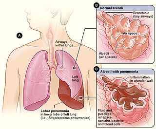
Lobar pneumonia is a form of pneumonia characterized by inflammatory exudate within the intra-alveolar space resulting in consolidation that affects a large and continuous area of the lobe of a lung.

In the anatomy of the human digestive tract, there are two colic flexures, or curvatures in the transverse colon. The right colic flexure is also known as the hepatic flexure, and the left colic flexure is also known as the splenic flexure. Note that "right" refers to the patient's anatomical right, which may be depicted on the left of a diagram.

Omental cake is a radiologic sign indicative of an abnormally thickened greater omentum. It refers to infiltration of the normal omental structure by other types of soft-tissue or chronic inflammation resulting in a thickened, or cake-like appearance.

High-resolution computed tomography (HRCT) is a type of computed tomography (CT) with specific techniques to enhance image resolution. It is used in the diagnosis of various health problems, though most commonly for lung disease, by assessing the lung parenchyma. On the other hand, HRCT of the temporal bone is used to diagnose various middle ear diseases such as otitis media, cholesteatoma, and evaluations after ear operations.

A lung nodule or pulmonary nodule is a relatively small focal density in the lung. A solitary pulmonary nodule (SPN) or coin lesion, is a mass in the lung smaller than three centimeters in diameter. A pulmonary micronodule has a diameter of less than three millimetres. There may also be multiple nodules.
Radiopaedia is a wiki-based international collaborative educational web resource containing a radiology encyclopedia and imaging case repository. It is currently the largest freely available radiology related resource in the world with more than 50,000 patient cases and over 16,000 reference articles on radiology-related topics. The open edit nature of articles allows radiologists, radiology trainees, radiographers, sonographers, and other healthcare professionals interested in medical imaging to refine most content through time. An editorial board peer reviews all contributions.

Magnetic resonance imaging (MRI) is a medical imaging technique mostly used in radiology and nuclear medicine in order to investigate the anatomy and physiology of the body, and to detect pathologies including tumors, inflammation, neurological conditions such as stroke, disorders of muscles and joints, and abnormalities in the heart and blood vessels among others. Contrast agents may be injected intravenously or into a joint to enhance the image and facilitate diagnosis. Unlike CT and X-ray, MRI uses no ionizing radiation and is, therefore, a safe procedure suitable for diagnosis in children and repeated runs. Patients with specific non-ferromagnetic metal implants, cochlear implants, and cardiac pacemakers nowadays may also have an MRI in spite of effects of the strong magnetic fields. This does not apply on older devices, and details for medical professionals are provided by the device's manufacturer.

An MRI pulse sequence in magnetic resonance imaging (MRI) is a particular setting of pulse sequences and pulsed field gradients, resulting in a particular image appearance.
Fogging phenomenon in computerized tomography (CT) scanning of the head is vanishing signs of an infarct on the serial CT imaging in a patient with a recent stroke. It is a reversal of the hypodensity on the CT after an acute ischemic stroke. This happens as a result of re-nourishment of the infarcted area in subacute phase about one to three weeks after the stroke. In fact, resolution of the edema, which was caused by the accident, leads to increased attenuation of infarcted area that may regain near-normal density and mask the stroke. However, in the third week, parenchymal volume loss commonly appears as a hypoattenuation with a negative mass effect (shrinkage).
The empty delta sign is a radiologic sign seen on brain imaging which is associated with cerebral venous sinus thrombosis. It is usually seen on magnetic resonance imaging (MRI) or computed tomography (CT) scans with contrast. It is seen as dural wall enhancement in the absence of intra-sinus enhancement. This is due to the presence of a blood clot in the dural venous sinuses. The dural venous sinuses drain blood from the brain to the internal jugular veins, which in turn drains blood to the heart. It has been proposed that the empty delta sign occurs in dural venous thromboses due to contrast material filling the dural venous collateral circulation immediately surrounding the dura whilst being unable to fill the intra-dural sinus space due to the presence of a blood clot. The superior sagittal sinus is most commonly affected, but the radiologic sign may also be seen in the transverse sinuses.