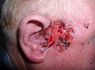
Basal-cell carcinoma (BCC), also known as basal-cell cancer, basalioma or rodent ulcer, is the most common type of skin cancer. It often appears as a painless raised area of skin, which may be shiny with small blood vessels running over it. It may also present as a raised area with ulceration. Basal-cell cancer grows slowly and can damage the tissue around it, but it is unlikely to spread to distant areas or result in death.

A skin condition, also known as cutaneous condition, is any medical condition that affects the integumentary system—the organ system that encloses the body and includes skin, nails, and related muscle and glands. The major function of this system is as a barrier against the external environment.

Nevus is a nonspecific medical term for a visible, circumscribed, chronic lesion of the skin or mucosa. The term originates from nævus, which is Latin for "birthmark"; however, a nevus can be either congenital or acquired. Common terms, including mole, birthmark, and beauty mark, are used to describe nevi, but these terms do not distinguish specific types of nevi from one another.

Actinic keratosis (AK), sometimes called solar keratosis or senile keratosis, is a pre-cancerous area of thick, scaly, or crusty skin. Actinic keratosis is a disorder of epidermal keratinocytes that is induced by ultraviolet (UV) light exposure. These growths are more common in fair-skinned people and those who are frequently in the sun. They are believed to form when skin gets damaged by UV radiation from the sun or indoor tanning beds, usually over the course of decades. Given their pre-cancerous nature, if left untreated, they may turn into a type of skin cancer called squamous cell carcinoma. Untreated lesions have up to a 20% risk of progression to squamous cell carcinoma, so treatment by a dermatologist is recommended.
Vulvitis is inflammation of the vulva, the external female mammalian genitalia that include the labia majora, labia minora, clitoris, and introitus. It may co-occur as vulvovaginitis with vaginitis, inflammation of the vagina, and may have infectious or non-infectious causes. The warm and moist conditions of the vulva make it easily affected. Vulvitis is prone to occur in any female especially those who have certain sensitivities, infections, allergies, or diseases that make them likely to have vulvitis. Postmenopausal women and prepubescent girls are more prone to be affected by it, as compared to women in their menstruation period. It is so because they have low estrogen levels which makes their vulvar tissue thin and dry. Women having diabetes are also prone to be affected by vulvitis due to the high sugar content in their cells, increasing their vulnerability. Vulvitis is not a disease, it is just an inflammation caused by an infection, allergy or injury. Vulvitis may also be symptom of any sexually transmitted infection or a fungal infection.

Ochronosis is a syndrome caused by the accumulation of homogentisic acid in connective tissues. The condition was named after the yellowish (ocher-like) discoloration of the tissue seen on microscopic examination. Macroscopically, though, the affected tissues appear bluish-grey because of a light-scattering phenomenon known as the Tyndall effect. The condition is most often associated with alkaptonuria, but can occur from exogenous administration of phenol complexes such as hydroquinone. It was first described by Rudolf Virchow in 1865.

Erythema ab igneEAI, also known as hot water bottle rash, is a skin condition caused by long-term exposure to heat. Prolonged thermal radiation exposure to the skin can lead to the development of reticulated erythema, hyperpigmentation, scaling, and telangiectasias in the affected area. Some people may complain of mild itchiness and a burning sensation, but often, unless a change in pigmentation is seen, it can go unnoticed.

Granuloma annulare (GA) is a common, sometimes chronic skin condition which presents as reddish bumps on the skin arranged in a circle or ring. It can initially occur at any age, though two-thirds of patients are under 30 years old, and it is seen most often in children and young adults. Females are two times as likely to have it as males.
Purpura fulminans is an acute, often fatal, thrombotic disorder which manifests as blood spots, bruising and discolouration of the skin resulting from coagulation in small blood vessels within the skin and rapidly leads to skin necrosis and disseminated intravascular coagulation.

Actinic cheilitis is cheilitis caused by long term sunlight exposure. Essentially it is a burn, and a variant of actinic keratosis which occurs on the lip. It is a premalignant condition, as it can develop into squamous cell carcinoma.
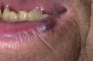
A venous lake is a generally solitary, soft, compressible, dark blue to violaceous, 0.2- to 1-cm papule commonly found on sun-exposed surfaces of the vermilion border of the lip, face and ears. Lesions generally occur among the elderly.

Schamberg's disease, is a chronic discoloration of the skin found in people of all ages, usually only affecting the feet, legs or thighs or a combination. It may occur as a single event or subsequent bouts may cause further spread. It is most common in males. It is named after Jay Frank Schamberg, who described it in 1901. There is no known cure for this disease but it is not a life-threatening condition and is mainly of cosmetic concern, although, because it can appear so suddenly, so extensively and because it usually leaves permanent discoloration of the skin, it can cause understandable psychological concern. The skin lesions sometimes cause itching, which can be treated by applying cortisone cream. The cortisone cream will only help with the itching and does not improve the discoloration of the skin. Schamberg's disease causes no other symptoms beside skin discoloration and itching. The condition is caused by inflammation of capillaries near the surface of skin and subsequent leaking of red blood cells into surrounding tissues. As the red blood cells break down and get mostly resorbed, some of the iron released by the red blood cells remains in the skin and causes the characteristic rust-colored appearance. The cause of the capillary inflammation is usually unknown.

Acrokeratoelastoidosis of Costa or Acrokeratoelastoidosis is a hereditary form of marginal keratoderma, and can be defined as a palmoplantar keratoderma. It is distinguished by tiny, firm pearly or warty papules on the sides of the hands and, occasionally, the feet. It is less common than the hereditary type of marginal keratoderma, keratoelastoidosis marginalis.

Cutaneous small-vessel vasculitis (CSVV), is inflammation of small blood vessels, usually accompanied by small lumps beneath the skin. The condition is also known as hypersensitivity vasculitis, cutaneous leukocytoclastic vasculitis, hypersensitivity angiitis, cutaneous leukocytoclastic angiitis, cutaneous necrotizing vasculitis and cutaneous necrotizing venulitis,
Photoaging or photoageing is a term used for the characteristic changes to skin induced by chronic UVA and UVB exposure. Tretinoin is the best studied retinoid in the treatment of photoaging.
Annular elastolytic giant-cell granuloma is a cutaneous condition characterized histologically by a dermal infiltrate of macrophages.
Actinic granuloma (AG) was first described by O'Brien in 1975 as a rare granulomatous disease. Lesions appear on sun-exposed areas, usually on the face, neck, and scalp, with a slight preference for middle-aged women. They are typically asymptomatic, single or multiple, annular or polycyclic lesions measuring up to 6 cm in diameter, with slow centrifugal expansion, an erythematous elevated edge, and a hypopigmented, atrophic center.

Livedoid vasculopathy(LV) is an uncommon thrombotic dermal vasculopathy that is characterized by excruciating, recurrent ulcers on the lower limbs. Livedo racemosa, a painful ulceration in the distal regions of the lower extremities, is the characteristic clinical appearance. It heals to form porcelain-white, atrophic scars, also known as Atrophie blanche.
Retiform purpura is the result of total vascular blockage and damage to the skin's blood vessels. The skin then shows lesions, appearing due to intravascular issues where clots, proteins, or emboli block skin vessels. They can also result from direct harm to the vessel walls, as seen in conditions like vasculitis, calciphylaxis, and certain severe opportunistic infections.
