Disseminated intravascular coagulation

Last updated
Disseminated intravascular coagulation
Other namesDisseminated intravascular coagulopathy, consumptive coagulopathy, defibrination syndrome [1]
Acute thrombotic microangiopathy - pas - very high mag.jpg
Micrograph showing acute thrombotic microangiopathy due to DIC in a kidney biopsy. A clot is present in the hilum of the glomerulus (center of image).
Specialty Hematology
Symptoms Chest pain, shortness of breath, leg pain, problems speaking, problems moving part of the body, bleeding [1]
Complications Organ failure [2]
TypesAcute, chronic [1]
Causes Sepsis, surgery, major trauma, cancer, complications of pregnancy, snake bites, frostbite, burns [1]
Diagnostic method Blood tests [2]
Differential diagnosis Thrombotic thrombocytopenic purpura, hemolytic-uremic syndrome [1]
TreatmentDirected at the underlying condition [3]
Medication Platelets, cryoprecipitate, fresh frozen plasma, heparin [2]
Prognosis 20–50% risk of death [4]
Frequency1% of people admitted to hospital [4]

Disseminated intravascular coagulation (DIC) is a condition in which blood clots form throughout the body, blocking small blood vessels. [1] Symptoms may include chest pain, shortness of breath, leg pain, problems speaking, or problems moving parts of the body. [1] As clotting factors and platelets are used up, bleeding may occur. [1] This may include blood in the urine, blood in the stool, or bleeding into the skin. [1] Complications may include organ failure. [2]

Contents

Relatively common causes include sepsis, surgery, major trauma, cancer, and complications of pregnancy. [1] Less common causes include snake bites, frostbite, and burns. [1] There are two main types: acute (rapid onset) and chronic (slow onset). [1] Diagnosis is typically based on blood tests. [2] Findings may include low platelets, low fibrinogen, high INR, or high D-dimer. [2]

Treatment is mainly directed towards the underlying condition. [2] [3] Other measures may include giving platelets, cryoprecipitate, or fresh frozen plasma. [2] Evidence to support these treatments, however, is poor. [2] Heparin may be useful in the slowly developing form. [2] About 1% of people admitted to hospital are affected by the condition. [4] In those with sepsis, rates are between 20% and 50%. [4] The risk of death among those affected varies from 20% to 50%. [4]

Signs and symptoms

In DIC, the underlying cause usually leads to symptoms and signs, and DIC is discovered on laboratory testing. The onset of DIC can be sudden, as in endotoxic shock or amniotic fluid embolism, or it may be insidious and chronic, as in cancer. DIC can lead to multiorgan failure and widespread bleeding. [5]

Causes

DIC can occur in the following conditions: [5] [6] [7] [8]

Liver disease, HELLP syndrome, thrombotic thrombocytopenic purpura, hemolytic uremic syndrome, and malignant hypertension may mimic DIC but originate via other pathways. [9] It is not the same as conditions where the blood vessels leak, such as capillary leak syndrome.[ citation needed ]

Pathophysiology

The coagulation cascade of secondary hemostasis. Coagulation full.svg
The coagulation cascade of secondary hemostasis.

Under homeostatic conditions, the body is maintained in a finely tuned balance of coagulation and fibrinolysis. The activation of the coagulation cascade yields thrombin that converts fibrinogen to fibrin; the stable fibrin clot being the final product of hemostasis. The fibrinolytic system then functions to break down fibrinogen and fibrin. Activation of the fibrinolytic system generates plasmin (in the presence of thrombin), which is responsible for the lysis of fibrin clots. The breakdown of fibrinogen and fibrin results in polypeptides called fibrin degradation products (FDPs) or fibrin split products (FSPs). In a state of homeostasis, the presence of plasmin is critical, as it is the central proteolytic enzyme of coagulation and is also necessary for the breakdown of clots, or fibrinolysis.[ citation needed ]

In DIC, the processes of coagulation and fibrinolysis are dysregulated, and the result is widespread clotting with resultant bleeding. Regardless of the triggering event of DIC, once initiated, the pathophysiology of DIC is similar in all conditions. One critical mediator of DIC is the release of a transmembrane glycoprotein called tissue factor (TF). TF is present on the surface of many cell types (including endothelial cells, macrophages, and monocytes) and is not normally in contact with the general circulation, but is exposed to the circulation after vascular damage. For example, TF is released in response to exposure to cytokines (particularly interleukin 1), tumor necrosis factor, and endotoxin. [10] This plays a major role in the development of DIC in septic conditions. TF is also abundant in tissues of the lungs, brain, and placenta. This helps to explain why DIC readily develops in patients with extensive trauma. Upon exposure to blood and platelets, TF binds with activated factor VIIa (normally present in trace amounts in the blood), forming the extrinsic tenase complex. This complex further activates factor IX and X to IXa and Xa, respectively, leading to the common coagulation pathway and the subsequent formation of thrombin and fibrin. [7]

The release of endotoxin is the mechanism by which Gram-negative sepsis provokes DIC. In acute promyelocytic leukemia, treatment causes the destruction of leukemic granulocyte precursors, resulting in the release of large amounts of proteolytic enzymes from their storage granules, causing microvascular damage. Other malignancies may enhance the expression of various oncogenes that result in the release of TF and plasminogen activator inhibitor-1 (PAI-1), which prevents fibrinolysis. [11]

Excess circulating thrombin results from the excess activation of the coagulation cascade. The excess thrombin cleaves fibrinogen, which ultimately leaves behind multiple fibrin clots in the circulation. These excess clots trap platelets to become larger clots, which leads to microvascular and macrovascular thrombosis. This lodging of clots in the microcirculation, in the large vessels, and in the organs is what leads to the ischemia, impaired organ perfusion, and end-organ damage that occurs with DIC. [12] [13]

Coagulation inhibitors are also consumed in this process. Decreased inhibitor levels will permit more clotting so that a positive feedback loop develops in which increased clotting leads to more clotting. At the same time, thrombocytopenia occurs and this has been attributed to the entrapment and consumption of platelets. Clotting factors are consumed in the development of multiple clots, which contributes to the bleeding seen with DIC. [12] [13]

Simultaneously, excess circulating thrombin assists in the conversion of plasminogen to plasmin, resulting in fibrinolysis. The breakdown of clots results in an excess of FDPs, which have powerful anticoagulant properties, contributing to hemorrhage. The excess plasmin also activates the complement and kinin systems. Activation of these systems leads to many of the clinical symptoms that patients experiencing DIC exhibits, such as shock, hypotension, and increased vascular permeability. The acute form of DIC is considered an extreme expression of the intravascular coagulation process with a complete breakdown of the normal homeostatic boundaries. DIC is associated with a poor prognosis and a high mortality rate. [12] [13]

There has been a recent challenge however to the basic assumptions and interpretations of the pathophysiology of DIC. A study of sepsis and DIC in animal models has shown that a highly expressed receptor on the surface of hepatocytes, termed the Ashwell-Morell receptor, is responsible for thrombocytopenia in bacteremia and sepsis due to Streptococcus pneumoniae (SPN) and possibly other pathogens. The thrombocytopenia observed in SPN sepsis was not due to increased consumption of coagulation factors such as platelets, but instead was the result of this receptor's activity, enabling hepatocytes to ingest and rapidly clear platelets from circulation. [14] By removing prothrombotic components before they participate in the coagulopathy of DIC, the Ashwell-Morell receptor lessens the severity of DIC, reducing thrombosis and tissue necrosis, and promoting survival. The hemorrhage observed in DIC and among some tissues lacking this receptor may therefore be secondary to increased thrombosis with loss of the mechanical vascular barrier. [12] [13]

Activation of the intrinsic and extrinsic coagulation pathways causes excess thrombus formation in the blood vessels. Consumption of coagulation factors due to extensive coagulation in turn causes bleeding. [12] [13]

Diagnosis

Blood film showing red blood cell fragments (schistocytes) Schizocyte smear 2009-12-22.JPG
Blood film showing red blood cell fragments (schistocytes)

The diagnosis of DIC is not made on a single laboratory value, but rather the constellation of laboratory markers and a consistent history of an illness known to cause DIC. Laboratory markers consistent with DIC include: [3] [7] [15]

A diagnostic algorithm has been proposed by the International Society of Thrombosis and Haemostasis. This algorithm appears to be 91% sensitive and 97% specific for the diagnosis of overt DIC. A score of 5 or higher is compatible with DIC and it is recommended that the score is repeated daily, while a score below 5 is suggestive but not affirmative for DIC and it is recommended that it is repeated only occasionally: [15] [16] It has been recommended that a scoring system be used in the diagnosis and management of DIC in terms of improving outcome. [17]

Treatment

Treatment of DIC is centered on treating the underlying condition. Transfusions of platelets or fresh frozen plasma can be considered in cases of significant bleeding, or those with a planned invasive procedure. The target goal of such transfusion depends on the clinical situation. Cryoprecipitate can be considered in those with a low fibrinogen level. Treatment of thrombosis with anticoagulants such as heparin is rarely used due to the risk of bleeding.[ citation needed ]

Recombinant human activated protein C was previously recommended in those with severe sepsis and DIC, but drotrecogin alfa has been shown to confer no benefit and was withdrawn from the market in 2011. [19]

Recombinant factor VII has been proposed as a "last resort" in those with severe hemorrhage due to obstetric or other causes, but conclusions about its use are still insufficient. [20]

Prognosis

Prognosis varies depending on the underlying disorder, and the extent of the intravascular thrombosis (clotting). The prognosis for those with DIC, regardless of cause, is often grim: between 20% and 50% of patients will die. [21] DIC with sepsis (infection) has a significantly higher rate of death than DIC associated with trauma. [21]

Epidemiology

DIC is observed in approximately 1% of academic hospital admissions. [22] DIC occurs at higher rates in people with bacterial sepsis (83%), [23] severe trauma (31%), [24] and cancer (6.8%). [25]

Related Research Articles

<span class="mw-page-title-main">Thrombus</span> Blood clot

A thrombus, colloquially called a blood clot, is the final product of the blood coagulation step in hemostasis. There are two components to a thrombus: aggregated platelets and red blood cells that form a plug, and a mesh of cross-linked fibrin protein. The substance making up a thrombus is sometimes called cruor. A thrombus is a healthy response to injury intended to stop and prevent further bleeding, but can be harmful in thrombosis, when a clot obstructs blood flow through healthy blood vessels in the circulatory system.

<span class="mw-page-title-main">Platelet</span> Component of blood aiding in coagulation

Platelets or thrombocytes are a component of blood whose function is to react to bleeding from blood vessel injury by clumping, thereby initiating a blood clot. Platelets have no cell nucleus; they are fragments of cytoplasm derived from the megakaryocytes of the bone marrow or lung, which then enter the circulation. Platelets are found only in mammals, whereas in other vertebrates, thrombocytes circulate as intact mononuclear cells.

<span class="mw-page-title-main">Coagulation</span> Process of formation of blood clots

Coagulation, also known as clotting, is the process by which blood changes from a liquid to a gel, forming a blood clot. It potentially results in hemostasis, the cessation of blood loss from a damaged vessel, followed by repair. The mechanism of coagulation involves activation, adhesion and aggregation of platelets, as well as deposition and maturation of fibrin.

<span class="mw-page-title-main">Fibrin</span> Fibrous protein involved in blood coagulation

Fibrin is a fibrous, non-globular protein involved in the clotting of blood. It is formed by the action of the protease thrombin on fibrinogen, which causes it to polymerize. The polymerized fibrin, together with platelets, forms a hemostatic plug or clot over a wound site.

<span class="mw-page-title-main">Fibrinogen</span> Soluble protein complex in blood plasma and involved in clot formation

Fibrinogen is a glycoprotein complex, produced in the liver, that circulates in the blood of all vertebrates. During tissue and vascular injury, it is converted enzymatically by thrombin to fibrin and then to a fibrin-based blood clot. Fibrin clots function primarily to occlude blood vessels to stop bleeding. Fibrin also binds and reduces the activity of thrombin. This activity, sometimes referred to as antithrombin I, limits clotting. Fibrin also mediates blood platelet and endothelial cell spreading, tissue fibroblast proliferation, capillary tube formation, and angiogenesis and thereby promotes revascularization and wound healing.

<span class="mw-page-title-main">Thrombin</span> Enzyme involved in blood coagulation in humans

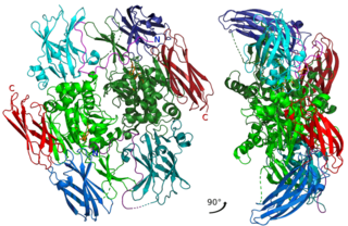
Thrombin is a serine protease, an enzyme that, in humans, is encoded by the F2 gene. During the clotting process, prothrombin is proteolytically cleaved by the prothrombinase enzyme complex to form thrombin. Thrombin in turn acts as a serine protease that converts soluble fibrinogen into insoluble strands of fibrin, as well as catalyzing many other coagulation-related reactions.

Fibrinolysis is a process that prevents blood clots from growing and becoming problematic. Primary fibrinolysis is a normal body process, while secondary fibrinolysis is the breakdown of clots due to a medicine, a medical disorder, or some other cause.

D-dimer is a dimer that is a fibrin degradation product, a small protein fragment present in the blood after a blood clot is degraded by fibrinolysis. It is so named because it contains two D fragments of the fibrin protein joined by a cross-link, hence forming a protein dimer.

<span class="mw-page-title-main">Purpura</span> Hemorrhagic lesion of the skin caused by bleeding underneath the skin

Purpura is a condition of red or purple discolored spots on the skin that do not blanch on applying pressure. The spots are caused by bleeding underneath the skin secondary to platelet disorders, vascular disorders, coagulation disorders, or other causes. They measure 3–10 mm, whereas petechiae measure less than 3 mm, and ecchymoses greater than 1 cm.

<span class="mw-page-title-main">Factor XIII</span> Class of enzymes

Factor XIII or fibrin stabilizing factor is a zymogen found in blood of humans and some other animals. It is activated by thrombin to factor XIIIa. Factor XIIIa is an enzyme of the blood coagulation system that crosslinks fibrin. Deficiency of XIII worsens clot stability and increases bleeding tendency.

<span class="mw-page-title-main">Schistocyte</span> Fragmented portion of a red blood cell

A schistocyte or schizocyte is a fragmented part of a red blood cell. Schistocytes are typically irregularly shaped, jagged, and have two pointed ends.

Thromboelastography (TEG) is a method of testing the efficiency of blood coagulation. It is a test mainly used in surgery and anesthesiology, although increasingly used in resuscitations in emergency departments, intensive care units, and labor and delivery suites. More common tests of blood coagulation include prothrombin time (PT) and partial thromboplastin time (aPTT) which measure coagulation factor function, but TEG also can assess platelet function, clot strength, and fibrinolysis which these other tests cannot.

<span class="mw-page-title-main">Hypoprothrombinemia</span> Medical condition

Hypoprothrombinemia is a rare blood disorder in which a deficiency in immunoreactive prothrombin, produced in the liver, results in an impaired blood clotting reaction, leading to an increased physiological risk for spontaneous bleeding. This condition can be observed in the gastrointestinal system, cranial vault, and superficial integumentary system, affecting both the male and female population. Prothrombin is a critical protein that is involved in the process of hemostasis, as well as illustrating procoagulant activities. This condition is characterized as an autosomal recessive inheritance congenital coagulation disorder affecting 1 per 2,000,000 of the population, worldwide, but is also attributed as acquired.

Purpura fulminans is an acute, often fatal, thrombotic disorder which manifests as blood spots, bruising and discolouration of the skin resulting from coagulation in small blood vessels within the skin and rapidly leads to skin necrosis and disseminated intravascular coagulation.

The dysfibrinogenemias consist of three types of fibrinogen disorders in which a critical blood clotting factor, fibrinogen, circulates at normal levels but is dysfunctional. Congenital dysfibrinogenemia is an inherited disorder in which one of the parental genes produces an abnormal fibrinogen. This fibrinogen interferes with normal blood clotting and/or lysis of blood clots. The condition therefore may cause pathological bleeding and/or thrombosis. Acquired dysfibrinogenemia is a non-hereditary disorder in which fibrinogen is dysfunctional due to the presence of liver disease, autoimmune disease, a plasma cell dyscrasias, or certain cancers. It is associated primarily with pathological bleeding. Hereditary fibrinogen Aα-Chain amyloidosis is a sub-category of congenital dysfibrinogenemia in which the dysfunctional fibrinogen does not cause bleeding or thrombosis but rather gradually accumulates in, and disrupts the function of, the kidney.

<span class="mw-page-title-main">Quebec platelet disorder</span> Medical condition

Quebec platelet disorder (QPD) is a rare autosomal dominant bleeding disorder first described in a family from the province of Quebec in Canada. The disorder is characterized by large amounts of the fibrinolytic enzyme urokinase-type plasminogen activator (uPA) in platelets. This causes accelerated fibrinolysis (blood clot breakdown) which can result in bleeding.

The fibrinolysis system is responsible for removing blood clots. Hyperfibrinolysis describes a situation with markedly enhanced fibrinolytic activity, resulting in increased, sometimes catastrophic bleeding. Hyperfibrinolysis can be caused by acquired or congenital reasons. Among the congenital conditions for hyperfibrinolysis, deficiency of alpha-2-antiplasmin or plasminogen activator inhibitor type 1 (PAI-1) are very rare. The affected individuals show a hemophilia-like bleeding phenotype. Acquired hyperfibrinolysis is found in liver disease, in patients with severe trauma, during major surgical procedures, and other conditions. A special situation with temporarily enhanced fibrinolysis is thrombolytic therapy with drugs which activate plasminogen, e.g. for use in acute ischemic events or in patients with stroke. In patients with severe trauma, hyperfibrinolysis is associated with poor outcome. Moreover, hyperfibrinolysis may be associated with blood brain barrier impairment, a plasmin-dependent effect due to an increased generation of bradykinin.

Intravascular immunity describes the immune response in the bloodstream, and its role is to fight and prevent the spread of pathogens. Components of intravascular immunity include the cellular immune response and the macromolecules secreted by these cells. It can result in responses such as inflammation and immunothrombosis. Dysregulated intravascular immune response or pathogen evasion can create conditions like thrombosis, sepsis, or disseminated intravascular coagulation.

Thrombodynamics test is a method for blood coagulation monitoring and anticoagulant control. This test is based on imitation of coagulation processes occurring in vivo, is sensitive both to pro- and anticoagulant changes in the hemostatic balance. Highly sensitive to thrombosis.

The platelet plug, also known as the hemostatic plug or platelet thrombus, is an aggregation of platelets formed during early stages of hemostasis in response to one or more injuries to blood vessel walls. After platelets are recruited and begin to accumulate around the breakage, their “sticky” nature allows them to adhere to each other. This forms a platelet plug, which prevents more blood from leaving the body as well as any outside contaminants from getting in. The plug provides a temporary blockage of the break in the vasculature. As such, platelet plug formation occurs after vasoconstriction of the blood vessels but before the creation of the fibrin mesh clot, which is the more permanent solution to the injury. The result of the platelet plug formation is the coagulation of blood. It can also be referred to as primary hemostasis.

References

  1. 1 2 3 4 5 6 7 8 9 10 11 12 "Disseminated Intravascular Coagulation | NHLBI, NIH". www.nhlbi.nih.gov. Retrieved 20 December 2017.
  2. 1 2 3 4 5 6 7 8 9 10 "Disseminated Intravascular Coagulation (DIC) - Hematology and Oncology". Merck Manuals Professional Edition. September 2016. Retrieved 20 December 2017.
  3. 1 2 3 Levi M (2007). "Disseminated Intravascular Coagulation". Critical Care Medicine. 35 (9): 2191–2195. doi:10.1097/01.CCM.0000281468.94108.4B. PMID   17855836. S2CID   7158989.
  4. 1 2 3 4 5 Gando S, Levi M, Toh C (2 June 2016). "Disseminated intravascular coagulation". Nature Reviews Disease Primers. 2: 16037. doi:10.1038/nrdp.2016.37. PMID   27250996. S2CID   4059451.
  5. 1 2 Robbins, Stanley L., Cotran, Ramzi S., Kumar, Vinay, Collins, Tucker (1999). Robbins' Pathologic Basis of Disease (6 ed.). Philadelphia: Saunders. ISBN   0-7216-7335-X.
  6. Davidson's Principles and Practice of Medicine (19 ed.). Churchill Livingstone. 2002. ISBN   0-443-07036-9.
  7. 1 2 3 Haematology: Basic Principles and Practice (6 ed.). Elsevier Saunders. 2012. ISBN   978-1437729283.
  8. Clark, Michael, Kumar, Parveen J. (1998). Clinical Medicine: A Textbook for Medical Students and Doctors (4 ed.). Philadelphia: W.B. Saunders. ISBN   0-7020-2458-9.
  9. "Disseminated Intravascular Coagulation". The Lecturio Medical Concept Library. Retrieved 12 July 2021.
  10. Kumar, Vinay; Abbas, Abul K.; Fausto, Nelson; & Mitchell, Richard N. (2007). Robbins Basic Pathology (8th ed.). Saunders Elsevier. pp. 469-471 ISBN   978-1-4160-2973-1
  11. Rak J, Yu JL, Luyendyk J, Mackman N (2006). "Oncogenes, trousseau syndrome, and cancer-related changes in the coagulome of mice and humans". Cancer Res. 66 (22): 10643–6. doi:10.1158/0008-5472.CAN-06-2350. PMID   17108099.
  12. 1 2 3 4 5 "Disseminated Intravascular Coagulation". National Heart, Lung And Blood Institute. Retrieved 12 July 2021.
  13. 1 2 3 4 5 "Disseminated Intravascular Coagulation (DIC)". Medscape. Retrieved 12 July 2021.
  14. Grewal PK, Uchiyama, S, Ditto, D, Varki, N, Le, DT, Nizet, V, Marth, JD (June 2008). "The Ashwell receptor mitigates the lethal coagulopathy of sepsis". Nature Medicine. 14 (6): 648–55. doi:10.1038/nm1760. PMC   2853759 . PMID   18488037.
  15. 1 2 Levi M, Toh, C-H, et al. (2009). "Guidelines for the diagnosis and management of disseminated intravascular coagulation". British Journal of Haematology. 145 (5): 24–33. doi:10.1111/j.1365-2141.2009.07600.x. PMID   19222477. S2CID   694153.
  16. Taylor F, Toh, C-h, et al. (2001). "Towards Definition, Clinical and Laboratory Criteria, and a Scoring System for Disseminated Intravascular Coagulation". Thrombosis and Haemostasis. 86 (5): 1327–30. doi:10.1055/s-0037-1616068. PMID   11816725. S2CID   39696424.
  17. Gando S (2012). "The Utility of a Diagnostic Scoring System for Disseminated Intravascular Coagulation". Critical Care Clinics . 28 (3): 378–88. doi:10.1016/j.ccc.2012.04.004. PMID   22713612.
  18. Levi M, Toh CH, Thachil J, Watson HG (2009). "Guidelines for the diagnosis and management of disseminated intravascular coagulation". British Journal of Haematology. 145 (1): 24–33. doi:10.1111/j.1365-2141.2009.07600.x. PMID   19222477. S2CID   694153.
  19. Armstrong D (October 25, 2011). "Lilly Pulls Xigris Off Markets After Sepsis Drug Fails Study". Bloomberg News . Retrieved June 26, 2020.
  20. Franchini M, Manzato, F, Salvagno GL, et al. (2007). "Potential role of recombinant activated factor VII for the treatment of severe bleeding associated with disseminated intravascular coagulation: a systematic review". Blood Coagul Fibrinolysis. 18 (7): 589–93. doi:10.1097/MBC.0b013e32822d2a3c. PMID   17890943. S2CID   37247533.
  21. 1 2 Becker, Joseph U and Charles R Wira. Disseminated intravascular coagulation Archived 2010-01-30 at the Wayback Machine at eMedicine, 10 September 2009
  22. Matsuda T (Jan–Feb 1996). "Clinical aspects of DIC--disseminated intravascular coagulation". Pol J Pharmacol. 48 (1): 73–5. PMID   9112631.
  23. Smith OP (1997). "Use of protein-C concentrate, heparin, and haemodiafiltration in meningococcus-induced purpura fulminans". Lancet. 350 (9091): 1590–1593. doi:10.1016/s0140-6736(97)06356-3. PMID   9393338. S2CID   38614343.
  24. Gando S (1999). "Disseminated intravascular coagulation and sustained systemic inflammatory response syndrome predict organ dysfunctions after trauma: application of clinical decision analysis". Ann Surg. 229 (1): 121–127. doi:10.1097/00000658-199901000-00016. PMC   1191617 . PMID   9923809.
  25. Sallah S (2001). "Disseminated intravascular coagulation in solid tumors: clinical and pathologic study". Thromb. Haemost. 86 (3): 828–833. doi:10.1055/s-0037-1616139. PMID   11583315. S2CID   11667422.