Trousseau sign of malignancy

Last updated
Trousseau sign of malignancy
Differential diagnosis thrombophlebitis migrans

The Trousseau sign of malignancy or Trousseau's syndrome is a medical sign involving episodes of vessel inflammation due to blood clot (thrombophlebitis) which are recurrent or appearing in different locations over time (thrombophlebitis migrans or migratory thrombophlebitis). The location of the clot is tender and the clot can be felt as a nodule under the skin. [1] Trousseau's syndrome is a rare variant of venous thrombosis that is characterized by recurrent, migratory thrombosis in superficial veins and in uncommon sites, such as the chest wall and arms. This syndrome is particularly associated with pancreatic, gastric and lung cancer and Trousseau's syndrome can be an early sign of cancer [2] [3] sometimes appearing months to years before the tumor would be otherwise detected. [4] Heparin therapy is recommended to prevent future clots. [5] The Trousseau sign of malignancy should not be confused with the Trousseau sign of latent tetany caused by low levels of calcium in the blood.

Contents

History

Armand Trousseau first described this finding in the 1860s; he later found the same sign in himself, was subsequently diagnosed with gastric cancer and died soon thereafter. [6] Trousseau presciently attributed thromboembolism in malignancy to changes in blood composition rather than local inflammatory or mechanical forces. By correlating clinical observation with surgical and autopsy findings, Trousseau recognized that a localized cancer could induce a generalized hypercoagulable state in which thrombosis could occur elsewhere in the body, such as in extremities with visceral malignancy. Trousseau described several cases in which recurrent thrombosis was the presenting feature of visceral cancer, and his confidence in the utility of this connection led him to say, "So great, in my opinion, is the semiotic value of phlegmasia in the cancerous cachexia, that I regard this phlegmasia as a sign of the cancerous diathesis as certain as sanguinolent effusion into the serous cavities."

Pathophysiology

Some malignancies, especially gliomas (25%), as well as adenocarcinomas of the pancreas and lung, are associated with hypercoagulability (the tendency to form blood clots) for reasons that are incompletely understood, but may be related to factors secreted by the tumors, in particular a circulating pool of cell-derived tissue factor-containing microvesicles. [7] Some adenocarcinomas secrete mucin that can interact with selectin found on platelets, thereby causing small clots to form. [8] [9] Moreover, most malignant tumors overexpress and secrete heparanase, [10] an enzyme that degrade heparan sulfate [11] and endogenous heparin, [12] and thus contribute to the hypercoagulable state in cancer patients. [13] [14] [15]

Potential Mechanisms of Cancer-Related Hypercoagulability: Cancer-associated thrombosis can result from: (1) stasis, i.e., direct pressure on blood vessels by the tumor mass, poor performance status, and bed rest following surgical procedures; (2) iatrogenic, due to treatment with antineoplastic medications; and (3) secretion of heparanase from malignant tumors that results in degradation of endogenous heparin. Nasser NJ, Fox J, Agbarya A. Cancers (Basel). 2020 Feb 29;12(3):566. https://doi.org/10.3390/cancers12030566 Potential Mechanisms of Cancer-Related Hypercoagulability.png
Potential Mechanisms of Cancer-Related Hypercoagulability: Cancer-associated thrombosis can result from: (1) stasis, i.e., direct pressure on blood vessels by the tumor mass, poor performance status, and bed rest following surgical procedures; (2) iatrogenic, due to treatment with antineoplastic medications; and (3) secretion of heparanase from malignant tumors that results in degradation of endogenous heparin. Nasser NJ, Fox J, Agbarya A. Cancers (Basel). 2020 Feb 29;12(3):566. https://doi.org/10.3390/cancers12030566

In patients with malignancy-associated hypercoagulable states, the blood may spontaneously form clots in the portal vessels (portal vein thrombosis), the deep veins of the limbs (deep vein thrombosis), or the superficial veins (superficial vein thrombosis) anywhere on the body. These clots present as visibly swollen blood vessels (thrombophlebitis), especially the veins, or as intermittent pain in the affected areas.

Related Research Articles

<span class="mw-page-title-main">Thrombosis</span> Medical condition caused by blood clots

Thrombosis is the formation of a blood clot inside a blood vessel, obstructing the flow of blood through the circulatory system. When a blood vessel is injured, the body uses platelets (thrombocytes) and fibrin to form a blood clot to prevent blood loss. Even when a blood vessel is not injured, blood clots may form in the body under certain conditions. A clot, or a piece of the clot, that breaks free and begins to travel around the body is known as an embolus.

<span class="mw-page-title-main">Venous thrombosis</span> Blood clot (thrombus) that forms within a vein

Venous thrombosis is the blockage of a vein caused by a thrombus. A common form of venous thrombosis is deep vein thrombosis (DVT), when a blood clot forms in the deep veins. If a thrombus breaks off (embolizes) and flows to the lungs to lodge there, it becomes a pulmonary embolism (PE), a blood clot in the lungs. The conditions of DVT only, DVT with PE, and PE only, are all captured by the term venous thromboembolism (VTE).

<span class="mw-page-title-main">Disseminated intravascular coagulation</span> Medical condition where blood clots block small blood vessels

Disseminated intravascular coagulation (DIC) is a condition in which blood clots form throughout the body, blocking small blood vessels. Symptoms may include chest pain, shortness of breath, leg pain, problems speaking, or problems moving parts of the body. As clotting factors and platelets are used up, bleeding may occur. This may include blood in the urine, blood in the stool, or bleeding into the skin. Complications may include organ failure.

<span class="mw-page-title-main">Antiphospholipid syndrome</span> Medical condition

Antiphospholipid syndrome, or antiphospholipid antibody syndrome, is an autoimmune, hypercoagulable state caused by antiphospholipid antibodies. APS can lead to blood clots (thrombosis) in both arteries and veins, pregnancy-related complications, and other symptoms like low platelets, kidney disease, heart disease, and rash. Although the exact etiology of APS is still not clear, genetics is believed to play a key role in the development of the disease. Diagnosis is made based on symptoms and testing, but sometimes research criteria are used to aid in diagnosis. The research criteria for definite APS requires one clinical event and two positive blood test results spaced at least three months apart that detect lupus anticoagulant, anti-apolipoprotein antibodies, and/or anti-cardiolipin antibodies.

<span class="mw-page-title-main">Deep vein thrombosis</span> Formation of a blood clot (thrombus) in a deep vein

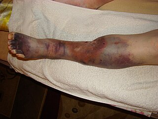
Deep vein thrombosis (DVT) is a type of venous thrombosis involving the formation of a blood clot in a deep vein, most commonly in the legs or pelvis. A minority of DVTs occur in the arms. Symptoms can include pain, swelling, redness, and enlarged veins in the affected area, but some DVTs have no symptoms.

<span class="mw-page-title-main">Budd–Chiari syndrome</span> Medical condition

Budd–Chiari syndrome is a very rare condition, affecting one in a million adults. The condition is caused by occlusion of the hepatic veins that drain the liver. The symptoms are non-specific and vary widely, but it may present with the classical triad of abdominal pain, ascites, and liver enlargement. It is usually seen in younger adults, with the median age at diagnosis between the ages of 35 and 40, and it has a similar incidence in males and females. The syndrome can be fulminant, acute, chronic, or asymptomatic. Subacute presentation is the most common form.

<span class="mw-page-title-main">Thrombophilia</span> Abnormality of blood coagulation

Thrombophilia is an abnormality of blood coagulation that increases the risk of thrombosis. Such abnormalities can be identified in 50% of people who have an episode of thrombosis that was not provoked by other causes. A significant proportion of the population has a detectable thrombophilic abnormality, but most of these develop thrombosis only in the presence of an additional risk factor.

<span class="mw-page-title-main">Thrombophlebitis</span> Medical condition

Thrombophlebitis is a phlebitis related to a thrombus. When it occurs repeatedly in different locations, it is known as thrombophlebitis migrans.

<span class="mw-page-title-main">Paget–Schroetter disease</span> Medical condition

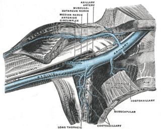
Paget–Schroetter disease is a form of upper extremity deep vein thrombosis (DVT), a medical condition in which blood clots form in the deep veins of the arms. These DVTs typically occur in the axillary and/or subclavian veins.

<span class="mw-page-title-main">Renal vein thrombosis</span> Medical condition

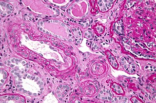
Renal vein thrombosis (RVT) is the formation of a clot in the vein that drains blood from the kidneys, ultimately leading to a reduction in the drainage of one or both kidneys and the possible migration of the clot to other parts of the body. First described by German pathologist Friedrich Daniel von Recklinghausen in 1861, RVT most commonly affects two subpopulations: newly born infants with blood clotting abnormalities or dehydration and adults with nephrotic syndrome.

<span class="mw-page-title-main">P-selectin</span> Type-1 transmembrane protein

P-selectin is a type-1 transmembrane protein that in humans is encoded by the SELP gene.

<span class="mw-page-title-main">Activated protein C resistance</span> Medical condition

Activated protein C resistance (APCR) is a hypercoagulability characterized by a lack of a response to activated protein C (APC), which normally helps prevent blood from clotting excessively. This results in an increased risk of venous thrombosis, which resulting in medical conditions such as deep vein thrombosis and pulmonary embolism. The most common cause of hereditary APC resistance is factor V Leiden mutation.

<span class="mw-page-title-main">Vascular disease</span> Medical condition

Vascular disease is a class of diseases of the vessels of the circulatory system in the body, including blood vessels – the arteries and veins, and the lymphatic vessels. Vascular disease is a subgroup of cardiovascular disease. Disorders in this vast network of blood and lymph vessels can cause a range of health problems that can sometimes become severe, and fatal. Coronary heart disease for example, is the leading cause of death for men and women in the United States.

<span class="mw-page-title-main">Post-thrombotic syndrome</span> Medical condition

Post-thrombotic syndrome (PTS), also called postphlebitic syndrome and venous stress disorder is a medical condition that may occur as a long-term complication of deep vein thrombosis (DVT).

Hypercoagulability in pregnancy is the propensity of pregnant women to develop thrombosis. Pregnancy itself is a factor of hypercoagulability, as a physiologically adaptive mechanism to prevent post partum bleeding. However, when combined with an additional underlying hypercoagulable states, the risk of thrombosis or embolism may become substantial.

<span class="mw-page-title-main">Cerebral venous sinus thrombosis</span> Presence of a blood clot in the dural venous sinuses or cerebral veins

Cerebral venous sinus thrombosis (CVST), cerebral venous and sinus thrombosis or cerebral venous thrombosis (CVT), is the presence of a blood clot in the dural venous sinuses, the cerebral veins, or both. Symptoms may include severe headache, visual symptoms, any of the symptoms of stroke such as weakness of the face and limbs on one side of the body, and seizures, which occur in around 40% of patients.

<span class="mw-page-title-main">Heparanase</span> Mammalian protein found in Homo sapiens

Heparanase, also known as HPSE, is an enzyme that acts both at the cell-surface and within the extracellular matrix to degrade polymeric heparan sulfate molecules into shorter chain length oligosaccharides.

<span class="mw-page-title-main">Superficial thrombophlebitis</span> Medical condition

Superficial thrombophlebitis is a thrombosis and inflammation of superficial veins which presents as a painful induration (thickening) with erythema, often in a linear or branching configuration; forming a cord-like appearance.

Septic pelvic thrombophlebitis (SPT), also known as suppurative pelvic thrombophlebitis, is a rare postpartum complication which consists of a persistent postpartum fever that is not responsive to broad-spectrum antibiotics, in which pelvic infection leads to infection of the vein wall and intimal damage leading to thrombogenesis in the ovarian veins. The thrombus is then invaded by microorganisms. Ascending infections cause 99% of postpartum SPT.

<span class="mw-page-title-main">Superficial vein thrombosis</span> Medical condition

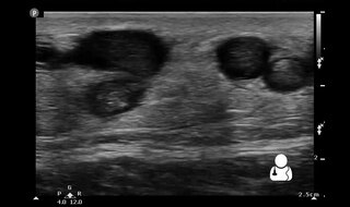
Superficial vein thrombosis (SVT) is a blood clot formed in a superficial vein, a vein near the surface of the body. Usually there is thrombophlebitis, which is an inflammatory reaction around a thrombosed vein, presenting as a painful induration with redness. SVT itself has limited significance when compared to a deep vein thrombosis (DVT), which occurs deeper in the body at the deep venous system level. However, SVT can lead to serious complications, and is therefore no longer regarded as a benign condition. If the blood clot is too near the saphenofemoral junction there is a higher risk of pulmonary embolism, a potentially life-threatening complication.

References

  1. Trousseau's sign of visceral malignancy in GPnotebook, retrieved November 2012
  2. Caine, Graham (Nov 2002). "The Hypercoagulable State of Malignancy: Pathogenesis and Current Debate". Neoplasia. 4 (6): 465–473. doi:10.1038/sj.neo.7900263. PMC   1550339 . PMID   12407439.
  3. Callander, N; S I Rapaport (1993). "Trousseau's syndrome". Western Journal of Medicine. 158 (4): 364–371. ISSN   0093-0415. PMC   1022062 . PMID   8317122.
  4. "Trousseau sign" "at Dorland's Medical Dictionary
  5. Callander, N; S I Rapaport (1993). "Trousseau's syndrome". Western Journal of Medicine. 158 (4): 364–371. ISSN   0093-0415. PMC   1022062 . PMID   8317122.
  6. Samuels MA, King ME, Balis U (2002). "Case records of the Massachusetts General Hospital. Weekly clinicopathological exercises. Case 31-2002. A 61-year-old man with headache and multiple infarcts". N. Engl. J. Med. 347 (15): 1187–94. doi:10.1056/NEJMcpc020117. PMID   12374880.
  7. Del Conde I, Bharwani LD, Dietzen DJ, Pendurthi U, Thiagarajan P, López JA (2007). "Microvesicle-associated tissue factor and Trousseau's syndrome". J Thromb Haemost. 5 (1): 70–4. doi:10.1111/j.1538-7836.2006.02301.x. PMC   3410746 . PMID   17239164.{{cite journal}}: CS1 maint: multiple names: authors list (link)
  8. Wahrenbrock M, Borsig L, Le D, Varki N, Varki A (2003). "Selectin-mucin interactions as a probable molecular explanation for the association of Trousseau syndrome with mucinous adenocarcinomas". J Clin Invest. 112 (6): 853–862. doi:10.1172/jci200318882. PMC   193671 . PMID   12975470.
  9. Varki, Ajit (2007). "Trousseau's syndrome: multiple definitions and multiple mechanisms". Blood. 110 (6): 1723–1729. doi:10.1182/blood-2006-10-053736. ISSN   0006-4971. PMC   1976377 . PMID   17496204.
  10. Edovitsky, Evgeny; Elkin, Michael; Zcharia, Eyal; Peretz, Tamar; Vlodavsky, Israel (2004-08-18). "Heparanase gene silencing, tumor invasiveness, angiogenesis, and metastasis". Journal of the National Cancer Institute. 96 (16): 1219–1230. doi: 10.1093/jnci/djh230 . ISSN   1460-2105. PMID   15316057.
  11. Vlodavsky, I.; Friedmann, Y.; Elkin, M.; Aingorn, H.; Atzmon, R.; Ishai-Michaeli, R.; Bitan, M.; Pappo, O.; Peretz, T.; Michal, I.; Spector, L. (July 1999). "Mammalian heparanase: gene cloning, expression and function in tumor progression and metastasis". Nature Medicine. 5 (7): 793–802. doi:10.1038/10518. ISSN   1078-8956. PMID   10395325. S2CID   38895589.
  12. Nasser, N. J.; Sarig, G.; Brenner, B.; Nevo, E.; Goldshmidt, O.; Zcharia, E.; Li, J. P.; Vlodavsky, I. (March 2006). "Heparanase neutralizes the anticoagulation properties of heparin and low-molecular-weight heparin". Journal of Thrombosis and Haemostasis. 4 (3): 560–565. doi: 10.1111/j.1538-7836.2006.01792.x . ISSN   1538-7933. PMID   16460439.
  13. Nasser, Nicola J.; Na'amad, Mira; Weinberg, Ido; Gabizon, Alberto A. (January 2015). "Pharmacokinetics of low molecular weight heparin in patients with malignant tumors". Anti-Cancer Drugs. 26 (1): 106–111. doi:10.1097/CAD.0000000000000176. ISSN   1473-5741. PMID   25280062. S2CID   6639067.
  14. Rickles, F. R. (March 2006). "If heparanase is the answer, what is the question?". Journal of Thrombosis and Haemostasis. 4 (3): 557–559. doi: 10.1111/j.1538-7836.2006.01828.x . ISSN   1538-7933. PMID   16460438. S2CID   5631803.
  15. Nasser, Nicola J.; Fox, Jana; Agbarya, Abed (2020-02-29). "Potential Mechanisms of Cancer-Related Hypercoagulability". Cancers. 12 (3): 566. doi: 10.3390/cancers12030566 . ISSN   2072-6694. PMC   7139427 . PMID   32121387.