Related Research Articles

An X-ray, or, much less commonly, X-radiation, is a penetrating form of high-energy electromagnetic radiation. Most X-rays have a wavelength ranging from 10 picometers to 10 nanometers, corresponding to frequencies in the range 30 petahertz to 30 exahertz (3×10¹⁶ Hz to 3×10¹⁹ Hz) and energies in the range 145 eV to 124 keV. X-ray wavelengths are shorter than those of UV rays and typically longer than those of gamma rays. In many languages, X-radiation is referred to as Röntgen radiation, after the German scientist Wilhelm Conrad Röntgen, who discovered it on November 8, 1895. He named it X-radiation to signify an unknown type of radiation. Spellings of X-ray(s) in English include the variants x-ray(s), xray(s), and X ray(s). The most familiar use of X-rays is checking for fractures (broken bones), but X-rays are also used in other ways. For example, chest X-rays can spot pneumonia. Mammograms use X-rays to look for breast cancer.

Radiography is an imaging technique using X-rays, gamma rays, or similar ionizing radiation and non-ionizing radiation to view the internal form of an object. Applications of radiography include medical radiography and industrial radiography. Similar techniques are used in airport security. To create an image in conventional radiography, a beam of X-rays is produced by an X-ray generator and is projected toward the object. A certain amount of the X-rays or other radiation is absorbed by the object, dependent on the object's density and structural composition. The X-rays that pass through the object are captured behind the object by a detector. The generation of flat two dimensional images by this technique is called projectional radiography. In computed tomography an X-ray source and its associated detectors rotate around the subject which itself moves through the conical X-ray beam produced. Any given point within the subject is crossed from many directions by many different beams at different times. Information regarding attenuation of these beams is collated and subjected to computation to generate two dimensional images in three planes which can be further processed to produce a three dimensional image.

A synchrotron light source is a source of electromagnetic radiation (EM) usually produced by a storage ring, for scientific and technical purposes. First observed in synchrotrons, synchrotron light is now produced by storage rings and other specialized particle accelerators, typically accelerating electrons. Once the high-energy electron beam has been generated, it is directed into auxiliary components such as bending magnets and insertion devices in storage rings and free electron lasers. These supply the strong magnetic fields perpendicular to the beam which are needed to convert high energy electrons into photons.
The Canadian Light Source (CLS) is Canada's national synchrotron light source facility, located on the grounds of the University of Saskatchewan in Saskatoon, Saskatchewan, Canada. The CLS has a third-generation 2.9 GeV storage ring, and the building occupies a footprint the size of a Canadian football field. It opened in 2004 after a 30-year campaign by the Canadian scientific community to establish a synchrotron radiation facility in Canada. It has expanded both its complement of beamlines and its building in two phases since opening. As a national synchrotron facility with over 1000 individual users, it hosts scientists from all regions of Canada and around 20 other countries. Research at the CLS has ranged from viruses to superconductors to dinosaurs, and it has also been noted for its industrial science and its high school education programs.

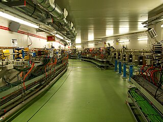
The European Synchrotron Radiation Facility (ESRF) is a joint research facility situated in Grenoble, France, supported by 22 countries.
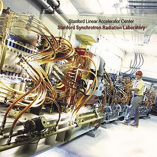
The Stanford Synchrotron Radiation Lightsource, a division of SLAC National Accelerator Laboratory, is operated by Stanford University for the Department of Energy. SSRL is a National User Facility which provides synchrotron radiation, a name given to electromagnetic radiation in the x-ray, ultraviolet, visible and infrared realms produced by electrons circulating in a storage ring at nearly the speed of light. The extremely bright light that is produced can be used to investigate various forms of matter ranging from objects of atomic and molecular size to man-made materials with unusual properties. The obtained information and knowledge is of great value to society, with impact in areas such as the environment, future technologies, health, biology, basic research, and education.
Phase-contrast imaging is a method of imaging that has a range of different applications. It measures differences in the refractive index of different materials to differentiate between structures under analysis. In conventional light microscopy, phase contrast can be employed to distinguish between structures of similar transparency, and to examine crystals on the basis of their double refraction. This has uses in biological, medical and geological science. In X-ray tomography, the same physical principles can be used to increase image contrast by highlighting small details of differing refractive index within structures that are otherwise uniform. In transmission electron microscopy (TEM), phase contrast enables very high resolution (HR) imaging, making it possible to distinguish features a few Angstrom apart.
The National Synchrotron Light Source (NSLS) at Brookhaven National Laboratory (BNL) in Upton, New York was a national user research facility funded by the U.S. Department of Energy (DOE). Built from 1978 through 1984, and officially shut down on September 30, 2014, the NSLS was considered a second-generation synchrotron.

The hard X-ray nanoprobe at the Center for Nanoscale Materials (CNM), Argonne National Lab advanced the state of the art by providing a hard X-ray microscopy beamline with the highest spatial resolution in the world. It provides for fluorescence, diffraction, and transmission imaging with hard X-rays at a spatial resolution of 30 nm or better. A dedicated source, beamline, and optics form the basis for these capabilities. This unique instrument is not only key to the specific research areas of the CNM; it will also be a general utility, available to the broader nanoscience community in studying nanomaterials and nanostructures, particularly for embedded structures.
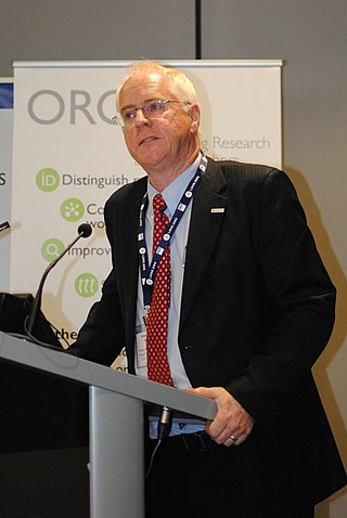
Keith Alexander Nugent FAA is an Australian physicist. He is the Deputy Vice-Chancellor of the Australian National University (ANU) in Canberra.
ALBA is a third-generation synchrotron light source facility located in the Barcelona Synchrotron Park in Cerdanyola del Vallès near Barcelona, in Catalonia (Spain). It was constructed and is operated by CELLS, and co-financed by the Spanish central administration and regional Catalan Government.
ANSTO's Australian Synchrotron is a 3 GeV national synchrotron radiation facility located in Clayton, in the south-eastern suburbs of Melbourne, Victoria, which opened in 2007.
The European X-Ray Free-Electron Laser Facility is an X-ray research laser facility commissioned during 2017. The first laser pulses were produced in May 2017 and the facility started user operation in September 2017. The international project with twelve participating countries; nine shareholders at the time of commissioning, later joined by three other partners, is located in the German federal states of Hamburg and Schleswig-Holstein. A free-electron laser generates high-intensity electromagnetic radiation by accelerating electrons to relativistic speeds and directing them through special magnetic structures. The European XFEL is constructed such that the electrons produce X-ray light in synchronisation, resulting in high-intensity X-ray pulses with the properties of laser light and at intensities much brighter than those produced by conventional synchrotron light sources.

The Extreme Light Infrastructure (ELI) is an international series of physics laboratories for generating and studying intense laser light. It is part of the European ESFRI Roadmap. ELI hosts the most intense beamline system worldwide, develop new interdisciplinary research opportunities with light from these lasers and secondary radiation derived from them, and make them available to the international scientific user community. ELI aims to be the world's biggest and first international user facility in beamline and laser research.
ANKA is a synchrotron light source facility at the Karlsruhe Institute of Technology (KIT). The KIT runs ANKA as a national synchrotron light source and as a large scale user facility for the international science community. Being a large scale machine of the performance category LK II of the Helmholtz Association, ANKA is part of a national and European infrastructure offering research services to scientific and commercial users for their purposes in research and development. The facility was opened to external users in 2003.

The National Synchrotron Light Source II (NSLS-II) at Brookhaven National Laboratory (BNL) in Upton, New York is a national user research facility funded primarily by the U.S. Department of Energy's (DOE) Office of Science. NSLS-II is one of the world's most advanced synchrotron light sources, designed to produce x-rays 10,000 times brighter than BNL's original light source, the National Synchrotron Light Source (NSLS). NSLS-II supports basic and applied research in energy security, advanced materials synthesis and manufacturing, environment, and human health.
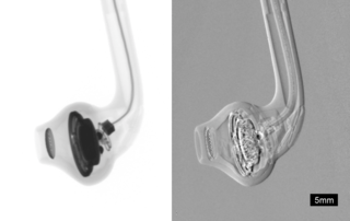
Phase-contrast X-ray imaging or phase-sensitive X-ray imaging is a general term for different technical methods that use information concerning changes in the phase of an X-ray beam that passes through an object in order to create its images. Standard X-ray imaging techniques like radiography or computed tomography (CT) rely on a decrease of the X-ray beam's intensity (attenuation) when traversing the sample, which can be measured directly with the assistance of an X-ray detector. However, in phase contrast X-ray imaging, the beam's phase shift caused by the sample is not measured directly, but is transformed into variations in intensity, which then can be recorded by the detector.
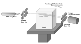
Energy-dispersive X-ray diffraction (EDXRD) is an analytical technique for characterizing materials. It differs from conventional X-ray diffraction by using polychromatic photons as the source and is usually operated at a fixed angle. With no need for a goniometer, EDXRD is able to collect full diffraction patterns very quickly. EDXRD is almost exclusively used with synchrotron radiation which allows for measurement within real engineering materials.
Franz Pfeiffer is a German physicist known for his contributions to the development of Phase-contrast X-ray imaging and its applications in biomedical research.

Solaris is the only synchrotron in Central-Eastern Europe. Built in Poland in 2015, under the auspices of the Jagiellonian University, it is located on the Campus of the 600th Anniversary of the Jagiellonian University Revival, in the southern part of Krakow. It is the central facility of the National Center of Synchrotron Radiation SOLARIS.
References
- ↑ Varghese, Jose (2013). "Steve Wilkins (1946–2013)". Acta Crystallographica Section A. 69 (5): 528–529. doi: 10.1107/S0108767313017650 . Retrieved 7 February 2022.
- ↑ Ward, Colin (13 January 2015). "Stephen William Wilkins (1946 – 2013)". CSRIOpedia. CSIRO. Retrieved 7 February 2022.
- ↑ Robinson, Rob (2013). "Tough times: Mourning the loss of friends and preparing for the September federal election" (PDF). Australian Physics. 50 (3): 79. Retrieved 7 February 2022.
- ↑ Wilkins SW, Gureyev TE, Gao D, Pogany A, Stevenson AW (1996). "Phase-contrast imaging using polychromatic hard X-rays". Nature. 383 (6607): 335–338. Bibcode:1996Natur.384..335W. doi:10.1038/384335a0. S2CID 4273199 . Retrieved 7 February 2022.
- ↑ "Phase-contrast imaging using polychromatic hard X-rays". Google Scholar. Retrieved 7 February 2022.
- ↑ Creagh, Dudley (March 2013). "THE DECOMMISSIONING OF THE AUSTRALIAN NATIONAL BEAMLINE (BL20B) AT THE PHOTON FACTORY KEK TSUKUBA JAPAN" (PDF). IRPS Bulletin - Newsletter of the International Radiation Physics Society. 27 (1): 9–15. Retrieved 7 February 2022.
- ↑ Barnea Z, Creagh DC, Davis TJ, Garrett RF, Janky S, Stevenson AW, Wilkins SW (1992). "The Australian diffractometer at the Photon Factory". Review of Scientific Instruments. 63 (1): 1069–1072. Bibcode:1992RScI...63.1069B. doi:10.1063/1.1143202 . Retrieved 7 February 2022.