Stereo microscope

Last updated
Stereo microscope Optical stereo microscope nikon smz10.jpg
Stereo microscope
Modern stereomicroscope optical design.
A - Objective B - Galilean telescopes (rotating objectives) C - Zoom control D - Internal objective E - Prism F - Relay lens G - Reticle H - Eyepiece Sztereomik.png
Modern stereomicroscope optical design.
A - Objective B - Galilean telescopes (rotating objectives) C - Zoom control D - Internal objective E - Prism F - Relay lens G - Reticle H - Eyepiece

The stereo, stereoscopic or dissecting microscope is an optical microscope variant designed for low magnification observation of a sample, typically using light reflected from the surface of an object rather than transmitted through it. The instrument uses two separate optical paths with two objectives and eyepieces to provide slightly different viewing angles to the left and right eyes. This arrangement produces a three-dimensional visualization of the sample being examined. [1] Stereomicroscopy overlaps macrophotography for recording and examining solid samples with complex surface topography, where a three-dimensional view is needed for analyzing the detail.

Contents

The stereo microscope is often used to study the surfaces of solid specimens or to carry out close work such as dissection, microsurgery, watch-making, circuit board manufacture or inspection, and fracture surfaces as in fractography and forensic engineering. They are thus widely used in manufacturing industry for manufacture, inspection and quality control. Stereo microscopes are essential tools in entomology.

The stereo microscope should not be confused with a compound microscope equipped with double eyepieces and a binoviewer. In such a microscope, both eyes see the same image, with the two eyepieces serving to provide greater viewing comfort. However, the image in such a microscope is no different from that obtained with a single monocular eyepiece.

History

The first optically feasible stereomicroscope was invented in 1892 and became commercially available in 1896, produced by Zeiss AG in Jena, Germany. [2]

1896 Greenough Stereo Microscope by Carl Zeiss Jena 1896 Greenough-type Stereo Microscope by Carl Zeiss Jena (6892932332).gif
1896 Greenough Stereo Microscope by Carl Zeiss Jena

American zoologist Horatio Saltonstall Greenough grew up in the elite of Boston, Massachusetts, the son of the famous sculptor Horatio Greenough Sr. Without the pressures of having to make a living, he instead pursued a career in science and relocated to France. At the marine observatory in Concarneau on the Bretton coast, lead by the former director of the Muséum national d'histoire naturelle, Georges Pouchet, he was influenced by the new scientific ideals of the day, namely experimentation. While dissection of dead and prepared specimens had been the main concern for zoologists, anatomists and morphologists, during Greenough’s stay at Concarneau interest was revived in experimenting on live and developing organisms. This way scientists could study embryonic development in action rather than as a series of petrified, two-dimensional specimens. In order to yield images that would do justice to the three-dimensionality and relative size of developing invertebrate marine embryos, a new microscope was needed. While there had been attempts to build stereomicroscopes before, by for example Chérubin d’Orleans and Pieter Harting, none had been optically sophisticated. Furthermore, up until the 1880s no scientist needed a microscope with such low resolution.

Greenough took action and, influenced by his Concarneau colleague Laurent Chabry’s attempts to construct intricate mechanisms to turn and manipulate the live embryo, conceived of his own instrument. Building on the recent discovery of binocularity as the cause of depth perception by Charles Wheatstone, Greenough designed his instrument with the phenomenon of stereopsis in mind. [2]

Differences from normal optical microscopes

Unlike a compound light microscope, illumination in a stereo microscope most often uses reflected illumination rather than transmitted (diascopic) illumination, that is, light reflected from the surface of an object rather than light transmitted through an object. Use of reflected light from the object allows examination of specimens that would be too thick or otherwise opaque for compound microscopy. Some stereo microscopes are also capable of transmitted light illumination as well, typically by having a bulb or mirror beneath a transparent stage underneath the object, though unlike a compound microscope, transmitted illumination is not focused through a condenser in most systems. [3] Stereoscopes with specially-equipped illuminators can be used for dark field microscopy, using either reflected or transmitted light. [4]

Scientist using a stereo microscope outfitted with a digital imaging pick-up and fibre-optic illumination Manusingmicroscope.jpg
Scientist using a stereo microscope outfitted with a digital imaging pick-up and fibre-optic illumination

Great working distance and depth of field are important qualities for this type of microscope. Both qualities are inversely correlated with resolution: the higher the resolution (i.e. the greater the distance at which two adjacent points can be distinguished as separate), the smaller the depth of field and working distance. Some stereo microscopes can deliver a useful magnification up to 100×, comparable to a 10× objective and 10× eyepiece in a normal compound microscope, although the magnification is often much lower. This is around one tenth the useful resolution of a normal compound optical microscope.

The large working distance at low magnification is useful in examining large solid objects such as fracture surfaces, especially using fibre-optic illumination as discussed below. Such samples can also be manipulated easily so as to determine the points of interest.

Magnification

There are two major types of magnification systems in stereo microscopes. One type is fixed magnification in which primary magnification is achieved by a paired set of objective lenses with a set degree of magnification. The other is zoom or pancratic magnification, which are capable of a continuously variable degree of magnification across a set range. Zoom systems can achieve further magnification through the use of auxiliary objectives that increase total magnification by a set factor. Also, total magnification in both fixed and zoom systems can be varied by changing eyepieces. [1]

Intermediate between fixed magnification and zoom magnification systems is a system attributed to Galileo as the "Galilean optical system" ; here an arrangement of fixed-focus convex lenses is used to provide a fixed magnification, but with the crucial distinction that the same optical components in the same spacing will, if physically inverted, result in a different, though still fixed, magnification. This allows one set of lenses to provide two different magnifications ; two sets of lenses to provide four magnifications on one turret ; three sets of lenses provide six magnifications and will still fit into one turret. Practical experience shows that such Galilean optics systems are as useful as a considerably more expensive zoom system, with the advantage of knowing the magnification in use as a set value without having to read analogue scales. (In remote locations, the robustness of the systems is also a non-trivial advantage.)

Illumination

Stereomicroscope with an illuminated butterfly specimen Looking at the butterfly.jpg
Stereomicroscope with an illuminated butterfly specimen

Small specimens necessarily require intense illumination, especially at high magnifications, and this is usually provided by a fibre-optic light source. Fiber optics utilize halogen lamps which provide high light output for a given power input. The lamps are small enough to be fitted easily near the microscope, although they often need cooling to ameliorate high temperatures from the bulb. The fibre-optic stalk gives the operator much freedom in choosing appropriate lighting conditions for the sample. The stalk is encased in a sheath that is easy to move and manipulate to any desired position. The stalk is normally unobtrusive when the lit end is near the specimen, so usually does not interfere with the image in the microscope. Examination of fracture surfaces frequently need oblique lighting so as to highlight surface features during fractography, and fibre-optic lights are ideal for this purpose. Several such light stalks can be used for the same specimen, so increasing the illumination yet further.

More recent developments in the lighting for dissecting microscopes include the use of high-power LEDs which are much more energy efficient than halogens and are able to produce a spectrum of colors of light, making them useful for fluorophore analysis of biological samples (impossible with a halogen or mercury vapor light source).

Digital display

Labomed LB-343 5.0 MP digital stereo microscope with 9 inch HD LCD screen, HDMI video output, X/Y digital micrometer and moving stage Labomed Inc LB-343 Digital LCD Stereo Measuring Microscope.jpg
Labomed LB-343 5.0 MP digital stereo microscope with 9 inch HD LCD screen, HDMI video output, X/Y digital micrometer and moving stage

Video cameras are integrated into some stereo microscopes, allowing the magnified images to be displayed on a high resolution monitor. The large display helps to reduce the eye fatigue that would result from using a conventional microscope for extended periods.

In some units, a built-in computer converts the images from two cameras (one per eyepiece) to a 3D anaglyph image for viewing with red/cyan glasses, or to the cross converged process[ clarify ] for clear glasses and improved color accuracy. The results are viewable by a group wearing the glasses. More typically, a 2D image is displayed from a single camera attached to one of the eyepieces.

See also

Related Research Articles

Microscopy Viewing of objects which are too small to be seen with the naked eye

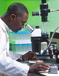
Microscopy is the technical field of using microscopes to view objects and areas of objects that cannot be seen with the naked eye. There are three well-known branches of microscopy: optical, electron, and scanning probe microscopy, along with the emerging field of X-ray microscopy.

Microscope Scientific instrument

A microscope is a laboratory instrument used to examine objects that are too small to be seen by the naked eye. Microscopy is the science of investigating small objects and structures using a microscope. Microscopic means being invisible to the eye unless aided by a microscope.

Scanning electron microscope Type of electron microscope

A scanning electron microscope (SEM) is a type of electron microscope that produces images of a sample by scanning the surface with a focused beam of electrons. The electrons interact with atoms in the sample, producing various signals that contain information about the surface topography and composition of the sample. The electron beam is scanned in a raster scan pattern, and the position of the beam is combined with the intensity of the detected signal to produce an image. In the most common SEM mode, secondary electrons emitted by atoms excited by the electron beam are detected using a secondary electron detector. The number of secondary electrons that can be detected, and thus the signal intensity, depends, among other things, on specimen topography. Some SEMs can achieve resolutions better than 1 nanometer.

Optical microscope Microscope that uses visible light

The optical microscope, also referred to as a light microscope, is a type of microscope that commonly uses visible light and a system of lenses to generate magnified images of small objects. Optical microscopes are the oldest design of microscope and were possibly invented in their present compound form in the 17th century. Basic optical microscopes can be very simple, although many complex designs aim to improve resolution and sample contrast.

Transmission electron microscopy Technique in microscopy

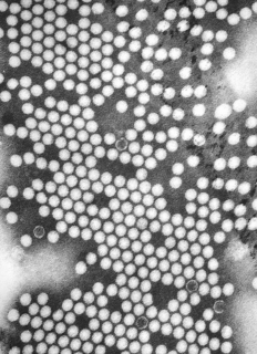
Transmission electron microscopy (TEM) is a microscopy technique in which a beam of electrons is transmitted through a specimen to form an image. The specimen is most often an ultrathin section less than 100 nm thick or a suspension on a grid. An image is formed from the interaction of the electrons with the sample as the beam is transmitted through the specimen. The image is then magnified and focused onto an imaging device, such as a fluorescent screen, a layer of photographic film, or a sensor such as a scintillator attached to a charge-coupled device.

Objective (optics)

In optical engineering, the objective is the optical element that gathers light from the object being observed and focuses the light rays to produce a real image. Objectives can be a single lens or mirror, or combinations of several optical elements. They are used in microscopes, binoculars, telescopes, cameras, slide projectors, CD players and many other optical instruments. Objectives are also called object lenses, object glasses, or objective glasses.

Total internal reflection fluorescence microscope

A total internal reflection fluorescence microscope (TIRFM) is a type of microscope with which a thin region of a specimen, usually less than 200 nanometers can be observed.

Fluorescence microscope

A fluorescence microscope is an optical microscope that uses fluorescence instead of, or in addition to, scattering, reflection, and attenuation or absorption, to study the properties of organic or inorganic substances. "Fluorescence microscope" refers to any microscope that uses fluorescence to generate an image, whether it is a simple set up like an epifluorescence microscope or a more complicated design such as a confocal microscope, which uses optical sectioning to get better resolution of the fluorescence image.

Confocal microscopy

Confocal microscopy, most frequently confocal laser scanning microscopy (CLSM) or laser confocal scanning microscopy (LCSM), is an optical imaging technique for increasing optical resolution and contrast of a micrograph by means of using a spatial pinhole to block out-of-focus light in image formation. Capturing multiple two-dimensional images at different depths in a sample enables the reconstruction of three-dimensional structures within an object. This technique is used extensively in the scientific and industrial communities and typical applications are in life sciences, semiconductor inspection and materials science.

Metallography

Metallography is the study of the physical structure and components of metals, by using microscopy.

Dark-field microscopy Laboratory technique

Dark-field microscopy describes microscopy methods, in both light and electron microscopy, which exclude the unscattered beam from the image. As a result, the field around the specimen is generally dark.

Bright-field microscopy

Bright-field microscopy (BF) is the simplest of all the optical microscopy illumination techniques. Sample illumination is transmitted white light, and contrast in the sample is caused by attenuation of the transmitted light in dense areas of the sample. Bright-field microscopy is the simplest of a range of techniques used for illumination of samples in light microscopes, and its simplicity makes it a popular technique. The typical appearance of a bright-field microscopy image is a dark sample on a bright background, hence the name.

Köhler illumination is a method of specimen illumination used for transmitted and reflected light optical microscopy. Köhler illumination acts to generate an even illumination of the sample and ensures that an image of the illumination source is not visible in the resulting image. Köhler illumination is the predominant technique for sample illumination in modern scientific light microscopy. It requires additional optical elements which are more expensive and may not be present in more basic light microscopes.

Harold Hopkins (physicist) British physicist

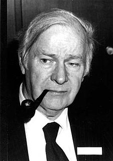
Harold Horace Hopkins FRS was a British physicist. His Wave Theory of Aberrations,, is central to all modern optical design and provides the mathematical analysis which enables the use of computers to create the wealth of high quality lenses available today. In addition to his theoretical work, his many inventions are in daily use throughout the world. These include zoom lenses, coherent fibre-optics and more recently the rod-lens endoscopes which 'opened the door' to modern key-hole surgery. He was the recipient of many of the world's most prestigious awards and was twice nominated for a Nobel Prize. His citation on receiving the Rumford Medal from the Royal Society in 1984 stated: "In recognition of his many contributions to the theory and design of optical instruments, especially of a wide variety of important new medical instruments which have made a major contribution to clinical diagnosis and surgery."

Digital microscope

A digital microscope is a variation of a traditional optical microscope that uses optics and a digital camera to output an image to a monitor, sometimes by means of software running on a computer. A digital microscope often has its own in-built LED light source, and differs from an optical microscope in that there is no provision to observe the sample directly through an eyepiece. Since the image is focused on the digital circuit, the entire system is designed for the monitor image. The optics for the human eye are omitted.

Optical sectioning Imaging of focal planes within a thick sample

Optical sectioning is the process by which a suitably designed microscope can produce clear images of focal planes deep within a thick sample. This is used to reduce the need for thin sectioning using instruments such as the microtome. Many different techniques for optical sectioning are used and several microscopy techniques are specifically designed to improve the quality of optical sectioning.

USB microscope

A USB microscope is a low-powered digital microscope which connects to a computer's USB port. Microscopes essentially the same as USB models are also available with other interfaces either in addition to or instead of USB, such as via WiFi. They are widely available at low cost for use at home or in commerce. Their cost varies in the range of tens to thousands of dollars. In essence, a USB microscope is a webcam with a high-powered macro lens, and generally uses reflected rather than transmitted light, using built-in LED light sources surrounding the lens. The camera is usually sensitive enough not to need additional illumination beyond normal ambient lighting. The camera attaches directly to the USB port of a computer without the need for an eyepiece, and the images are shown directly on the computer's display.

Condenser (optics) type of optical lens

A condenser is an optical lens which renders a divergent beam from a point source into a parallel or converging beam to illuminate an object.

Microscopy with UV Surface Excitation (MUSE) is a novel microscopy method that utilize the shallow penetration of UV photons excitation. Compared to conventional microscopes, which usually require sectioning to exclude blurred signals from outside of the focal plane, MUSE's low penetration depth limits the excitation volume to a thin layer, and removes the tissue sectioning requirement. The entire signal collected is the desired light, and all photons collected contribute to the image formation.

A macroscope is a type of optical microscope developed and named by Swiss microscope manufacturers Wild Heerbrugg and later, after that company's merger with Leica in 1987, by Leica Microsystems of Germany, optimised for high quality macro photography and/or viewing using a single objective lens and light path, rather than stereoscopic viewing of specimens, at magnifications up to around x40.

References

  1. 1 2 "Introduction to Stereomicroscopy" by Paul E. Nothnagle, William Chambers, and Michael W. Davidson, Nikon MicroscopyU.
  2. 1 2 Simon-Stickley, Anna (2019). "Image and Imagination. The Stereomicroscope on the Cusp of Modern Biology". NTM Journal of the History of Science, Technology and Medicine. 27 (2): 109–144. doi: 10.1007/s00048-019-00211-0 . PMID   31062033. S2CID   146809758.
  3. "Illumination for Stereomicroscopy: Reflected (Episcopic) Light" by Paul E. Nothnagle, William Chambers, Thomas J. Fellers, and Michael W. Davidson , Nikon MicroscopyU.
  4. "Illumination for Stereomicroscopy: Darkfield Illumination" by William Chambers, Thomas J. Fellers, and Michael W. Davidson , Nikon MicroscopyU.