Related Research Articles

Collagen is the main structural protein in the extracellular matrix found in the body's various connective tissues. As the main component of connective tissue, it is the most abundant protein in mammals, making up from 25% to 35% of the whole-body protein content. Collagen consists of amino acids bound together to form a triple helix of elongated fibril known as a collagen helix. It is mostly found in connective tissue such as cartilage, bones, tendons, ligaments, and skin. Vitamin C is vital for collagen synthesis, and Vitamin E improves the production of collagen.

The endoplasmic reticulum (ER) is a part of a transportation system of the eukaryotic cell, and has many other important functions such as protein folding. It is a type of organelle made up of two subunits – rough endoplasmic reticulum (RER), and smooth endoplasmic reticulum (SER). The endoplasmic reticulum is found in most eukaryotic cells and forms an interconnected network of flattened, membrane-enclosed sacs known as cisternae, and tubular structures in the SER. The membranes of the ER are continuous with the outer nuclear membrane. The endoplasmic reticulum is not found in red blood cells, or spermatozoa.

The endomembrane system is composed of the different membranes (endomembranes) that are suspended in the cytoplasm within a eukaryotic cell. These membranes divide the cell into functional and structural compartments, or organelles. In eukaryotes the organelles of the endomembrane system include: the nuclear membrane, the endoplasmic reticulum, the Golgi apparatus, lysosomes, vesicles, endosomes, and plasma (cell) membrane among others. The system is defined more accurately as the set of membranes that forms a single functional and developmental unit, either being connected directly, or exchanging material through vesicle transport. Importantly, the endomembrane system does not include the membranes of plastids or mitochondria, but might have evolved partially from the actions of the latter.

In biology, tissue is a historically derived biological organizational level between cells and a complete organ. A tissue is therefore often thought of as an assembly of similar cells and their extracellular matrix from the same embryonic origin that together carry out a specific function. Organs are then formed by the functional grouping together of multiple tissues.

A tendon or sinew is a tough band of dense fibrous connective tissue that connects muscle to bone. It sends the mechanical forces of muscle contraction to the skeletal system, while withstanding tension.

A hepatocyte is a cell of the main parenchymal tissue of the liver. Hepatocytes make up 80% of the liver's mass. These cells are involved in:

A muscle cell, also known as a myocyte, is a mature contractile cell in the muscle of an animal. In humans and other vertebrates there are three types: skeletal, smooth, and cardiac (cardiomyocytes). A skeletal muscle cell is long and threadlike with many nuclei and is called a muscle fiber. Muscle cells develop from embryonic precursor cells called myoblasts.
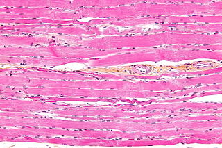
Striated muscle tissue is a muscle tissue that features repeating functional units called sarcomeres. The presence of sarcomeres manifests as a series of bands visible along the muscle fibers, which is responsible for the striated appearance observed in microscopic images of this tissue. There are two types of striated muscle:

Sarcoplasm is the cytoplasm of a muscle cell. It is comparable to the cytoplasm of other cells, but it contains unusually large amounts of glycogen (a polymer of glucose), myoglobin, a red-colored protein necessary for binding oxygen molecules that diffuse into muscle fibers, and mitochondria. The calcium ion concentration in sarcoplasma is also a special element of the muscle fiber; it is the means by which muscle contractions take place and are regulated. The sarcoplasm plays a critical role in muscle contraction as an increase in Ca2+ concentration in the sarcoplasm begins the process of filament sliding. A decrease in Ca2+ in the sarcoplasm subsequently ceases filament sliding. The sarcoplasm also aids in pH and ion balance within muscle cells.

Hemidesmosomes are very small stud-like structures found in keratinocytes of the epidermis of skin that attach to the extracellular matrix. They are similar in form to desmosomes when visualized by electron microscopy, however, desmosomes attach to adjacent cells. Hemidesmosomes are also comparable to focal adhesions, as they both attach cells to the extracellular matrix. Instead of desmogleins and desmocollins in the extracellular space, hemidesmosomes utilize integrins. Hemidesmosomes are found in epithelial cells connecting the basal epithelial cells to the lamina lucida, which is part of the basal lamina. Hemidesmosomes are also involved in signaling pathways, such as keratinocyte migration or carcinoma cell intrusion.

The sarcolemma also called the myolemma, is the cell membrane surrounding a skeletal muscle fiber or a cardiomyocyte. It consists of a lipid bilayer and a thin outer coat of polysaccharide material (glycocalyx) that contacts the basement membrane. The basement membrane contains numerous thin collagen fibrils and specialized proteins such as laminin that provide a scaffold to which the muscle fiber can adhere. Through transmembrane proteins in the plasma membrane, the actin skeleton inside the cell is connected to the basement membrane and the cell's exterior. At each end of the muscle fiber, the surface layer of the sarcolemma fuses with a tendon fiber, and the tendon fibers, in turn, collect into bundles to form the muscle tendons that adhere to bones.
Endoplasm generally refers to the inner, dense part of a cell's cytoplasm. This is opposed to the ectoplasm which is the outer (non-granulated) layer of the cytoplasm, which is typically watery and immediately adjacent to the plasma membrane. The nucleus is separated from the endoplasm by the nuclear envelope. The different makeups/viscosities of the endoplasm and ectoplasm contribute to the amoeba's locomotion through the formation of a pseudopod. However, other types of cells have cytoplasm divided into endo- and ectoplasm. The endoplasm, along with its granules, contains water, nucleic acids, amino acids, carbohydrates, inorganic ions, lipids, enzymes, and other molecular compounds. It is the site of most cellular processes as it houses the organelles that make up the endomembrane system, as well as those that stand alone. The endoplasm is necessary for most metabolic activities, including cell division.

The operculum is a corneous or calcareous anatomical structure like a trapdoor that exists in many groups of sea snails and freshwater snails, and also in a few groups of land snails; the structure is found in some marine and freshwater gastropods, and in a minority of terrestrial gastropods, including the families Helicinidae, Cyclophoridae, Aciculidae, Maizaniidae, Pomatiidae, etc.
A biological system is a complex network which connects several biologically relevant entities. Biological organization spans several scales and are determined based different structures depending on what the system is. Examples of biological systems at the macro scale are populations of organisms. On the organ and tissue scale in mammals and other animals, examples include the circulatory system, the respiratory system, and the nervous system. On the micro to the nanoscopic scale, examples of biological systems are cells, organelles, macromolecular complexes and regulatory pathways. A biological system is not to be confused with a living system, such as a living organism.
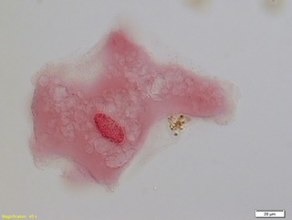
A fibrocyte is an inactive mesenchymal cell, that is, a cell showing minimal cytoplasm, limited amounts of rough endoplasmic reticulum and lacks biochemical evidence of protein synthesis.
The junctional epithelium (JE) is that epithelium which lies at, and in health also defines, the base of the gingival sulcus. The probing depth of the gingival sulcus is measured by a calibrated periodontal probe. In a healthy-case scenario, the probe is gently inserted, slides by the sulcular epithelium (SE), and is stopped by the epithelial attachment (EA). However, the probing depth of the gingival sulcus may be considerably different from the true histological gingival sulcus depth.
In cell biology, membrane bound polyribosomes are attached to a cell's endoplasmic reticulum. When certain proteins are synthesized by a ribosome they can become "membrane-bound". The newly produced polypeptide chains are inserted directly into the endoplasmic reticulum by the ribosome and are then transported to their destinations. Bound ribosomes usually produce proteins that are used within the cell membrane or are expelled from the cell via exocytosis.
Histology is the study of the minute structure, composition, and function of tissues. Mature human vocal cords are composed of layered structures which are quite different at the histological level.
O-linked glycosylation is the attachment of a sugar molecule to the oxygen atom of serine (Ser) or threonine (Thr) residues in a protein. O-glycosylation is a post-translational modification that occurs after the protein has been synthesised. In eukaryotes, it occurs in the endoplasmic reticulum, Golgi apparatus and occasionally in the cytoplasm; in prokaryotes, it occurs in the cytoplasm. Several different sugars can be added to the serine or threonine, and they affect the protein in different ways by changing protein stability and regulating protein activity. O-glycans, which are the sugars added to the serine or threonine, have numerous functions throughout the body, including trafficking of cells in the immune system, allowing recognition of foreign material, controlling cell metabolism and providing cartilage and tendon flexibility. Because of the many functions they have, changes in O-glycosylation are important in many diseases including cancer, diabetes and Alzheimer's. O-glycosylation occurs in all domains of life, including eukaryotes, archaea and a number of pathogenic bacteria including Burkholderia cenocepacia, Neisseria gonorrhoeae and Acinetobacter baumannii.
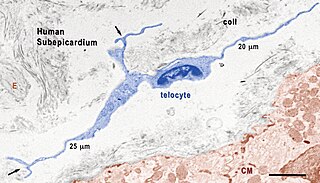
Telocytes are a type of interstitial (stromal) cells with very long and very thin prolongations called telopodes.
References
- Krstić RV (1984). Illustrated Encyclopedia of Human Histology. Berlin: Springer Verlag.
- Tompa AS, Watabe N (July 1976). "Ultrastructural investigation of the mechanism of muscle attachment to the gastropod shell". Journal of Morphology. 149 (3): 339–52. doi:10.1002/jmor.1051490304. PMID 957445. S2CID 35530166.
| | This cell biology article is a stub. You can help Wikipedia by expanding it. |