Related Research Articles
Crystallography is the experimental science of determining the arrangement of atoms in crystalline solids. Crystallography is a fundamental subject in the fields of materials science and solid-state physics. The word crystallography is derived from the Ancient Greek word κρύσταλλος, with its meaning extending to all solids with some degree of transparency, and γράφειν. In July 2012, the United Nations recognised the importance of the science of crystallography by proclaiming that 2014 would be the International Year of Crystallography.
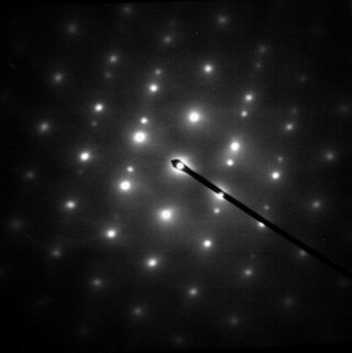
Electron diffraction is a generic term for phenomena associated with changes in the direction of electron beams due to elastic interactions with atoms. It occurs due to elastic scattering, when there is no change in the energy of the electrons. The negatively charged electrons are scattered due to Coulomb forces when they interact with both the positively charged atomic core and the negatively charged electrons around the atoms. The resulting map of the directions of the electrons far from the sample is called a diffraction pattern, see for instance Figure 1. Beyond patterns showing the directions of electrons, electron diffraction also plays a major role in the contrast of images in electron microscopes.

Neutron diffraction or elastic neutron scattering is the application of neutron scattering to the determination of the atomic and/or magnetic structure of a material. A sample to be examined is placed in a beam of thermal or cold neutrons to obtain a diffraction pattern that provides information of the structure of the material. The technique is similar to X-ray diffraction but due to their different scattering properties, neutrons and X-rays provide complementary information: X-Rays are suited for superficial analysis, strong x-rays from synchrotron radiation are suited for shallow depths or thin specimens, while neutrons having high penetration depth are suited for bulk samples.

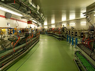
A synchrotron light source is a source of electromagnetic radiation (EM) usually produced by a storage ring, for scientific and technical purposes. First observed in synchrotrons, synchrotron light is now produced by storage rings and other specialized particle accelerators, typically accelerating electrons. Once the high-energy electron beam has been generated, it is directed into auxiliary components such as bending magnets and insertion devices in storage rings and free electron lasers. These supply the strong magnetic fields perpendicular to the beam that are needed to stimulate the high energy electrons to emit photons.

An X-ray microscope uses electromagnetic radiation in the X-ray band to produce magnified images of objects. Since X-rays penetrate most objects, there is no need to specially prepare them for X-ray microscopy observations.
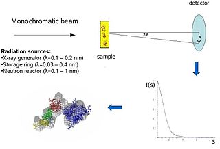
Biological small-angle scattering is a small-angle scattering method for structure analysis of biological materials. Small-angle scattering is used to study the structure of a variety of objects such as solutions of biological macromolecules, nanocomposites, alloys, and synthetic polymers. Small-angle X-ray scattering (SAXS) and small-angle neutron scattering (SANS) are the two complementary techniques known jointly as small-angle scattering (SAS). SAS is an analogous method to X-ray and neutron diffraction, wide angle X-ray scattering, as well as to static light scattering. In contrast to other X-ray and neutron scattering methods, SAS yields information on the sizes and shapes of both crystalline and non-crystalline particles. When used to study biological materials, which are very often in aqueous solution, the scattering pattern is orientation averaged.
In physics, the phase problem is the problem of loss of information concerning the phase that can occur when making a physical measurement. The name comes from the field of X-ray crystallography, where the phase problem has to be solved for the determination of a structure from diffraction data. The phase problem is also met in the fields of imaging and signal processing. Various approaches of phase retrieval have been developed over the years.
Electron crystallography is a method to determine the arrangement of atoms in solids using a transmission electron microscope (TEM). It can involve the use of high-resolution transmission electron microscopy images, electron diffraction patterns including convergent-beam electron diffraction or combinations of these. It has been successful in determining some bulk structures, and also surface structures. Two related methods are low-energy electron diffraction which has solved the structure of many surfaces, and reflection high-energy electron diffraction which is used to monitor surfaces often during growth.
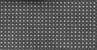
Powder diffraction is a scientific technique using X-ray, neutron, or electron diffraction on powder or microcrystalline samples for structural characterization of materials. An instrument dedicated to performing such powder measurements is called a powder diffractometer.
Selected area (electron) diffraction is a crystallographic experimental technique typically performed using a transmission electron microscope (TEM). It is a specific case of electron diffraction used primarily in material science and solid state physics as one of the most common experimental techniques. Especially with appropriate analytical software, SAD patterns (SADP) can be used to determine crystal orientation, measure lattice constants or examine its defects.
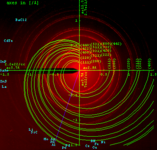
High-energy X-rays or HEX-rays are very hard X-rays, with typical energies of 80–1000 keV (1 MeV), about one order of magnitude higher than conventional X-rays used for X-ray crystallography. They are produced at modern synchrotron radiation sources such as the beamlines ID15 and BM18 at the European Synchrotron Radiation Facility (ESRF). The main benefit is the deep penetration into matter which makes them a probe for thick samples in physics and materials science and permits an in-air sample environment and operation. Scattering angles are small and diffraction directed forward allows for simple detector setups.
ALBA is a third-generation synchrotron light source facility located in the Barcelona Synchrotron Park in Cerdanyola del Vallès near Barcelona, in Catalonia (Spain). It was constructed and is operated by CELLS, and co-financed by the Spanish central administration and regional Catalan Government.
An X-ray microscope uses electromagnetic radiation in the soft X-ray band to produce images of very small objects.
Diffraction topography is a imaging technique based on Bragg diffraction. Diffraction topographic images ("topographies") record the intensity profile of a beam of X-rays diffracted by a crystal. A topography thus represents a two-dimensional spatial intensity mapping of reflected X-rays, i.e. the spatial fine structure of a Laue reflection. This intensity mapping reflects the distribution of scattering power inside the crystal; topographs therefore reveal the irregularities in a non-ideal crystal lattice. X-ray diffraction topography is one variant of X-ray imaging, making use of diffraction contrast rather than absorption contrast which is usually used in radiography and computed tomography (CT). Topography is exploited to a lesser extends with neutrons, and has similarities to dark field imaging in the electron microscope community.
McXtrace is an open source software package for performing Monte Carlo simulations of X-ray scattering experiments. While its chief objective is to aid in the optimization of beamlines at e.g. synchrotrons, it may also be used for data analysis and at laboratory sources and beamlines. McXtrace is free software released under the GNU GPL.
A materials oscilloscope is a time-resolved synchrotron high-energy X-ray technique to study rapid phase composition and microstructural related changes in a polycrystalline sample. Such device has been developed for in-situ studies of specimens undergoing physical thermo-mechanical simulation.
Microcrystal electron diffraction, or MicroED, is a CryoEM method that was developed by the Gonen laboratory in late 2013 at the Janelia Research Campus of the Howard Hughes Medical Institute. MicroED is a form of electron crystallography where thin 3D crystals are used for structure determination by electron diffraction. Prior to this demonstration, macromolecular (protein) electron crystallography was only used on 2D crystals, for example.

SOLARIS is the only synchrotron in Central-Eastern Europe. Built in Poland in 2015, under the auspices of the Jagiellonian University, it is located on the Campus of the 600th Anniversary of the Jagiellonian University Revival, in the southern part of Krakow. It is the central facility of the National Synchrotron Radiation Centre SOLARIS.
Dark-field X-ray microscopy is an imaging technique used for multiscale structural characterisation. It is capable of mapping deeply embedded structural elements with nm-resolution using synchrotron X-ray diffraction-based imaging. The technique works by using scattered X-rays to create a high degree of contrast, and by measuring the intensity and spatial distribution of the diffracted beams, it is possible to obtain a three-dimensional map of the sample's structure, orientation, and local strain.
X-ray diffraction computed tomography is an experimental technique that combines X-ray diffraction with the computed tomography data acquisition approach. X-ray diffraction (XRD) computed tomography (CT) was first introduced in 1987 by Harding et al. using a laboratory diffractometer and a monochromatic X-ray pencil beam. The first implementation of the technique at synchrotron facilities was performed in 1998 by Kleuker et al.
References
- ↑ Poulsen, H. F.; Nielsen, S. F.; Lauridsen, E. M.; Schmidt, S.; Suter, R. M.; Lienert, U.; Margulies, L.; Lorentzen, T.; Juul Jensen, D. (2001). "Three-dimensional maps of grain boundaries and the stress state of individual grains in polycrystals and powders" (PDF). Journal of Applied Crystallography. 34 (6): 751–756. doi: 10.1107/s0021889801014273 .
- ↑ Poulsen, Henning (2004). Three-Dimensional X-Ray Diffraction Microscopy. Springer Tracts in Modern Physics. Vol. 205. doi:10.1007/b97884. ISBN 978-3-540-22330-6.
- ↑ Ludwig, Wolfgang; Schmidt, Søren; Lauridsen, Erik Mejdal; Poulsen, Henning Friis (2008-04-01). "X-ray diffraction contrast tomography: a novel technique for three-dimensional grain mapping of polycrystals. I. Direct beam case". Journal of Applied Crystallography. 41 (2): 302–309. doi:10.1107/s0021889808001684. ISSN 0021-8898.
- ↑ Suter, RM; Hennessy, D; Xiao, C; Lienert, U (2006-12-01). "Forward modeling method for microstructure reconstruction using x-ray diffraction microscopy: Single-crystal verification". Review of Scientific Instruments. 77 (12): 123905–123905–12. Bibcode:2006RScI...77l3905S. doi:10.1063/1.2400017. ISSN 0034-6748. S2CID 6298472.
- ↑ Zaefferer, S.; Wright, S. I.; Raabe, D. (2008-02-01). "Three-Dimensional Orientation Microscopy in a Focused Ion Beam–Scanning Electron Microscope: A New Dimension of Microstructure Characterization". Metallurgical and Materials Transactions A. 39 (2): 374–389. Bibcode:2008MMTA...39..374Z. doi: 10.1007/s11661-007-9418-9 . ISSN 1073-5623.
- ↑ Poulsen, Henning Friis (2012-12-01). "An introduction to three-dimensional X-ray diffraction microscopy". Journal of Applied Crystallography. 45 (6): 1084–1097. doi:10.1107/s0021889812039143. ISSN 0021-8898.
- ↑ "ID11 - Materials science beamline". www.esrf.eu. Retrieved 2017-04-13.
- ↑ "fable". SourceForge. Retrieved 2017-04-13.
- ↑ Schmidt, Søren (2014-02-01). "GrainSpotter: a fast and robust polycrystalline indexing algorithm" (PDF). Journal of Applied Crystallography. 47 (1): 276–284. doi:10.1107/s1600576713030185. ISSN 1600-5767. S2CID 36349208.
- ↑ Staron, Peter; Schreyer, Andreas; Clemens, Helmut; Mayer, Svea (2017-06-19). Neutrons and Synchrotron Radiation in Engineering Materials Science: From Fundamentals to Applications, 2nd Edition. John Wiley & Sons. ISBN 978-3-527-33592-3.
- ↑ Sedmák, P.; Pilch, J.; Heller, L.; Kopeček, J.; Wright, J.; Sedlák, P.; Frost, M.; Šittner, P. (2016-08-05). "Grain-resolved analysis of localized deformation in nickel-titanium wire under tensile load". Science. 353 (6299): 559–562. Bibcode:2016Sci...353..559S. doi:10.1126/science.aad6700. ISSN 0036-8075. PMID 27493178. S2CID 206644339.
- ↑ Liu, H. H.; Schmidt, S.; Poulsen, H. F.; Godfrey, A.; Liu, Z. Q.; Sharon, J. A.; Huang, X. (2011-05-13). "Three-Dimensional Orientation Mapping in the Transmission Electron Microscope". Science. 332 (6031): 833–834. Bibcode:2011Sci...332..833L. doi:10.1126/science.1202202. ISSN 0036-8075. PMID 21566190. S2CID 206532089.
- ↑ Cereser, Alberto (2016-01-01). Time-of-flight 3D Neutron Diffraction for Multigrain Crystallography. Department of Physics, Technical University of Denmark.
- ↑ Cereser, Alberto; Strobl, Markus; Hall, Stephen A.; Steuwer, Axel; Kiyanagi, Ryoji; Tremsin, Anton S.; Knudsen, Erik B.; Shinohara, Takenao; Willendrup, Peter K. (2017-08-25). "Time-of-Flight Three Dimensional Neutron Diffraction in Transmission Mode for Mapping Crystal Grain Structures". Scientific Reports. 7 (1): 9561. doi:10.1038/s41598-017-09717-w. ISSN 2045-2322. PMC 5572055 . PMID 28842660.
- ↑ Raventos, M.; Tovar, M.; Medarde, M.; Shang, T.; Strobl, M.; Samothrakitis, S.; Pomjakushina, E.; Gruenzweig, C.; Schmidt, S. (2019-03-18). "Laue three dimensional neutron diffraction". Scientific Reports. 9 (1): 4798. doi: 10.1038/s41598-019-41071-x . PMC 6423297 . PMID 30886172.
- ↑ Simons, H.; King, A.; Ludwig, W.; Detlefs, C.; Pantleon, W.; Schmidt, S.; Snigireva, I.; Snigirev, A.; Poulsen, H. F. (2015-01-14). "Dark-field X-ray microscopy for multiscale structural characterization". Nature Communications. 6: 6098. Bibcode:2015NatCo...6.6098S. doi:10.1038/ncomms7098. ISSN 2041-1723. PMC 4354092 . PMID 25586429.
- ↑ "Research centre for the application of steel dives into the dark-field microscopy at ESRF". www.esrf.eu. European Synchrotron Radiation Facility . Retrieved 2022-01-28.
- ↑ McDonald, S. A.; Holzner, C.; Lauridsen, E. M.; Reischig, P.; Merkle, A. P.; Withers, P. J. (2017-07-12). "Microstructural evolution during sintering of copper particles studied by laboratory diffraction contrast tomography (LabDCT)". Scientific Reports. 7 (1): 5251. doi:10.1038/s41598-017-04742-1. ISSN 2045-2322. PMC 5507940 . PMID 28701768.