Related Research Articles
Crystallography is the experimental science of determining the arrangement of atoms in crystalline solids. Crystallography is a fundamental subject in the fields of materials science and solid-state physics. The word crystallography is derived from the Ancient Greek word κρύσταλλος, with its meaning extending to all solids with some degree of transparency, and γράφειν. In July 2012, the United Nations recognised the importance of the science of crystallography by proclaiming that 2014 would be the International Year of Crystallography.

X-ray crystallography is the experimental science determining the atomic and molecular structure of a crystal, in which the crystalline structure causes a beam of incident X-rays to diffract into many specific directions. By measuring the angles and intensities of these diffracted beams, a crystallographer can produce a three-dimensional picture of the density of electrons within the crystal. From this electron density, the mean positions of the atoms in the crystal can be determined, as well as their chemical bonds, their crystallographic disorder, and various other information.

A computed tomography scan is a medical imaging technique used to obtain detailed internal images of the body. The personnel that perform CT scans are called radiographers or radiology technologists.

Tomography is imaging by sections or sectioning that uses any kind of penetrating wave. The method is used in radiology, archaeology, biology, atmospheric science, geophysics, oceanography, plasma physics, materials science, cosmochemistry, astrophysics, quantum information, and other areas of science. The word tomography is derived from Ancient Greek τόμος tomos, "slice, section" and γράφω graphō, "to write" or, in this context as well, "to describe." A device used in tomography is called a tomograph, while the image produced is a tomogram.
Tomographic reconstruction is a type of multidimensional inverse problem where the challenge is to yield an estimate of a specific system from a finite number of projections. The mathematical basis for tomographic imaging was laid down by Johann Radon. A notable example of applications is the reconstruction of computed tomography (CT) where cross-sectional images of patients are obtained in non-invasive manner. Recent developments have seen the Radon transform and its inverse used for tasks related to realistic object insertion required for testing and evaluating computed tomography use in airport security.
Electron crystallography is a method to determine the arrangement of atoms in solids using a transmission electron microscope (TEM). It can involve the use of high-resolution transmission electron microscopy images, electron diffraction patterns including convergent-beam electron diffraction or combinations of these. It has been successful in determining some bulk structures, and also surface structures. Two related methods are low-energy electron diffraction which has solved the structure of many surfaces, and reflection high-energy electron diffraction which is used to monitor surfaces often during growth.
A diffractometer is a measuring instrument for analyzing the structure of a material from the scattering pattern produced when a beam of radiation or particles interacts with it.

In radiography, X-ray microtomography uses X-rays to create cross-sections of a physical object that can be used to recreate a virtual model without destroying the original object. It is similar to tomography and X-ray computed tomography. The prefix micro- is used to indicate that the pixel sizes of the cross-sections are in the micrometre range. These pixel sizes have also resulted in creation of its synonyms high-resolution X-ray tomography, micro-computed tomography, and similar terms. Sometimes the terms high-resolution computed tomography (HRCT) and micro-CT are differentiated, but in other cases the term high-resolution micro-CT is used. Virtually all tomography today is computed tomography.
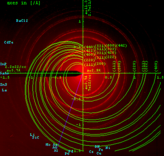
Powder diffraction is a scientific technique using X-ray, neutron, or electron diffraction on powder or microcrystalline samples for structural characterization of materials. An instrument dedicated to performing such powder measurements is called a powder diffractometer.
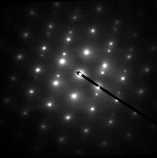
Selected area (electron) diffraction is a crystallographic experimental technique typically performed using a transmission electron microscope (TEM). It is a specific case of electron diffraction used primarily in material science and solid state physics as one of the most common experimental techniques. Especially with appropriate analytical software, SAD patterns (SADP) can be used to determine crystal orientation, measure lattice constants or examine its defects.
Hugo M. Rietveld was a Dutch crystallographer who is famous for his publication on the full profile refinement method in powder diffraction, which became later known as the Rietveld refinement method. The method is used for the characterisation of crystalline materials from X-ray powder diffraction data. The Rietveld refinement uses a least squares approach to refine a theoretical line profile until it matches the measured profile. The introduction of this technique which used the full profile instead of individual reflections was a significant step forward in the diffraction analysis of powder samples.
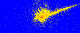
Diffraction topography is a imaging technique based on Bragg diffraction. Diffraction topographic images ("topographies") record the intensity profile of a beam of X-rays diffracted by a crystal. A topography thus represents a two-dimensional spatial intensity mapping of reflected X-rays, i.e. the spatial fine structure of a Laue reflection. This intensity mapping reflects the distribution of scattering power inside the crystal; topographs therefore reveal the irregularities in a non-ideal crystal lattice. X-ray diffraction topography is one variant of X-ray imaging, making use of diffraction contrast rather than absorption contrast which is usually used in radiography and computed tomography (CT). Topography is exploited to a lesser extends with neutrons, and has similarities to dark field imaging in the electron microscope community.
Coherent diffractive imaging (CDI) is a "lensless" technique for 2D or 3D reconstruction of the image of nanoscale structures such as nanotubes, nanocrystals, porous nanocrystalline layers, defects, potentially proteins, and more. In CDI, a highly coherent beam of X-rays, electrons or other wavelike particle or photon is incident on an object.

Industrial computed tomography (CT) scanning is any computer-aided tomographic process, usually X-ray computed tomography, that uses irradiation to produce three-dimensional internal and external representations of a scanned object. Industrial CT scanning has been used in many areas of industry for internal inspection of components. Some of the key uses for industrial CT scanning have been flaw detection, failure analysis, metrology, assembly analysis and reverse engineering applications. Just as in medical imaging, industrial imaging includes both nontomographic radiography and computed tomographic radiography.

Cone beam computed tomography is a medical imaging technique consisting of X-ray computed tomography where the X-rays are divergent, forming a cone.
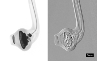
Phase-contrast X-ray imaging or phase-sensitive X-ray imaging is a general term for different technical methods that use information concerning changes in the phase of an X-ray beam that passes through an object in order to create its images. Standard X-ray imaging techniques like radiography or computed tomography (CT) rely on a decrease of the X-ray beam's intensity (attenuation) when traversing the sample, which can be measured directly with the assistance of an X-ray detector. However, in phase contrast X-ray imaging, the beam's phase shift caused by the sample is not measured directly, but is transformed into variations in intensity, which then can be recorded by the detector.
Three-dimensional X-ray diffraction (3DXRD) is a microscopy technique using hard X-rays to investigate the internal structure of polycrystalline materials in three dimensions. For a given sample, 3DXRD returns the shape, juxtaposition, and orientation of the crystallites ("grains") it is made of. 3DXRD allows investigating micrometer- to millimetre-sized samples with resolution ranging from hundreds of nanometers to micrometers. Other techniques employing X-rays to investigate the internal structure of polycrystalline materials include X-ray diffraction contrast tomography (DCT) and high energy X-ray diffraction (HEDM).

X-ray computed tomography operates by using an X-ray generator that rotates around the object; X-ray detectors are positioned on the opposite side of the circle from the X-ray source.

Cryogenic electron microscopy (cryo-EM) is a cryomicroscopy technique applied on samples cooled to cryogenic temperatures. For biological specimens, the structure is preserved by embedding in an environment of vitreous ice. An aqueous sample solution is applied to a grid-mesh and plunge-frozen in liquid ethane or a mixture of liquid ethane and propane. While development of the technique began in the 1970s, recent advances in detector technology and software algorithms have allowed for the determination of biomolecular structures at near-atomic resolution. This has attracted wide attention to the approach as an alternative to X-ray crystallography or NMR spectroscopy for macromolecular structure determination without the need for crystallization.
Dark-field X-ray microscopy is an imaging technique used for multiscale structural characterisation. It is capable of mapping deeply embedded structural elements with nm-resolution using synchrotron X-ray diffraction-based imaging. The technique works by using scattered X-rays to create a high degree of contrast, and by measuring the intensity and spatial distribution of the diffracted beams, it is possible to obtain a three-dimensional map of the sample's structure, orientation, and local strain.
References
- 1 2 Harding, G.; Kosanetzky, J.; Neitzel, U. (July 1987). "X-ray diffraction computed tomography: X-ray diffraction computed tomography". Medical Physics. 14 (4): 515–525. doi:10.1118/1.596063. PMID 3626990.
- ↑ Kleuker, U; Suortti, P; Weyrich, W; Spanne, P (1998-10-01). "Feasibility study of x-ray diffraction computed tomography for medical imaging". Physics in Medicine and Biology. 43 (10): 2911–2923. Bibcode:1998PMB....43.2911K. doi:10.1088/0031-9155/43/10/017. ISSN 0031-9155. PMID 9814526. S2CID 250820853.
- ↑ Hayashi, Yujiro; Setoyama, Daigo; Hirose, Yoshiharu; Yoshida, Tomoyuki; Kimura, Hidehiko (2019-12-20). "Intragranular three-dimensional stress tensor fields in plastically deformed polycrystals". Science. 366 (6472): 1492–1496. Bibcode:2019Sci...366.1492H. doi:10.1126/science.aax9167. ISSN 0036-8075. PMID 31857480. S2CID 209424420.
- ↑ Poulsen, Henning (2004). Three-Dimensional X-Ray Diffraction Microscopy. Springer Tracts in Modern Physics. Vol. 205. Berlin, Heidelberg: Springer Berlin Heidelberg. doi:10.1007/b97884. ISBN 978-3-540-22330-6.
- ↑ Ludwig, Wolfgang; Schmidt, Søeren; Lauridsen, Erik Mejdal; Poulsen, Henning Friis (2008-04-01). "X-ray diffraction contrast tomography: a novel technique for three-dimensional grain mapping of polycrystals. I. Direct beam case". Journal of Applied Crystallography. 41 (2): 302–309. doi:10.1107/S0021889808001684. ISSN 0021-8898.
- ↑ Suter, R. M.; Hennessy, D.; Xiao, C.; Lienert, U. (2006-12-01). "Forward modeling method for microstructure reconstruction using x-ray diffraction microscopy: Single-crystal verification". Review of Scientific Instruments. 77 (12): 123905–123905–12. Bibcode:2006RScI...77l3905S. doi:10.1063/1.2400017. ISSN 0034-6748.
- ↑ Kochetov, Vladislav; Mühlbauer, Martin J; Schökel, Alexander; Fischer, Torben; Müller, Timo; Hofmann, Michael; Staron, Peter; Lienert, Ulrich; Petry, Winfried; Senyshyn, Anatoliy (2021-03-10). "Powder diffraction computed tomography: a combined synchrotron and neutron study". Journal of Physics: Condensed Matter. 33 (10): 105901. Bibcode:2021JPCM...33j5901K. doi: 10.1088/1361-648X/abcdb0 . ISSN 0953-8984. PMID 33237884. S2CID 227176156.
- ↑ Birkbak, M. E.; Leemreize, H.; Frølich, S.; Stock, S. R.; Birkedal, H. (2015). "Diffraction scattering computed tomography: a window into the structures of complex nanomaterials". Nanoscale. 7 (44): 18402–18410. Bibcode:2015Nanos...718402B. doi:10.1039/C5NR04385A. ISSN 2040-3364. PMC 4727839 . PMID 26505175.
- ↑ Bleuet, Pierre; Welcomme, Eléonore; Dooryhée, Eric; Susini, Jean; Hodeau, Jean-Louis; Walter, Philippe (June 2008). "Probing the structure of heterogeneous diluted materials by diffraction tomography". Nature Materials. 7 (6): 468–472. Bibcode:2008NatMa...7..468B. doi:10.1038/nmat2168. ISSN 1476-4660. PMID 18425135.
- ↑ Bracewell, R. N.; Riddle, A. C. (November 1967). "Inversion of Fan-Beam Scans in Radio Astronomy". The Astrophysical Journal. 150: 427. Bibcode:1967ApJ...150..427B. doi:10.1086/149346. ISSN 0004-637X.
- 1 2 Vamvakeros, A.; Coelho, A. A.; Matras, D.; Dong, H.; Odarchenko, Y.; Price, S. W. T.; Butler, K. T.; Gutowski, O.; Dippel, A.-C.; Zimmermann, M.; Martens, I.; Drnec, J.; Beale, A. M.; Jacques, S. D. M. (2020-12-01). "DLSR: a solution to the parallax artefact in X-ray diffraction computed tomography data". Journal of Applied Crystallography. 53 (6): 1531–1541. doi:10.1107/S1600576720013576. ISSN 1600-5767. S2CID 229431294.
- ↑ Vamvakeros, A.; Jacques, S. D. M.; Di Michiel, M.; Middelkoop, V.; Egan, C. K.; Cernik, R. J.; Beale, A. M. (2015-12-01). "Removing multiple outliers and single-crystal artefacts from X-ray diffraction computed tomography data". Journal of Applied Crystallography. 48 (6): 1943–1955. doi:10.1107/S1600576715020701. ISSN 1600-5767.
- ↑ Kaestner, Anders P.; Munch, Beat; Trtik, Pavel (December 2011). "Spatiotemporal computed tomography of dynamic processes". Optical Engineering. 50 (12): 123201–123201–9. Bibcode:2011OptEn..50l3201K. doi:10.1117/1.3660298. ISSN 0091-3286. S2CID 121903995.
- ↑ Vamvakeros, A.; Jacques, S. D. M.; Di Michiel, M.; Senecal, P.; Middelkoop, V.; Cernik, R. J.; Beale, A. M. (2016-04-01). "Interlaced X-ray diffraction computed tomography". Journal of Applied Crystallography. 49 (2): 485–496. doi:10.1107/S160057671600131X. ISSN 1600-5767. PMC 4815873 . PMID 27047305.
- ↑ Wragg, D. S.; O'Brien, M. G.; Di Michiel, M.; Lønstad-Bleken, F. (2015-12-01). "Rietveld analysis of computed tomography and its application to methanol to olefin reactor beds". Journal of Applied Crystallography. 48 (6): 1719–1728. doi:10.1107/S1600576715017288. hdl: 10852/47665 . ISSN 1600-5767.