Thromboelastography

Last updated
Thromboelastography
MeSH D013916
LOINC 67790-6. Io

Thromboelastography (TEG) is a method of testing the efficiency of blood coagulation. It is a test mainly used in surgery and anesthesiology, although increasingly used in resuscitations in emergency departments, intensive care units, and labor and delivery suites. More common tests of blood coagulation include prothrombin time (PT) and partial thromboplastin time (aPTT) which measure coagulation factor function, but TEG also can assess platelet function, clot strength, and fibrinolysis which these other tests cannot. [1] [2]

Contents

Thromboelastometry (TEM), previously named rotational thromboelastography (ROTEG) or rotational thromboelastometry (ROTEM), is another version of TEG in which it is the sensor shaft, rather than the cup, that rotates.

Mechanics

Thromboelastography mechanics. Thromboelastography mechanics.jpg
Thromboelastography mechanics.

A small sample of blood is taken from the selected person and rotated gently through 4º 45', six times a minute, to imitate sluggish venous flow and activate coagulation. The clot forms around a thin wire probe used for measurement. The speed and strength of clot formation is measured in various ways, typically by computer. The speed at which the sample coagulates depends on the activity of the plasma coagulation system, platelet function, fibrinolysis and other factors which can be affected by genetics, illness, environment, and medications. The patterns of changes in strength and elasticity in the clot provide information about how well the blood can perform hemostasis and how well or poorly different factors are contributing to clot formation. [2]

Four values that represent clot formation are determined by this test: the reaction time (R value), the K value, the angle and the maximum amplitude (MA). The R value represents the time until the first evidence of a clot is detected. The K value is the time from the end of R until the clot reaches 20mm and this represents the speed of clot formation. The angle is the tangent of the curve made as the K is reached and offers similar information to K. The MA is a reflection of clot strength. A mathematical formula determined by the manufacturer can be used to determine a Coagulation Index (CI) (or overall assessment of coagulability) which takes into account the relative contribution of each of these 4 values into 1 equation. The G-value is a log-derivation of the MA and is meant to also represent the clot strength using dynes/sec as its units. There are some studies which suggest that an elevated G-value is associated with a hypercoagulable state and therefore increases the risk for venous thromboembolic disease. However, there are no studies dosing of prophylactic heparin products based on the G-value. TEG also measures clot lysis which is reported as both the estimated percent lysis (EPL) and the percentage of clot which has actually lysed after 30 minutes (LY 30,%). Although a normal EPL can be as high as 15% and a normal LY 30 can be as high as 8%, some studies in the trauma population suggest that a LY30 greater than 3% is associated with risk of hemorrhage. [2] [3]

Thromboelastometry (TEM), previously named rotational thromboelastography (ROTEG) or rotational thromboelastometry (ROTEM), is another version of TEG in which it is the sensor shaft, rather than the cup, that rotates. Blood (300 µl, anticoagulated with citrate) is placed into the disposable cuvette using an electronic pipette. A disposable pin is attached to a shaft which is connected with a thin spring (the equivalent to Hartert's torsion wire in thrombelastography) and slowly oscillates back and forth. The signal of the pin suspended in the blood sample is transmitted via an optical detector system. The test is started by adding appropriate reagents. The instrument measures and graphically displays the changes in elasticity at all stages of the developing and resolving clot. The typical test temperature is 37 °C, but different temperatures can be selected, e.g. for patients with hypothermia. [4]

Parameters

Normal thromboelastogram with parameters. Thromboelastography parameters.png
Normal thromboelastogram with parameters.

Parameters derived from thromboelastography are mainly: [5]

Interpretation

Following are examples of thromboelastography patterns and recommended treatments. [6] [7]

ConditionAppearanceMain treatment
Normal Normal thromboelastography.png
Hemodilution or clotting factor deficiency Thromboelastography in hemodilution or clotting factor deficiency.png Fresh frozen plasma
Fibrinogen deficiency Thromboelastography in fibrinogen deficiency.png Cryoprecipitate
Low or dysfunctional platelets Thromboelastography in low or dysfunctional platelets.png Platelets
Thrombosis Thromboelastography in thrombosis.png Anticoagulant
Primary fibrinolysis Thromboelastography in primary fibrinolysis.png Antifibrinolytics or tranexamic acid
Secondary fibrinolysis Thromboelastography in secondary fibrinolysis.png Treating disseminated intravascular coagulation

Assay types

There are several types of assays that can be run using TEG: Standard (kaolin), RapidTEG, heparinase, Functional Fibrinogen and PlateletMapping. A standard TEG is the most commonly ordered test and includes the parameters noted above. A RapidTEG uses tissue factor in addition to kaolin thereby further speeding up the reaction. In this assay, the R-value is replaced by the TEG-ACT value which is measured in seconds rather than in minutes. The remainder of the TEG parameters do not differ between a standard and RapidTEG. A heparinase TEG is used to assess for heparin-associated anticoagulation as the cause of hemorrhage. It is used most commonly following cardiopulmonary bypass procedures where heparin is reversed using protamine intraoperatively. In instances where a patient develops bleeding due to recurrent coagulopathy (usually shortly after arrival to the ICU), the heparinase TEG can help quickly discern patients who can be treated with additional dosing of protamine versus those who need to be taken back to the operating room for re-exploration. In this assay, a standard TEG is run twice – once using the patient's blood only and another time using the patient's blood plus added heparinase. If the two graphs are nearly the same, the cause of bleeding is not related to heparin rebound. However, if the R-time associated with the heparinase-added specimen is significantly shorter than the R-time of the patient's blood without added heparinase, the bleeding is likely due to heparin rebound and should respond to administration of protamine. Lastly, the platelet map TEG aims to determine to what degree platelet function may be inhibited due to pharmacologic inhibition of either the arachidonic acid (AA) or adenosine diphosphate (ADP) pathways. Aspirin inhibits platelet function via the AA pathway while clopidogrel inhibits platelet function via the ADP pathway; thus, this test can be used to determine the degree to which a patient is anticoagulated due to either medication. In this assay, a standard TEG is run using patient's whole blood. Then, separate assays are run using the patient's blood with added AA or ADP. The contribution of fibrin to the MA is subtracted using a mathematical formula. This allows determination of the MA (AA) and MA (ADP), respectively. The difference between the patient's whole blood result and AA/ADP added results are used to calculate the percent inhibition.

Use in treatment

Because the R value on the TEG represents the time it takes for clot formation to start, it is a reflection of coagulation factor activity. Coagulation factors are essentially enzymes that drive clot formation. Thus, a significantly prolonged R time could be treated with frozen plasma. The alpha angle represents the thrombin burst and conversion of fibrinogen to fibrin. Thus, a depressed alpha angle could be treated with cryoprecipitate. 80% of the MA is derived from platelet function whereas the remaining 20% is derived from fibrin. Thus, a significantly depressed MA could be treated with platelet transfusion or medications that improve platelet function, such as DDAVP. An elevated EPL or LY30 suggests fibrinolysis and may be treated with an antifibrinolytic, such as tranexamic acid or aminocaproic acid, in the appropriate clinical setting. A single, modified TEG assay with exogenous tissue plasminogen activator (tPA) demonstrated remarkable efficiency in unmasking patients' impending risk for massive transfusion in trauma patients. [8]

Clinical studies of thromboelastography during elective surgery (cardiac and liver surgery) and emergency resuscitation have shown improvements in clinical outcomes. [9] In elective surgery there was a decreased need for blood products (platelets and plasma) and reduced operating room length of stay as well as duration of intensive care admission and bleeding rates; mortality was not affected. In emergency settings, mortality was reduced with an associated decrease in the need for platelets and plasma. [9]

Additional studies show thromboelastography may be used to characterize COVID-19-associated coagulopathy. TEG with platelet mapping may be used to guide use of anticoagulant and antiplatelet medications. When using a TEG-guided strategy hospital length of stay, intensive care unit length of stay, mortality, acute kidney injury, intensive care unit admissions and need for mechanical ventilation may be reduced. [10]

Related Research Articles

<span class="mw-page-title-main">Thrombus</span> Blood clot

A thrombus, colloquially called a blood clot, is the final product of the blood coagulation step in hemostasis. There are two components to a thrombus: aggregated platelets and red blood cells that form a plug, and a mesh of cross-linked fibrin protein. The substance making up a thrombus is sometimes called cruor. A thrombus is a healthy response to injury intended to stop and prevent further bleeding, but can be harmful in thrombosis, when a clot obstructs blood flow through healthy blood vessels in the circulatory system.

<span class="mw-page-title-main">Platelet</span> Component of blood aiding in coagulation

Platelets or thrombocytes are a component of blood whose function is to react to bleeding from blood vessel injury by clumping, thereby initiating a blood clot. Platelets have no cell nucleus; they are fragments of cytoplasm derived from the megakaryocytes of the bone marrow or lung, which then enter the circulation. Platelets are found only in mammals, whereas in other vertebrates, thrombocytes circulate as intact mononuclear cells.

<span class="mw-page-title-main">Coagulation</span> Process of formation of blood clots

Coagulation, also known as clotting, is the process by which blood changes from a liquid to a gel, forming a blood clot. It potentially results in hemostasis, the cessation of blood loss from a damaged vessel, followed by repair. The mechanism of coagulation involves activation, adhesion and aggregation of platelets, as well as deposition and maturation of fibrin.

<span class="mw-page-title-main">Heparin</span> Anticoagulant

Heparin, also known as unfractionated heparin (UFH), is a medication and naturally occurring glycosaminoglycan. Since heparins depend on the activity of antithrombin, they are considered anticoagulants. Specifically it is also used in the treatment of heart attacks and unstable angina. It is given intravenously or by injection under the skin. Other uses for its anticoagulant properties include inside blood specimen test tubes and kidney dialysis machines.

<span class="mw-page-title-main">Disseminated intravascular coagulation</span> Medical condition where blood clots block small blood vessels

Disseminated intravascular coagulation (DIC) is a condition in which blood clots form throughout the body, blocking small blood vessels. Symptoms may include chest pain, shortness of breath, leg pain, problems speaking, or problems moving parts of the body. As clotting factors and platelets are used up, bleeding may occur. This may include blood in the urine, blood in the stool, or bleeding into the skin. Complications may include organ failure.

<span class="mw-page-title-main">Antiphospholipid syndrome</span> Medical condition

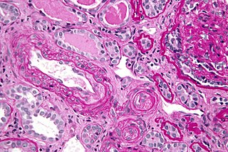
Antiphospholipid syndrome, or antiphospholipid antibody syndrome, is an autoimmune, hypercoagulable state caused by antiphospholipid antibodies. APS can lead to blood clots (thrombosis) in both arteries and veins, pregnancy-related complications, and other symptoms like low platelets, kidney disease, heart disease, and rash. Although the exact etiology of APS is still not clear, genetics is believed to play a key role in the development of the disease. Diagnosis is made based on symptoms and testing, but sometimes research criteria are used to aid in diagnosis. The research criteria for definite APS requires one clinical event and two positive blood test results spaced at least three months apart that detect lupus anticoagulant, anti-apolipoprotein antibodies, and/or anti-cardiolipin antibodies.

Fibrinolysis is a process that prevents blood clots from growing and becoming problematic. Primary fibrinolysis is a normal body process, while secondary fibrinolysis is the breakdown of clots due to a medicine, a medical disorder, or some other cause.

<span class="mw-page-title-main">Cardiopulmonary bypass</span> Technique that temporarily takes over the function of the heart and lungs during surgery

Cardiopulmonary bypass (CPB) is a technique in which a machine temporarily takes over the function of the heart and lungs during cardiac surgery, maintaining the circulation of blood and oxygen to the body. The CPB pump itself is often referred to as a heart-lung machine or "the pump". Cardiopulmonary bypass pumps are operated by perfusionists. CPB is a form of extracorporeal circulation. Extracorporeal membrane oxygenation (ECMO) is generally used for longer-term treatment.

In biology, hemostasis or haemostasis is a process to prevent and stop bleeding, meaning to keep blood within a damaged blood vessel. It is the first stage of wound healing. This involves coagulation, which changes blood from a liquid to a gel. Intact blood vessels are central to moderating blood's tendency to form clots. The endothelial cells of intact vessels prevent blood clotting with a heparin-like molecule and thrombomodulin, and prevent platelet aggregation with nitric oxide and prostacyclin. When endothelium of a blood vessel is damaged, the endothelial cells stop secretion of coagulation and aggregation inhibitors and instead secrete von Willebrand factor, which initiate the maintenance of hemostasis after injury. Hemostasis involves three major steps:

Low-molecular-weight heparin (LMWH) is a class of anticoagulant medications. They are used in the prevention of blood clots and treatment of venous thromboembolism and in the treatment of myocardial infarction.

D-dimer is a dimer that is a fibrin degradation product, a small protein fragment present in the blood after a blood clot is degraded by fibrinolysis. It is so named because it contains two D fragments of the fibrin protein joined by a cross-link, hence forming a protein dimer.

<span class="mw-page-title-main">Hypovolemic shock</span> Medical condition

Hypovolemic shock is a form of shock caused by severe hypovolemia. It could be the result of severe dehydration through a variety of mechanisms or blood loss. Hypovolemic shock is a medical emergency; if left untreated, the insufficient blood flow can cause damage to organs, leading to multiple organ failure.

<span class="mw-page-title-main">Heparin-induced thrombocytopenia</span> Low platelet count due to heparin, associated with a risk of thrombosis

Heparin-induced thrombocytopenia (HIT) is the development of thrombocytopenia, due to the administration of various forms of heparin, an anticoagulant. HIT predisposes to thrombosis. When thrombosis is identified the condition is called heparin-induced thrombocytopenia and thrombosis (HITT). HIT is caused by the formation of abnormal antibodies that activate platelets, which release microparticles that activate thrombin, leading to thrombosis. If someone receiving heparin develops new or worsening thrombosis, or if the platelet count falls, HIT can be confirmed with specific blood tests.

An antihemorrhagic agent is a substance that promotes hemostasis. It may also be known as a hemostatic agent.

<span class="mw-page-title-main">Thrombin time</span>

The thrombin time (TT), also known as the thrombin clotting time (TCT), is a blood test that measures the time it takes for a clot to form in the plasma of a blood sample containing anticoagulant, after an excess of thrombin has been added. It is used to diagnose blood coagulation disorders and to assess the effectiveness of fibrinolytic therapy. This test is repeated with pooled plasma from normal patients. The difference in time between the test and the 'normal' indicates an abnormality in the conversion of fibrinogen to fibrin, an insoluble protein.

Clotting time is a general term for the time required for a sample of blood to form a clot, or, in medical terms, coagulate. The term "clotting time" is often used when referring to tests such as the prothrombin time (PT), activated partial thromboplastin time, activated clotting time (ACT), thrombin time (TT), or Reptilase time. These tests are coagulation studies performed to assess the natural clotting ability of a sample of blood. In a clinical setting, healthcare providers will order one of these tests to evaluate a patient's blood for any abnormalities in the time it takes for their blood to clot. Each test involves adding a specific substance to the blood and measuring the time until the blood forms fibrin which is one of the first signs of clotted blood. Each test points to a different component of the clotting sequence which is made up of coagulation factors that help form clots. Abnormal results could be due to a number of reasons including, but, not limited to, deficiency in clotting factors, dysfunction of clotting factors, blood-thinning medications, medication side-effects, platelet deficiency, inherited bleeding or clotting disorders, liver disease, or advanced illness resulting in a medical emergency known as disseminated intravascular coagulation (DIC).

The fibrinolysis system is responsible for removing blood clots. Hyperfibrinolysis describes a situation with markedly enhanced fibrinolytic activity, resulting in increased, sometimes catastrophic bleeding. Hyperfibrinolysis can be caused by acquired or congenital reasons. Among the congenital conditions for hyperfibrinolysis, deficiency of alpha-2-antiplasmin or plasminogen activator inhibitor type 1 (PAI-1) are very rare. The affected individuals show a hemophilia-like bleeding phenotype. Acquired hyperfibrinolysis is found in liver disease, in patients with severe trauma, during major surgical procedures, and other conditions. A special situation with temporarily enhanced fibrinolysis is thrombolytic therapy with drugs which activate plasminogen, e.g. for use in acute ischemic events or in patients with stroke. In patients with severe trauma, hyperfibrinolysis is associated with poor outcome. Moreover, hyperfibrinolysis may be associated with blood brain barrier impairment, a plasmin-dependent effect due to an increased generation of bradykinin.

Thromboelastometry (TEM), previously named rotational thromboelastography (ROTEG) or rotational thromboelastometry (ROTEM), is an established viscoelastic method for hemostasis testing in whole blood. It is a modification of traditional thromboelastography (TEG).

Thrombodynamics test is a method for blood coagulation monitoring and anticoagulant control. This test is based on imitation of coagulation processes occurring in vivo, is sensitive both to pro- and anticoagulant changes in the hemostatic balance. Highly sensitive to thrombosis.

Blood clotting tests are the tests used for diagnostics of the hemostasis system. Coagulometer is the medical laboratory analyzer used for testing of the hemostasis system. Modern coagulometers realize different methods of activation and observation of development of blood clots in blood or in blood plasma.

References

  1. Da Luz, Luis Teodoro; Nascimento, Bartolomeu; Rizoli, Sandro (2013). "Thrombelastography (TEG®): practical considerations on its clinical use in trauma resuscitation". Scandinavian Journal of Trauma, Resuscitation and Emergency Medicine. 21: 29. doi: 10.1186/1757-7241-21-29 . PMC   3637505 . PMID   23587157.
  2. 1 2 3 Hartmann, Jan; Walsh, Mark; Grisoli, Anne; Thomas, Anthony V.; Shariff, Faisal; McCauley, Ross; Vande Lune, Stefani; Zackariya, Nuha; Patel, Shivani; Farrell, Michael S.; Sixta, Sherry; March, Robert; Evans, Edward; Tracy, Rebecca; Campello, Elena; Scărlătescu, Ecaterina; Agostini, Vanessa; Dias, João; Greve, Sarah; Thomas, Scott (March 2020). "Diagnosis and Treatment of Trauma-Induced Coagulopathy by Viscoelastography". Seminars in Thrombosis and Hemostasis. 46 (2): 134–146. doi:10.1055/s-0040-1702171. PMID   32160640. S2CID   212679113.
  3. Donahue SM, Otto CM, Thromboelastography: a tool for measuring hypercoagulability, hypocoagulability, and fibrinolysis, Journal of Veterinary Emergency and Critical Care:15(1), March 2005, Pages: 9-16
  4. Dirkmann D, Hanke AA, Görlinger K, Peters J. Hypothermia and acidosis synergistically impair coagulation in human whole blood.Anesth Analg. 2008;106:1627-32
  5. Tyler PD, Yang LM, Snider SB, Lerner AB, Aird WC, Shapiro NI (2021). "New Uses for Thromboelastography and Other Forms of Viscoelastic Monitoring in the Emergency Department: A Narrative Review". Ann Emerg Med. 77 (3): 357–366. doi: 10.1016/j.annemergmed.2020.07.026 . PMID   32988649.{{cite journal}}: CS1 maint: multiple names: authors list (link)
  6. Collins S, MacIntyre C, Hewer I (2016). "Thromboelastography: Clinical Application, Interpretation, and Transfusion Management". AANA J. 84 (2): 129–34. PMID   27311154.{{cite journal}}: CS1 maint: multiple names: authors list (link)
  7. Kreitzer NP, Bonomo J, Kanter D, Zammit C (2015). "Review of Thromboelastography in Neurocritical Care". Neurocrit Care. 23 (3): 427–33. doi:10.1007/s12028-015-0187-9. PMID   26275677. S2CID   29854774.{{cite journal}}: CS1 maint: multiple names: authors list (link)
  8. Moore, Hunter B.; Moore, Ernest E.; Chapman, Michael P.; Huebner, Benjamin R.; Einersen, Peter M.; Oushy, Solimon; Silliman, Christopher C.; Banerjee, Anirban; Sauaia, Angela (July 2017). "Viscoelastic Tissue Plasminogen Activator Challenge Predicts Massive Transfusion in 15 Minutes". Journal of the American College of Surgeons. 225 (1): 138–147. doi:10.1016/j.jamcollsurg.2017.02.018. ISSN   1879-1190. PMC   5527680 . PMID   28522144.
  9. 1 2 Dias, João D.; Sauaia, Angela; Achneck, Hardean E.; Hartmann, Jan; Moore, Ernest E. (4 April 2019). "Thromboelastography-guided therapy improves patient blood management and certain clinical outcomes in elective cardiac and liver surgery and emergency resuscitation: a systematic review and analysis". Journal of Thrombosis and Haemostasis. Online first (6): 984–994. doi: 10.1111/jth.14447 . PMC   6852204 . PMID   30947389.
  10. Hranjec T, Estreicher M, Rogers B, et al. Integral Use of Thromboelastography With Platelet Mapping to Guide Appropriate Treatment, Avoid Complications, and Improve Survival of Patients With Coronavirus Disease 2019-Related Coagulopathy. Crit Care Explor. 2020;2(12):e0287. Published 2020 Dec 21. doi:10.1097/CCE.0000000000000287 PMCID: PMC7769351 PMID: 33381763