Related Research Articles

The peritoneum is the serous membrane forming the lining of the abdominal cavity or coelom in amniotes and some invertebrates, such as annelids. It covers most of the intra-abdominal organs, and is composed of a layer of mesothelium supported by a thin layer of connective tissue. This peritoneal lining of the cavity supports many of the abdominal organs and serves as a conduit for their blood vessels, lymphatic vessels, and nerves.

The abdominal cavity is a large body cavity in humans and many other animals that contains many organs. It is a part of the abdominopelvic cavity. It is located below the thoracic cavity, and above the pelvic cavity. Its dome-shaped roof is the thoracic diaphragm, a thin sheet of muscle under the lungs, and its floor is the pelvic inlet, opening into the pelvis.

The spermatic cord is the cord-like structure in males formed by the vas deferens and surrounding tissue that runs from the deep inguinal ring down to each testicle. Its serosal covering, the tunica vaginalis, is an extension of the peritoneum that passes through the transversalis fascia. Each testicle develops in the lower thoracic and upper lumbar region and migrates into the scrotum during its descent it carries along with it vas deferens, its vessels, nerves etc. There is one on each side.

Anders Adolph Retzius, was a Swedish professor of anatomy and a supervisor at the Karolinska Institute in Stockholm.

The retroperitoneal space (retroperitoneum) is the anatomical space in the abdominal cavity behind (retro) the peritoneum. It has no specific delineating anatomical structures. Organs are retroperitoneal if they have peritoneum on their anterior side only. Structures that are not suspended by mesentery in the abdominal cavity and that lie between the parietal peritoneum and abdominal wall are classified as retroperitoneal.

The mesentery is an organ that attaches the intestines to the posterior abdominal wall in humans and is formed by the double fold of peritoneum. It helps in storing fat and allowing blood vessels, lymphatics, and nerves to supply the intestines, among other functions.

The renal capsule is a tough fibrous layer surrounding the kidney and covered in a layer of perirenal fat known as the adipose capsule of kidney. The adipose capsule is sometimes included in the structure of the renal capsule. It provides some protection from trauma and damage. The renal capsule is surrounded by the renal fascia. Overlying the renal fascia and between this and the transverse fascia is a region of pararenal fat.

The lesser omentum is the double layer of peritoneum that extends from the liver to the lesser curvature of the stomach and the first part of the duodenum.

The ascending colon is the part of the colon located between the cecum and the transverse colon.

In human anatomy, the greater sac, also known as the general cavity or peritoneum of the peritoneal cavity proper, is the cavity in the abdomen that is inside the peritoneum but outside the lesser sac.
In anatomy, the abdominal wall represents the boundaries of the abdominal cavity. The abdominal wall is split into the anterolateral and posterior walls.

The left colic artery is a branch of the inferior mesenteric artery that runs to the left behind the peritoneum and in front of the psoas major muscle, and after a short, but variable, course divides into an ascending and a descending branch; the stem of the artery or its branches cross the left ureter and left internal spermatic vessels.
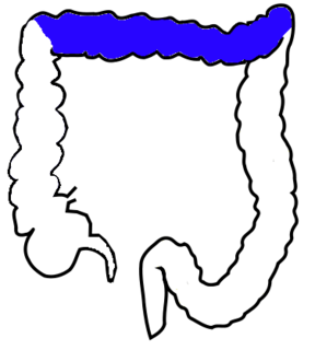
The transverse colon is the longest and most movable part of the colon. It crosses the abdomen from the ascending colon at the hepatic or right colic flexure with a downward convexity to the descending colon where it curves sharply on itself beneath the lower end of the spleen forming the splenic or left colic flexure. In its course, it describes an arch, the concavity of which is directed backward and a little upward. Toward its splenic end there is often an abrupt U-shaped curve which may descend lower than the main curve.

The transversalis fascia is a thin aponeurotic membrane which lies between the inner surface of the transverse abdominal muscle and the parietal peritoneum.

The greater omentum is a large apron-like fold of visceral peritoneum that hangs down from the stomach. It extends from the greater curvature of the stomach, passing in front of the small intestines and doubles back to ascend to the transverse colon before reaching to the posterior abdominal wall. The greater omentum is larger than the lesser omentum, which hangs down from the liver to the lesser curvature. The common anatomical term "epiploic" derives from "epiploon", from the Greek epipleein, meaning to float or sail on, since the greater omentum appears to float on the surface of the intestines. It is the first structure observed when the abdominal cavity is opened anteriorly.

The rectus sheath, also called the rectus fascia, is formed by the aponeuroses of the transverse abdominal and the internal and external oblique muscles. It contains the rectus abdominis and pyramidalis muscles.

In the male, the peritoneum encircles the sigmoid colon, from which it is reflected to the posterior wall of the pelvis as a fold, the sigmoid mesocolon. It then leaves the sides and, finally, the front of the rectum, and is continued on to the upper ends of the seminal vesicles and the bladder; on either side of the rectum it forms a fossa, the pararectal fossa, which varies in size with the distension of the rectum.

The renal fascia is a layer of connective tissue encapsulating the kidneys and the adrenal glands. It can be divided into:

Carl Toldt was an Austrian anatomist who was a native of Bruneck, Tyrol.
Between the inner surface of the general layer of the fascia which lines the interior of the abdominal and pelvic cavities, and the peritoneum, there is a considerable amount of connective tissue, termed the extraperitoneal fat or subperitoneal connective tissue.
References
- ↑ Culligan K, Walsh S, Dunne C, et al. (January 2014). "The Mesocolon: A Histological and Electron Microscopic Characterization of the Mesenteric Attachment of the Colon Prior to and After Surgical Mobilization". Annals of Surgery. 260 (6): 1048–56. doi:10.1097/SLA.0000000000000323. hdl:10344/4895. PMID 24441808.
- ↑ Toldt C (1919). "Splanchology – general considerations". In Toldt C & Della Rossa A (ed.). An atlas of human anatomy for students and physicians. 4. New York: Rebman Company. p. 408.
| Animals | |
|---|---|
| Plants | |
| This anatomy article is a stub. You can help Wikipedia by expanding it. |