Tomosynthesis

Last updated
Tomosynthesis of a lung with chronic fibrosing pulmonary aspergillosis. Tomosynthesis of chronic fibrosing pulmonary aspergillosis.gif
Tomosynthesis of a lung with chronic fibrosing pulmonary aspergillosis.

Tomosynthesis, also digital tomosynthesis (DTS), is a method for performing high-resolution limited-angle tomography at radiation dose levels comparable with projectional radiography. It has been studied for a variety of clinical applications, including vascular imaging, dental imaging, orthopedic imaging, mammographic imaging, musculoskeletal imaging, and chest imaging. [1]

Contents

History

The concept of tomosynthesis was derived from the work of Ziedses des Plantes, who developed methods of reconstructing an arbitrary number of planes from a set of projections. Though this idea was displaced by the advent of computed tomography, tomosynthesis later gained interest as a low-dose tomographic alternative to CT. [2]

Reconstruction

Tomosynthesis reconstruction algorithms are similar to CT reconstructions, in that they are based on performing an inverse Radon transform. Due to partial data sampling with very few projections, approximation algorithms have to be used. Filtered back projection and iterative, expectation-maximization algorithms have both been used to reconstruct the data. [3]

Reconstruction algorithms for tomosynthesis are different from those of conventional CT because the conventional filtered back projection algorithm requires a complete set of data. Iterative algorithms based upon expectation maximization are most commonly used, but are computationally intensive. Some manufacturers have produced practical systems using off-the-shelf GPUs to perform the reconstruction in a few seconds.

Differences from other imaging modalities

Digital tomosynthesis combines digital image capture and processing with simple tube/detector motion as used in conventional computed tomography (CT). However, though there are some similarities to CT, it is a separate technique. In modern (helical) CT, the source/detector makes at least a complete 180-degree rotation about the subject obtaining a complete set of data from which images may be reconstructed. Digital tomosynthesis, on the other hand, only uses a limited rotation angle (e.g., 15-60 degrees) with a lower number of discrete exposures (e.g., 7-51) than CT. This incomplete set of projections is digitally processed to yield images similar to conventional tomography with a limited depth of field. Because the image processing is digital, a series of slices at different depths and with different thicknesses can be reconstructed from the same acquisition. However, since fewer projections are needed than CT to perform the reconstruction, radiation exposure and cost are both reduced. [4]

Applications

Breast

Digital breast tomosynthesis (DBT) [5] is Food and Drug Administration (FDA) approved for use in breast cancer screening. [6] The benefit for screening has been debated, [7] but consensus is being reached that the technology is improving sensitivity compared to two-view digital mammography at the cost of a slightly reduced specificity (increased recall rates). [8] Because the data acquired are 85 - 160 micron typical resolution, much higher than CT, DBT is unable to offer the narrow slice widths that CT offers (typically 1-1.5 mm). However, the higher resolution detectors permit very high in-plane resolution, even if the Z-axis resolution is less. Another interesting property of breast tomosynthesis is that image quality may vary substantially through the imaging volume. [9]

Photon-counting breast tomosynthesis has been investigated, [10] and spectral imaging applications, such as breast density measurement and lesion characterization, [11] [12] have been investigated on that platform.

Chest

Musculoskeletal imaging

Tomosynthesis has a much more limited depth of field than does CT. For this reason, it likely will not be able to replace CT for the evaluation of the deeper organs of the body. However, since bones are often near the skin, multiple musculoskeletal applications of tomosynthesis have been studied, most of which have mostly been used in research with limited use in everyday practice.

Evaluation of fractures

Tomosynthesis has been compared to both radiography and CT for the evaluation of healing fractures, especially in the presence of hardware. In a study of patients with wrist fractures, digital tomosynthesis was shown to enable detection of more fractures than radiography while simultaneously providing lower metal artifact than radiography. [4]

Evaluation of erosions in rheumatoid arthritis

Tomosynthesis has been compared to digital radiography, with CT as the standard, for the detection of erosions associated with rheumatoid arthritis. The radiation dose of digital tomosynthesis was very close to that of digital radiography. However, tomosynthesis showed sensitivity, specificity, accuracy, positive predictive value, and negative predictive value of 80%, 75%, 78%, 76%, and 80%, compared to digital radiography were 66%, 81%, 74%, 77%, and 71%. [13] The slight benefit digital tomosynthesis in this application may or may not justify the slightly increased cost of the modality compared to digital radiography.

Electronics

Tomosynthesis is also used for x-ray inspection of electronics, [14] particularly printed circuit board assemblies and electronic components. Tomosynthesis is usually used where a CT slice is required at high magnification, where conventional CT would not allow the sample to be located close enough to the x-ray source.

Related Research Articles

<span class="mw-page-title-main">CT scan</span> Medical imaging procedure using X-rays to produce cross-sectional images

A computed tomography scan is a medical imaging technique used to obtain detailed internal images of the body. The personnel that perform CT scans are called radiographers or radiology technologists.

<span class="mw-page-title-main">Radiography</span> Imaging technique using ionizing and non-ionizing radiation

Radiography is an imaging technique using X-rays, gamma rays, or similar ionizing radiation and non-ionizing radiation to view the internal form of an object. Applications of radiography include medical and industrial radiography. Similar techniques are used in airport security,. To create an image in conventional radiography, a beam of X-rays is produced by an X-ray generator and it is projected towards the object. A certain amount of the X-rays or other radiation are absorbed by the object, dependent on the object's density and structural composition. The X-rays that pass through the object are captured behind the object by a detector. The generation of flat two-dimensional images by this technique is called projectional radiography. In computed tomography, an X-ray source and its associated detectors rotate around the subject, which itself moves through the conical X-ray beam produced. Any given point within the subject is crossed from many directions by many different beams at different times. Information regarding the attenuation of these beams is collated and subjected to computation to generate two-dimensional images on three planes which can be further processed to produce a three-dimensional image.

<span class="mw-page-title-main">Mammography</span> Process of using low-energy X-rays to examine the human breast for diagnosis and screening

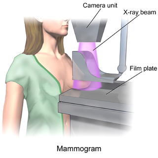
Mammography is the process of using low-energy X-rays to examine the human breast for diagnosis and screening. The goal of mammography is the early detection of breast cancer, typically through detection of characteristic masses or microcalcifications.

<span class="mw-page-title-main">Single-photon emission computed tomography</span> Nuclear medicine tomographic imaging technique

Single-photon emission computed tomography is a nuclear medicine tomographic imaging technique using gamma rays. It is very similar to conventional nuclear medicine planar imaging using a gamma camera, but is able to provide true 3D information. This information is typically presented as cross-sectional slices through the patient, but can be freely reformatted or manipulated as required.

<span class="mw-page-title-main">Tomography</span> Imaging by sections or sectioning using a penetrative wave

Tomography is imaging by sections or sectioning that uses any kind of penetrating wave. The method is used in radiology, archaeology, biology, atmospheric science, geophysics, oceanography, plasma physics, materials science, cosmochemistry, astrophysics, quantum information, and other areas of science. The word tomography is derived from Ancient Greek τόμος tomos, "slice, section" and γράφω graphō, "to write" or, in this context as well, "to describe." A device used in tomography is called a tomograph, while the image produced is a tomogram.

Computed tomography laser mammography (CTLM) is the trademark of Imaging Diagnostic Systems, Inc. for its optical tomographic technique for female breast imaging.

Willi A. Kalender is a German medical physicist and professor and former chairman of the Institute of Medical Physics of the University of Erlangen-Nuremberg. Kalender has produced several new technologies in the field of diagnostic radiology imaging.

<span class="mw-page-title-main">Projectional radiography</span> Formation of 2D images using X-rays

Projectional radiography, also known as conventional radiography, is a form of radiography and medical imaging that produces two-dimensional images by X-ray radiation. The image acquisition is generally performed by radiographers, and the images are often examined by radiologists. Both the procedure and any resultant images are often simply called 'X-ray'. Plain radiography or roentgenography generally refers to projectional radiography. Plain radiography can also refer to radiography without a radiocontrast agent or radiography that generates single static images, as contrasted to fluoroscopy, which are technically also projectional.

<span class="mw-page-title-main">Automatic exposure control</span>

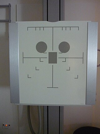
Automatic Exposure Control (AEC) is an X-ray exposure termination device. A medical radiographic exposure is always initiated by a human operator but an AEC detector system may be used to terminate the exposure when a predetermined amount of radiation has been received. The intention of AEC is to provide consistent x-ray image exposure, whether to film, a digital detector or a CT scanner. AEC systems may also automatically set exposure factors such as the X-ray tube current and voltage in a CT.

<span class="mw-page-title-main">Flat-panel detector</span> Class of solid-state x-ray digital radiography devices

Flat-panel detectors are a class of solid-state x-ray digital radiography devices similar in principle to the image sensors used in digital photography and video. They are used in both projectional radiography and as an alternative to x-ray image intensifiers (IIs) in fluoroscopy equipment.

<span class="mw-page-title-main">Cone beam computed tomography</span> Medical imaging technique

Cone beam computed tomography is a medical imaging technique consisting of X-ray computed tomography where the X-rays are divergent, forming a cone.

<span class="mw-page-title-main">Phase-contrast X-ray imaging</span> Imaging systems using changes in phase

Phase-contrast X-ray imaging or phase-sensitive X-ray imaging is a general term for different technical methods that use information concerning changes in the phase of an X-ray beam that passes through an object in order to create its images. Standard X-ray imaging techniques like radiography or computed tomography (CT) rely on a decrease of the X-ray beam's intensity (attenuation) when traversing the sample, which can be measured directly with the assistance of an X-ray detector. However, in phase contrast X-ray imaging, the beam's phase shift caused by the sample is not measured directly, but is transformed into variations in intensity, which then can be recorded by the detector.

<span class="mw-page-title-main">Photon counting</span> Counting photons using a single-photon detector

Photon counting is a technique in which individual photons are counted using a single-photon detector (SPD). A single-photon detector emits a pulse of signal for each detected photon. The counting efficiency is determined by the quantum efficiency and the system's electronic losses.

<span class="mw-page-title-main">Breast imaging</span>

In medicine, breast imaging is a sub-speciality of diagnostic radiology that involves imaging of the breasts for screening or diagnostic purposes. There are various methods of breast imaging using a variety of technologies as described in detail below. Traditional screening and diagnostic mammography uses x-ray technology and has been the mainstay of breast imaging for many decades. Breast tomosynthesis is a relatively new digital x-ray mammography technique that produces multiple image slices of the breast similar to, but distinct from, computed tomography (CT). Xeromammography and galactography are somewhat outdated technologies that also use x-ray technology and are now used infrequently in the detection of breast cancer. Breast ultrasound is another technology employed in diagnosis and screening that can help differentiate between fluid filled and solid lesions, an important factor to determine if a lesion may be cancerous. Breast MRI is a technology typically reserved for high-risk patients and patients recently diagnosed with breast cancer. Lastly, scintimammography is used in a subgroup of patients who have abnormal mammograms or whose screening is not reliable on the basis of using traditional mammography or ultrasound.

<span class="mw-page-title-main">4DCT</span>

Four-dimensional computed tomography (4DCT) is a type of CT scanning which records multiple images over time. It allows playback of the scan as a video, so that physiological processes can be observed and internal movement can be tracked. The name is derived from the addition of time to traditional 3D computed tomography. Alternatively, the phase of a particular process, such as respiration, may be considered the fourth dimension.

Computational imaging is the process of indirectly forming images from measurements using algorithms that rely on a significant amount of computing. In contrast to traditional imaging, computational imaging systems involve a tight integration of the sensing system and the computation in order to form the images of interest. The ubiquitous availability of fast computing platforms, the advances in algorithms and modern sensing hardware is resulting in imaging systems with significantly enhanced capabilities. Computational Imaging systems cover a broad range of applications include computational microscopy, tomographic imaging, MRI, ultrasound imaging, computational photography, Synthetic Aperture Radar (SAR), seismic imaging etc. The integration of the sensing and the computation in computational imaging systems allows for accessing information which was otherwise not possible. For example:

Jeffrey Harold Siewerdsen is an American physicist and biomedical engineer who is a Professor of Imaging Physics at The University of Texas MD Anderson Cancer Center as well as Biomedical Engineering, Computer Science, Radiology, and Neurosurgery at Johns Hopkins University.He is among the original inventors of cone-beam CT-guided radiotherapy as well as weight-bearing cone-beam CT for musculoskeletal radiology and orthopedic surgery. His work also includes the early development of flat-panel detectors on mobile C-arms for intraoperative cone-beam CT in image-guided surgery. He developed early models for the signal and noise performance of flat-panel detectors and later extended such analysis to dual-energy imaging and 3D imaging performance in cone-beam CT. He founded the ISTAR Lab in the Department of Biomedical Engineering, the Carnegie Center for Surgical Innovation at Johns Hopkins Hospital, and the Surgical Data Science Program at the Institute for Data Science in Oncology at The University of Texas MD Anderson Cancer Center.

Photon-counting computed tomography (PCCT) is a form of X-ray computed tomography (CT) in which X-rays are detected using a photon-counting detector (PCD) which registers the interactions of individual photons. By keeping track of the deposited energy in each interaction, the detector pixels of a PCD each record an approximate energy spectrum, making it a spectral or energy-resolved CT technique. In contrast, more conventional CT scanners use energy-integrating detectors (EIDs), where the total energy deposited in a pixel during a fixed period of time is registered. These EIDs thus register only photon intensity, comparable to black-and-white photography, whereas PCDs register also spectral information, similar to color photography.

Spectral imaging is an umbrella term for energy-resolved X-ray imaging in medicine. The technique makes use of the energy dependence of X-ray attenuation to either increase the contrast-to-noise ratio, or to provide quantitative image data and reduce image artefacts by so-called material decomposition. Dual-energy imaging, i.e. imaging at two energy levels, is a special case of spectral imaging and is still the most widely used terminology, but the terms "spectral imaging" and "spectral CT" have been coined to acknowledge the fact that photon-counting detectors have the potential for measurements at a larger number of energy levels.

Photon-counting mammography was introduced commercially in 2003 and was the first widely available application of photon-counting detector technology in medical x-ray imaging. Photon-counting mammography improves dose efficiency compared to conventional technologies, and enables spectral imaging.

References

  1. Dobbins, James; McAdams, H. Page (Nov 2009). "Chest Tomosynthesis: Technical Principles and Clinical Update". European Journal of Radiology. 72 (2): 244–251. doi:10.1016/j.ejrad.2009.05.054. PMC   3693857 . PMID   19616909.
  2. Dobbins JT, 3rd; Godfrey, DJ (7 October 2003). "Digital x-ray tomosynthesis: current state of the art and clinical potential". Physics in Medicine and Biology. 48 (19): R65–106. doi:10.1088/0031-9155/48/19/r01. PMID   14579853. S2CID   250737333.{{cite journal}}: CS1 maint: numeric names: authors list (link)
  3. Sechopoulos, Ioannis (2013). "A review of breast tomosynthesis. Part II. Image reconstruction, processing and analysis, and advanced applications". Medical Physics. 40 (1): 014302. Bibcode:2013MedPh..40a4302S. doi:10.1118/1.4770281. PMC   3548896 . PMID   23298127.
  4. 1 2 Ha, Alice; Lee, Amie; Hippe, Daniel; Chou, Shinn-Huey; Chew, Felix (July 2015). "Digital Tomosynthesis to Evaluate Fracture Healing: Prospective Comparison With Radiography and CT". American Journal of Roentgenology. 205 (1): 136–141. doi:10.2214/AJR.14.13833. PMID   26102392. S2CID   29516482.
  5. Smith, Andrew P.; Niklason, Loren; Ren, Baorui; Wu, Tao; Ruth, Chris; Jing, Zhenxue (2006). "Lesion Visibility in Low Dose Tomosynthesis". Digital Mammography. Lecture Notes in Computer Science. Vol. 4046. pp. 160–166. doi:10.1007/11783237_23. ISBN   978-3-540-35625-7.
  6. "Selenia Dimensions 3D System - P080003, US Food and Drug Administration (FDA), February 11, 2011
  7. Siu, Albert L. (12 January 2016). "Screening for Breast Cancer: U.S. Preventive Services Task Force Recommendation Statement". Annals of Internal Medicine. 164 (4): 279–96. doi: 10.7326/M15-2886 . PMID   26757170.
  8. Zackrisson, Sophia; Lång, Kristina; Rosso, Aldana; Johnson, Kristin; Dustler, Magnus; Förnvik, Daniel; Förnvik, Hannie; Sartor, Hanna; Timberg, Pontus; Tingberg, Anders; Andersson, Ingvar (November 2018). "One-view breast tomosynthesis versus two-view mammography in the Malmö Breast Tomosynthesis Screening Trial (MBTST): a prospective, population-based, diagnostic accuracy study". The Lancet Oncology. 19 (11): 1493–1503. doi:10.1016/s1470-2045(18)30521-7. ISSN   1470-2045. PMID   30322817. S2CID   53501736.
  9. Berggren, Karl; Cederström, Björn; Lundqvist, Mats; Fredenberg, Erik (2018). "Cascaded systems analysis of shift-variant image quality in slit-scanning breast tomosynthesis". Medical Physics. 45 (10): 4392–4401. Bibcode:2018MedPh..45.4392B. doi:10.1002/mp.13116. ISSN   2473-4209. PMID   30091470. S2CID   51941096.
  10. Berggren, Karl; Cederström, Björn; Lundqvist, Mats; Fredenberg, Erik (2018). "Characterization of photon-counting multislit breast tomosynthesis". Medical Physics. 45 (2): 549–560. Bibcode:2018MedPh..45..549B. doi:10.1002/mp.12684. ISSN   2473-4209. PMID   29159881. S2CID   26002936.
  11. Fredenberg, Erik; Berggren, Karl; Bartels, Matthias; Erhard, Klaus (2016), Tingberg, Anders; Lång, Kristina; Timberg, Pontus (eds.), "Volumetric Breast-Density Measurement Using Spectral Photon-Counting Tomosynthesis: First Clinical Results", Breast Imaging, vol. 9699, Cham: Springer International Publishing, pp. 576–584, arXiv: 2101.02758 , doi:10.1007/978-3-319-41546-8_72, ISBN   978-3-319-41545-1, S2CID   1257696 , retrieved 2020-12-30
  12. Cederström, Björn; Fredenberg, Erik; Berggren, Karl; Erhard, Klaus; Danielsson, Mats; Wallis, Matthew (2017-03-09). Flohr, Thomas G.; Lo, Joseph Y.; Gilat Schmidt, Taly (eds.). "Lesion characterization in spectral photon-counting tomosynthesis". Society of Photo-Optical Instrumentation Engineers (Spie) Conference Series. Medical Imaging 2017: Physics of Medical Imaging. 10132. Orlando, Florida, United States: 26–35. arXiv: 2102.00175 . Bibcode:2017SPIE10132E..05C. doi:10.1117/12.2253966. S2CID   125903314.
  13. Simon, Paolo; Gérard, Laurent; Kaiser, Marie-Joëlle; Ribbens, Clio; Rinkin, Charline; Malaise, Olivier; Malaise, Michecl (August 2016). "Use of Tomosynthesis for Detection of Bone Erosions of the Foot in Patients With Established Rheumatoid Arthritis: Comparison With Radiography and CT". American Journal of Roentgenology. 205 (2): 364–370. doi:10.2214/AJR.14.14120. PMID   26204289.
  14. A Practical Guide to X-ray Inspection Criteria and Common Defect Analysis (Second ed.). 2019. p. 83. ISBN   978-1527233614.