Related Research Articles

The trachea, also known as the windpipe, is a cartilaginous tube that connects the larynx to the bronchi of the lungs, allowing the passage of air, and so is present in almost all animals with lungs. The trachea extends from the larynx and branches into the two primary bronchi. At the top of the trachea, the cricoid cartilage attaches it to the larynx. The trachea is formed by a number of horseshoe-shaped rings, joined together vertically by overlying ligaments, and by the trachealis muscle at their ends. The epiglottis closes the opening to the larynx during swallowing.
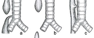
Esophageal atresia is a congenital medical condition that affects the alimentary tract. It causes the esophagus to end in a blind-ended pouch rather than connecting normally to the stomach. It comprises a variety of congenital anatomic defects that are caused by an abnormal embryological development of the esophagus. It is characterized anatomically by a congenital obstruction of the esophagus with interruption of the continuity of the esophageal wall.
Tracheomalacia is a condition or incident where the cartilage that keeps the airway (trachea) open is soft such that the trachea partly collapses especially during increased airflow. This condition is most commonly seen in infants and young children. The usual symptom is stridor when a person breathes out. This is usually known as a collapsed windpipe.

Esophageal varices are extremely dilated sub-mucosal veins in the lower third of the esophagus. They are most often a consequence of portal hypertension, commonly due to cirrhosis. People with esophageal varices have a strong tendency to develop severe bleeding which left untreated can be fatal. Esophageal varices are typically diagnosed through an esophagogastroduodenoscopy.

A tracheoesophageal fistula is an abnormal connection (fistula) between the esophagus and the trachea. TEF is a common congenital abnormality, but when occurring late in life is usually the sequela of surgical procedures such as a laryngectomy.
Atresia is a condition in which an orifice or passage in the body is closed or absent.

The cricothyroid ligament is a ligament in the neck. It connects the cricoid cartilage to the thyroid cartilage. It prevents these cartilages from moving too far apart. It is cut during an emergency cricothyrotomy to treat upper airway obstruction.

An imperforate anus or anorectal malformations (ARMs) are birth defects in which the rectum is malformed. ARMs are a spectrum of different congenital anomalies which vary from fairly minor lesions to complex anomalies. The cause of ARMs is unknown; the genetic basis of these anomalies is very complex because of their anatomical variability. In 8% of patients, genetic factors are clearly associated with ARMs. Anorectal malformation in Currarino syndrome represents the only association for which the gene HLXB9 has been identified.

The VACTERL association refers to a recognized group of birth defects which tend to co-occur. This pattern is a recognized association, as opposed to a syndrome, because there is no known pathogenetic cause to explain the grouped incidence.

In human anatomy, the fibularis tertius is a muscle in the anterior compartment of the leg. It acts to tilt the sole of the foot away from the midline of the body (eversion) and to pull the foot upward toward the body (dorsiflexion).

Beneath the neck of the radius, on the medial side, is an eminence, the radial tuberosity; its surface is divided into:
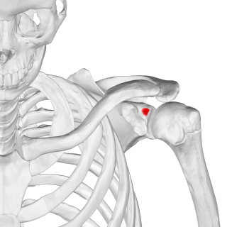
The supraglenoid tubercle is a region of the scapula from which the long head of the biceps brachii muscle originates. It is a small, rough projection superior to the glenoid cavity near the base of the coracoid process. The term supraglenoid is from the Latin supra, meaning above, and glenoid, meaning socket or cavity.

The linea semilunaris is a curved tendinous intersection found on either side of the rectus abdominis muscle.

The lung bud sometimes referred to as the respiratory bud forms from the respiratory diverticulum, an embryological endodermal structure that develops into the respiratory tract organs such as the larynx, trachea, bronchi and lungs. It arises from part of the laryngotracheal tube.
The trachealis muscle is a sheet of smooth muscle in the trachea.

Bronchomalacia is a term for weak cartilage in the walls of the bronchial tubes, often occurring in children under a day. Bronchomalacia means 'floppiness' of some part of the bronchi. Patients present with noisy breathing and/or wheezing. There is collapse of a main stem bronchus on exhalation. If the trachea is also involved the term tracheobronchomalacia (TBM) is used. If only the upper airway the trachea is involved it is called tracheomalacia (TM). There are two types of bronchomalacia. Primary bronchomalacia is due to a deficiency in the cartilaginous rings. Secondary bronchomalacia may occur by extrinsic compression from an enlarged vessel, a vascular ring or a bronchogenic cyst. Though uncommon, idiopathic tracheobronchomalacia has been described in older adults.
A laryngeal cleft or laryngotracheoesophageal cleft is a rare congenital abnormality in the posterior laryngo-tracheal wall. It occurs in approximately 1 in 10,000 to 20,000 births. It means there is a communication between the oesophagus and the trachea, which allows food or fluid to pass into the airway.
Tracheal agenesis is a rare birth defect with a prevalence of less than 1 in 50,000 in which the trachea fails to develop, resulting in an impaired communication between the larynx and the alveoli of the lungs. Although the defect is normally fatal, occasional cases have been reported of long-term survival following surgical intervention.

Pulmonary agenesis is an inborn lung underdevelopment that is rare and potentially lethal. The disorder is caused by a complete developmental arrest of the primitive lung during embryonic life, and it is often associated with other developmental defects. Bilateral and unilateral pulmonary agenesis are classified, depending on whether one side of the lung or both sides are affected. Bilateral pulmonary agenesis is lethal, while the mortality rate of unilateral pulmonary agenesis is higher than 50%. Depending on the severity, the symptom ranges from none to various respiratory complaints. It is detectable prenatally, however, its nonspecific clinical features act as the obstacle for diagnosing. The exact cause of pulmonary agenesis is still obscure. However, theories have been raised regarding the vascular, iatrogenic, viral and genetic causes of pulmonary agenesis in an attempt to explain the pathogenesis of the disorder. In most cases of pulmonary agenesis, surgical resection is performed to remove the malformed lobe or the entire defected lung of the patient depending on the severity of the respiratory impairment.

Mandibulofacial dysostosis with microcephaly syndrome, also known as growth delay-intellectual disability-mandibulofacial dysostosis-microcephaly-cleft palate syndrome, mandibulofacial dysostosis, guion-almeida type, or simply as MFDM syndrome is a rare genetic disorder which is characterized by developmental delays, intellectual disabilities, and craniofacial dysmorphisms.
References
- ↑ Respiratory System
- ↑ Bax, Klaas (2010). "27 - Esophageal Atresia and Tracheoesophageal Malformations". Ashcraft's Pediatric Surgery (5th ed.). Saunders. pp. 345–361. doi:10.1016/B978-1-4160-6127-4.00027-6. ISBN 978-1-4160-6127-4.
- 1 2 Sylvester, Karl G.; Ghole, Saif; Albanese, Craig T. (2010). "22 - Congenital Bronchopulmonary Malformations". Ashcraft's Pediatric Surgery (5th ed.). Saunders. pp. 279–289. doi:10.1016/B978-1-4160-6127-4.00022-7. ISBN 978-1-4160-6127-4.
- 1 2 3 Shojaie, Sharareh; Post, Martin (2017). "64 - Molecular Mechanisms of Lung Development and Lung Branching Morphogenesis". Fetal and Neonatal Physiology. Vol. 1 (5th ed.). Elsevier. pp. 658–666. doi:10.1016/B978-0-323-35214-7.00064-0. ISBN 978-0-323-35214-7.
- ↑ Development of the Digestive System Archived 2007-11-23 at the Wayback Machine
- ↑ "Esophageal Atresia and Tracheoesophageal Fistula - February 15, 1999 - American Academy of Family Physicians". Archived from the original on 2011-06-06. Retrieved 2007-11-11.
- ↑ Kulungowski, Ann M. (2018). "89 - Tracheoesophageal Malformations". Abernathy's Surgical Secrets (7th ed.). Elsevier. pp. 409–411. doi:10.1016/B978-0-323-47873-1.00089-9. ISBN 978-0-323-47873-1.
- ↑ Gulati, Reema; Radhakrishnan, Kadakkal; Thomson, Mike A. (2011). "20 - Developmental Anatomy and Physiology of the Esophagus". Pediatric Gastrointestinal and Liver Disease (4th ed.). Saunders. pp. 207–220. doi:10.1016/B978-1-4377-0774-8.10020-X. ISBN 978-1-4377-0774-8.
- ↑ Kumar, S.; Salib, R. (2006). "Upper Airway Obstruction". Encyclopedia of Respiratory Medicine. Academic Press. pp. 375–385. doi:10.1016/B0-12-370879-6/00415-4. ISBN 978-0-12-370879-3.