Type I topoisomerase

Last updated
DNA topoisomerase I, N-terminal (non-catalytic), viral
PDB 1vcc EBI.jpg
amino terminal 9kda domain of vaccinia virus dna topoisomerase i residues 1-77, experimental electron density for residues 1-77
Identifiers
SymbolVirDNA-topo-I_N
Pfam PF09266
InterPro IPR015346
SCOP2 1vcc / SCOPe / SUPFAM
Available protein structures:
Pfam   structures / ECOD  
PDB RCSB PDB; PDBe; PDBj
PDBsum structure summary

In molecular biology Type I topoisomerases are enzymes that cut one of the two strands of double-stranded DNA, relax the strand, and reanneal the strand. They are further subdivided into two structurally and mechanistically distinct topoisomerases: type IA and type IB.

Contents

Historically, type IA topoisomerases are referred to as prokaryotic topo I, while type IB topoisomerases are referred to as eukaryotic topoisomerase. This distinction, however, no longer applies as type IA and type IB topoisomerases exist in all domains of life.

Functionally, these subclasses perform very specialized functions. Prokaryotic topoisomerase I (topo IA) can only relax negative supercoiled DNA, whereas eukaryotic topoisomerase I (topo IB) can introduce positive supercoils, separating the DNA of daughter chromosomes after DNA replication, and relax DNA.

Function

These enzymes have several functions: to remove DNA supercoils during transcription and DNA replication; for strand breakage during recombination; for chromosome condensation; and to disentangle intertwined DNA during mitosis. [1] [2]

Structure

This domain assumes a beta(2)-alpha-beta-alpha-beta(2) fold, with a left-handed crossover between strands beta2 and beta3. It has a four criss-crossed beta-strands surrounded by four alpha-helices that are arranged in a Rossmann fold [3]

Mechanisms

Type I topoisomerases are ATP-independent enzymes (except for reverse gyrase), and can be subdivided according to their structure and reaction mechanisms: type IA (bacterial and archaeal topoisomerase I, topoisomerase III and reverse gyrase) and type IB (eukaryotic topoisomerase I and topoisomerase V). These enzymes are primarily responsible for relaxing positively and/or negatively supercoiled DNA, except for reverse gyrase, which can introduce positive supercoils into DNA.

DNA topoisomerases regulate the number of topological links between two DNA strands (i.e. change the number of superhelical turns) by catalysing transient single- or double-strand breaks, crossing the strands through one another, then resealing the breaks. [4]

Classes

DNA topoisomerases are divided into two classes: type I enzymes (EC; topoisomerases I, III and V) break single-strand DNA, and type II enzymes (EC; topoisomerases II, IV and VI) break double-strand DNA. [5]

Type IA topoisomerases

Structure of topo III bound to single stranded DNA (pdb ID 1I7D). Note that the DNA resembles B-form DNA TopoI.png
Structure of topo III bound to single stranded DNA (pdb ID 1I7D). Note that the DNA resembles B-form DNA

Introduction

Type IA topoisomerases, historically said to be found in prokaryotes, create a single break in DNA and pass a second strand or duplex through the break. This strand passage mechanism shares several features with type IIA topoisomerases. They both form a 5' phosphotyrosine intermediate, and require a divalent metal ion to perform its work. Unlike type II topoisomerases, type IA topoisomerases do not use energy to do its work (with the notable exception of reverse gyrase, see below).

Structure

Type IA topoisomerases have several domains, often number Domain 1-4. Domain I contains a Toprim domain (a Rossman fold known to coordinate Magnesium ions), domain IV and domain III each consist of a helix-turn-helix (HTH) domain; the catalytic tyrosine resides on the HTH of domain III. Domain II is a flexible bridge between domains III and IV. The structure of type IA topoisomerase resembles a lock, with Domains I, III and IV lying on the bottom of the structure. [6] The structure of topo III (see below) bound to single-stranded DNA [7] (pdb id = 1I7D) shows how the HTH and Toprim domain are coordinated about the DNA.

Type IA topoisomerase variants

There are several variants of Type IA topoisomerases, differing by appendages attached to the main core (sometimes referred to as the "topo-fold"). Members of this subclass include topo I, topo III (which contain additional Zinc-binding motifs), and reverse gyrase. Reverse gyrase is particularly interesting because an ATPase domain, which resembles the helicase-like domain of the Rho transcription factor, is attached (the structure of reverse gyrase was solved by Rodriguez and Stock, EMBO J 2002). The enzyme uses the hydrolysis of ATP to introduce positive supercoils and overwinds DNA, a feature attractive in hyperthermophiles, in which reverse gyrase is known to exist. Rodriguez and Stock have done further work to identify a "latch" that is involved in communicating the hydrolysis of ATP to the introduction of positive supercoils.

The topo III variant is likewise very interesting because it has zinc-binding motifs that is thought to bind single-stranded DNA. Topo III has been identified to be associated with the BLM (for Bloom Syndrome) helicase during recombination.

Mechanism

Type IA topoisomerases operate through a strand-passage mechanism, using a single gate (in contrast with type II topoisomerases). First, the single-stranded DNA binds domain III and I. The catalytic tyrosine cleaves the DNA backbone, creating a transient 5' phosphotyrosine intermediate. The break is then separated, using domain II as a hinge, and a second duplex or strand of DNA is passed through. Domain III and I close and the DNA is re-annealed.

Type IB topoisomerases

Important active site residues of Topoisomerase IB. DNA is colored raspberry. Topoisomerase IB Active Site.jpg
Important active site residues of Topoisomerase IB. DNA is colored raspberry.
Active site of Topoisomerase IB. DNA is colored raspberry with a light-blue backbone. (PDB ID = 1A36) 1a36 topoisomerase-IB-active-site.png
Active site of Topoisomerase IB. DNA is colored raspberry with a light-blue backbone. (PDB ID = 1A36)

Introduction

In contrast to type IA topoisomerases, type 1B Topoisomerase solves the problem of overwound and underwound (also referred to as positively or negatively supercoiled) DNA through a hindered rotary mechanism. Crystal structures, biochemistry, and single molecule experiments have contributed to a general mechanism. The enzyme first wraps around DNA and creates a single, 3' phosphotyrosine intermediate. The 5' end is then free to rotate, twisting it about the other strand, to relax DNA until the topoisomerase re-ligates the broken strands.

Structure

The structure of topo IB bound to DNA has been solved (pdb id = 1A36). Topo IB is composed of an NTD, a capping lobe, a catalytic lobe, and a C-terminal domain. The capping lobe and catalytic lobe wrap around the DNA.

Mechanism

Relaxation is not an active process and energy (in the form of ATP) is not spent during the nicking or ligation steps; this is because the reaction between the tyrosine residue at the active site of the enzyme with the phosphodiester DNA backbone simply replaces one phosphomonoester bond with another. The topoisomerase also does not use ATP during uncoiling of the DNA; rather, the torque present in the DNA drives the uncoiling and proceeds on average energetically downhill. Recent single molecule experiments have confirmed what bulk-plasmid relaxation experiments have proposed earlier, which is that uncoiling of the DNA is torque-driven and proceeds until religation occurs. No data suggest that Topo IB "controls" the swiveling insofar as that it has a mechanism in place that triggers religation after a specific number of supercoils removed. On the contrary, single-molecule experiments suggest that religation is a random process and has some probability of occurring each time the swiveling 5'-OH end comes in close proximity with the attachment site of the enzyme-linked 3'-end.

Type IB topoisomerases were originally identified in eukaryotes and in viruses. Viral topo I is unique because it binds DNA in a sequence-specific manner.

See the article TOP1 for further details on this well-studied type 1B topoisomerase.

Type IC topoisomerases

A third type of topoisomerase I was identified, topo V, in the archaeon Methanopyrus kandleri. Topo V is the founding member, and so far the only member, of the type IC topoisomerase, although some authors suggest it may have viral origins. [8] The crystal structure of topo V was solved. [9] Type IC topoisomerases work through a controlled rotary mechanism, much like type IB topoisomerases [10] (pdb ID = 2CSB and 2CSD), but the fold is unique.

Intermediates

All topoisomerases form a phosphotyrosine intermediate between the catalytic tyrosine of the enzyme and the scissile phosphoryl of the DNA backbone.

This intermediate is isoenergetic, meaning that the forward cleavage reaction and the backward religation reaction are both energetically equal. As such, no outside energy source is necessary to conduct this reaction.

Inhibition

As topoisomerases generate breaks in DNA, they are targets of small-molecule inhibitors that inhibit the enzyme. Type 1 topoisomerase is inhibited by irinotecan, topotecan, hexylresorcinol and camptothecin.

The human topoisomerase type IB enzyme forms a covalent 3'-phosphotyrosine intermediate, the topoisomerase 1-cleavage-complex (Top1cc). The active irinotecan metabolite, SN-38, acts by trapping (making a ternary complex with) a subset of Top1cc, those with a guanine +1 in the DNA sequence. [11] One irinotecan-derived SN-38 molecule stacks against the base pairs flanking the topoisomerase-induced cleavage site and poisons (inactivates) the topoisomerase 1 enzyme. [11]

Upon bacteriophage (phage) T4 infection of its bacterial host, Escherichia coli , the phage genome specifies a gene product (gp55.2) that inhibits the bacterial topoisomerase I. [12] Gp55.2 binds DNA and specifically blocks the relaxation of negatively supercoiled DNA by topoisomerase I. This inhibition appears to be an adaptation to subtly modulate host topoisomerase I activity during infection to ensure optimal phage yield.

Synthetic lethality

Synthetic lethality arises when a combination of deficiencies in the expression of two or more genes leads to cell death, whereas a deficiency in only one of these genes does not. The deficiencies can arise through mutation, epigenetic alteration or by inhibition of a gene's expression.

Topoisomerase 1 inhibition is synthetically lethal with deficiency of expression of certain DNA repair genes. In human patients the deficient DNA repair genes include WRN [13] and MRE11 . [14] In pre-clinical studies related to cancer, the deficient DNA repair genes include ATM [15] and NDRG1 . [16] [17]

Autoantibodies

Autoantibodies targeted against type I topoisomerase are called anti-Scl-70 antibodies, named by the association with scleroderma and the 70 kD extractable immunoreactive fragment that can be obtained from the otherwise larger (100-105 kD) target topoisomerase antigen (called the SCL-70 Antigen) of the antibodies. [18]

Related Research Articles

<span class="mw-page-title-main">DNA replication</span> Biological process

In molecular biology, DNA replication is the biological process of producing two identical replicas of DNA from one original DNA molecule. DNA replication occurs in all living organisms acting as the most essential part of biological inheritance. This is essential for cell division during growth and repair of damaged tissues, while it also ensures that each of the new cells receives its own copy of the DNA. The cell possesses the distinctive property of division, which makes replication of DNA essential.

Semiconservative replication describe the mechanism of DNA replication in all known cells. DNA replication occurs on multiple origins of replication along the DNA template strands. As the DNA double helix is unwound by helicase, replication occurs separately on each template strand in antiparallel directions. This process is known as semi-conservative replication because two copies of the original DNA molecule are produced, each copy conserving (replicating) the information from one half of the original DNA molecule. Each copy contains one original strand and one newly synthesized strand. The structure of DNA suggested that each strand of the double helix would serve as a template for synthesis of a new strand. It was not known how newly synthesized strands combined with template strands to form two double helical DNA molecules.

DNA topoisomerases are enzymes that catalyze changes in the topological state of DNA, interconverting relaxed and supercoiled forms, linked (catenated) and unlinked species, and knotted and unknotted DNA. Topological issues in DNA arise due to the intertwined nature of its double-helical structure, which, for example, can lead to overwinding of the DNA duplex during DNA replication and transcription. If left unchanged, this torsion would eventually stop the DNA or RNA polymerases involved in these processes from continuing along the DNA helix. A second topological challenge results from the linking or tangling of DNA during replication. Left unresolved, links between replicated DNA will impede cell division. The DNA topoisomerases prevent and correct these types of topological problems. They do this by binding to DNA and cutting the sugar-phosphate backbone of either one or both of the DNA strands. This transient break allows the DNA to be untangled or unwound, and, at the end of these processes, the DNA backbone is resealed. Since the overall chemical composition and connectivity of the DNA do not change, the DNA substrate and product are chemical isomers, differing only in their topology.

<span class="mw-page-title-main">Nucleoid</span> Region within a prokaryotic cell containing genetic material

The nucleoid is an irregularly shaped region within the prokaryotic cell that contains all or most of the genetic material. The chromosome of a typical prokaryote is circular, and its length is very large compared to the cell dimensions, so it needs to be compacted in order to fit. In contrast to the nucleus of a eukaryotic cell, it is not surrounded by a nuclear membrane. Instead, the nucleoid forms by condensation and functional arrangement with the help of chromosomal architectural proteins and RNA molecules as well as DNA supercoiling. The length of a genome widely varies and a cell may contain multiple copies of it.

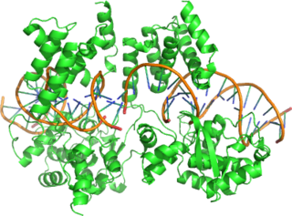
DNA gyrase, or simply gyrase, is an enzyme within the class of topoisomerase and is a subclass of Type II topoisomerases that reduces topological strain in an ATP dependent manner while double-stranded DNA is being unwound by elongating RNA-polymerase or by helicase in front of the progressing replication fork. It is the only known enzyme to actively contribute negative supercoiling to DNA, while it also is capable of relaxing positive supercoils. It does so by looping the template to form a crossing, then cutting one of the double helices and passing the other through it before releasing the break, changing the linking number by two in each enzymatic step. This process occurs in bacteria, whose single circular DNA is cut by DNA gyrase and the two ends are then twisted around each other to form supercoils. Gyrase is also found in eukaryotic plastids: it has been found in the apicoplast of the malarial parasite Plasmodium falciparum and in chloroplasts of several plants. Bacterial DNA gyrase is the target of many antibiotics, including nalidixic acid, novobiocin, albicidin, and ciprofloxacin.

<span class="mw-page-title-main">M13 bacteriophage</span> Species of virus

M13 is one of the Ff phages, a member of the family filamentous bacteriophage (inovirus). Ff phages are composed of circular single-stranded DNA (ssDNA), which in the case of the m13 phage is 6407 nucleotides long and is encapsulated in approximately 2700 copies of the major coat protein p8, and capped with about 5 copies each of four different minor coat proteins. The minor coat protein p3 attaches to the receptor at the tip of the F pilus of the host Escherichia coli. The life cycle is relatively short, with the early phage progeny exiting the cell ten minutes after infection. Ff phages are chronic phage, releasing their progeny without killing the host cells. The infection causes turbid plaques in E. coli lawns, of intermediate opacity in comparison to regular lysis plaques. However, a decrease in the rate of cell growth is seen in the infected cells. M13 plasmids are used for many recombinant DNA processes, and the virus has also been used for phage display, directed evolution, nanostructures and nanotechnology applications.

Topoisomerase IV is one of two Type II topoisomerases in bacteria, the other being DNA gyrase. Like gyrase, topoisomerase IV is able to pass one double-strand of DNA through another double-strand of DNA, thereby changing the linking number of DNA by two in each enzymatic step. Both share a hetero-4-mer structure formed by a symmetric homodimer of A/B heterodimers, usually named ParC and ParE.

<span class="mw-page-title-main">Replisome</span> Molecular complex

The replisome is a complex molecular machine that carries out replication of DNA. The replisome first unwinds double stranded DNA into two single strands. For each of the resulting single strands, a new complementary sequence of DNA is synthesized. The total result is formation of two new double stranded DNA sequences that are exact copies of the original double stranded DNA sequence.

<span class="mw-page-title-main">Cre recombinase</span> Genetic recombination enzyme

Cre recombinase is a tyrosine recombinase enzyme derived from the P1 bacteriophage. The enzyme uses a topoisomerase I-like mechanism to carry out site specific recombination events. The enzyme (38kDa) is a member of the integrase family of site specific recombinase and it is known to catalyse the site specific recombination event between two DNA recognition sites. This 34 base pair (bp) loxP recognition site consists of two 13 bp palindromic sequences which flank an 8bp spacer region. The products of Cre-mediated recombination at loxP sites are dependent upon the location and relative orientation of the loxP sites. Two separate DNA species both containing loxP sites can undergo fusion as the result of Cre mediated recombination. DNA sequences found between two loxP sites are said to be "floxed". In this case the products of Cre mediated recombination depends upon the orientation of the loxP sites. DNA found between two loxP sites oriented in the same direction will be excised as a circular loop of DNA whilst intervening DNA between two loxP sites that are opposingly orientated will be inverted. The enzyme requires no additional cofactors or accessory proteins for its function.

<span class="mw-page-title-main">DNA supercoil</span> Amount of twist in a particular DNA strand

DNA supercoiling refers to the amount of twist in a particular DNA strand, which determines the amount of strain on it. A given strand may be "positively supercoiled" or "negatively supercoiled". The amount of a strand’s supercoiling affects a number of biological processes, such as compacting DNA and regulating access to the genetic code. Certain enzymes, such as topoisomerases, change the amount of DNA supercoiling to facilitate functions such as DNA replication and transcription. The amount of supercoiling in a given strand is described by a mathematical formula that compares it to a reference state known as "relaxed B-form" DNA.

Topoisomerase inhibitors are chemical compounds that block the action of topoisomerases, which are broken into two broad subtypes: type I topoisomerases (TopI) and type II topoisomerases (TopII). Topoisomerase plays important roles in cellular reproduction and DNA organization, as they mediate the cleavage of single and double stranded DNA to relax supercoils, untangle catenanes, and condense chromosomes in eukaryotic cells. Topoisomerase inhibitors influence these essential cellular processes. Some topoisomerase inhibitors prevent topoisomerases from performing DNA strand breaks while others, deemed topoisomerase poisons, associate with topoisomerase-DNA complexes and prevent the re-ligation step of the topoisomerase mechanism. These topoisomerase-DNA-inhibitor complexes are cytotoxic agents, as the un-repaired single- and double stranded DNA breaks they cause can lead to apoptosis and cell death. Because of this ability to induce apoptosis, topoisomerase inhibitors have gained interest as therapeutics against infectious and cancerous cells.

<span class="mw-page-title-main">Type II topoisomerase</span>

Type II topoisomerases are topoisomerases that cut both strands of the DNA helix simultaneously in order to manage DNA tangles and supercoils. They use the hydrolysis of ATP, unlike Type I topoisomerase. In this process, these enzymes change the linking number of circular DNA by ±2. Topoisomerases are ubiquitous enzymes, found in all living organisms.

<span class="mw-page-title-main">Aminocoumarin</span> Class of antibiotic chemical compounds

Aminocoumarin is a class of antibiotics that act by an inhibition of the DNA gyrase enzyme involved in the cell division in bacteria. They are derived from Streptomyces species, whose best-known representative – Streptomyces coelicolor – was completely sequenced in 2002. The aminocoumarin antibiotics include:

<span class="mw-page-title-main">TOP1</span> DNA topoisomerase enzyme

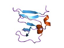
DNA topoisomerase 1 is an enzyme that in humans is encoded by the TOP1 gene. It is a DNA topoisomerase, an enzyme that catalyzes the transient breaking and rejoining of a single strand of DNA.

In enzymology, glutamate racemase is an enzyme that catalyzes the chemical reaction

<span class="mw-page-title-main">TOP3B</span> Protein-coding gene in the species Homo sapiens

DNA topoisomerase 3-beta-1 is an enzyme that in humans is encoded by the TOP3B gene.

<span class="mw-page-title-main">Circular chromosome</span> Type of chromosome

A circular chromosome is a chromosome in bacteria, archaea, mitochondria, and chloroplasts, in the form of a molecule of circular DNA, unlike the linear chromosome of most eukaryotes.

Saccharolobus solfataricus is a species of thermophilic archaeon. It was transferred from the genus Sulfolobus to the new genus Saccharolobus with the description of Saccharolobus caldissimus in 2018.

Elizabeth Lynn Zechiedrich is a professor in the department of Molecular Virology and Microbiology at Baylor College of Medicine. Her laboratory's research considers the structure-function properties of DNA and DNA topoisomerases. She was elected to the National Academy of Inventors in 2017.

<span class="mw-page-title-main">Reverse gyrase</span>

Reverse gyrase is a type I topoisomerase that introduces positive supercoils into DNA, contrary to the typical negative supercoils introduced by the type II topoisomerase DNA gyrase. These positive supercoils can be introduced to DNA that is either negatively supercoiled or fully relaxed. Where DNA gyrase forms a tetramer and is capable of cleaving a double-stranded region of DNA, reverse gyrase can only cleave single stranded DNA. More specifically, reverse gyrase is a member of the type IA topoisomerase class; along with the ability to relax negatively or positively supercoiled DNA, type IA enzymes also tend to have RNA-topoisomerase activities. These RNA topoisomerases help keep longer RNA strands from becoming tangled in what are referred to as "pseudoknots." Due to their ability to interact with RNA, it is thought that this is one of the most ancient class of enzymes found to date.

References

  1. Wang JC (June 2002). "Cellular roles of DNA topoisomerases: a molecular perspective". Nat. Rev. Mol. Cell Biol. 3 (6): 430–40. doi:10.1038/nrm831. PMID   12042765. S2CID   205496065.
  2. Champoux JJ (2001). "DNA topoisomerases: structure, function, and mechanism". Annu. Rev. Biochem. 70: 369–413. doi:10.1146/annurev.biochem.70.1.369. PMID   11395412.
  3. Sharma A; Hanai R; Mondragón A (August 1994). "Crystal structure of the amino-terminal fragment of vaccinia virus DNA topoisomerase I at 1.6 A resolution". Structure. 2 (8): 767–77. doi: 10.1016/s0969-2126(94)00077-8 . PMID   7994576.
  4. Roca J (April 1995). "The mechanisms of DNA topoisomerases". Trends Biochem. Sci. 20 (4): 156–60. doi:10.1016/s0968-0004(00)88993-8. PMID   7770916.
  5. Gadelle D, Filée J, Buhler C, Forterre P (March 2003). "Phylogenomics of type II DNA topoisomerases". BioEssays. 25 (3): 232–42. doi:10.1002/bies.10245. PMID   12596227. S2CID   4642743.
  6. Lima, C. D.; Wang, J. C.; Mondragón, A (1994). "Three-dimensional structure of the 67K N-terminal fragment of E. Coli DNA topoisomerase I". Nature. 367 (6459): 138–46. Bibcode:1994Natur.367..138L. doi:10.1038/367138a0. PMID   8114910. S2CID   4314431.
  7. Changela, A; Digate, R. J.; Mondragón, A (2001). "Crystal structure of a complex of a type IA DNA topoisomerase with a single-stranded DNA molecule". Nature. 411 (6841): 1077–81. Bibcode:2001Natur.411.1077C. doi:10.1038/35082615. PMID   11429611. S2CID   4426078.
  8. Forterre P (June 2006). "DNA topoisomerase V: a new fold of mysterious origin". Trends Biotechnol. 24 (6): 245–7. doi:10.1016/j.tibtech.2006.04.006. PMID   16650908.
  9. Taneja B, Patel A, Slesarev A, Mondragón A (January 2006). "Structure of the N-terminal fragment of topoisomerase V reveals a new family of topoisomerases". EMBO J. 25 (2): 398–408. doi:10.1038/sj.emboj.7600922. PMC   1383508 . PMID   16395333.
  10. Taneja, B; Schnurr, B; Slesarev, A; Marko, J. F.; Mondragón, A (2007). "Topoisomerase V relaxes supercoiled DNA by a constrained swiveling mechanism". Proceedings of the National Academy of Sciences. 104 (37): 14670–5. Bibcode:2007PNAS..10414670T. doi: 10.1073/pnas.0701989104 . PMC   1976220 . PMID   17804808.
  11. 1 2 Pommier Y (2013). "Drugging topoisomerases: lessons and challenges". ACS Chem. Biol. 8 (1): 82–95. doi:10.1021/cb300648v. PMC   3549721 . PMID   23259582.
  12. Mattenberger Y, Silva F, Belin D. 55.2, a phage T4 ORFan gene, encodes an inhibitor of Escherichia coli topoisomerase I and increases phage fitness. PLoS One. 2015 Apr 14;10(4):e0124309. doi: 10.1371/journal.pone.0124309. PMID: 25875362; PMCID: PMC4396842
  13. Agrelo R, Cheng WH, Setien F, Ropero S, Espada J, Fraga MF, Herranz M, Paz MF, Sanchez-Cespedes M, Artiga MJ, Guerrero D, Castells A, von Kobbe C, Bohr VA, Esteller M (2006). "Epigenetic inactivation of the premature aging Werner syndrome gene in human cancer". Proc. Natl. Acad. Sci. U.S.A. 103 (23): 8822–7. Bibcode:2006PNAS..103.8822A. doi: 10.1073/pnas.0600645103 . PMC   1466544 . PMID   16723399.
  14. Pavelitz T, Renfro L, Foster NR, Caracol A, Welsch P, Lao VV, Grady WB, Niedzwiecki D, Saltz LB, Bertagnolli MM, Goldberg RM, Rabinovitch PS, Emond M, Monnat RJ, Maizels N (2014). "MRE11-deficiency associated with improved long-term disease free survival and overall survival in a subset of stage III colon cancer patients in randomized CALGB 89803 trial". PLOS ONE. 9 (10): e108483. Bibcode:2014PLoSO...9j8483P. doi: 10.1371/journal.pone.0108483 . PMC   4195600 . PMID   25310185.
  15. Subhash VV, Tan SH, Yeo MS, Yan FL, Peethala PC, Liem N, Krishnan V, Yong WP (2016). "ATM expression predicts Veliparib and Irinotecan sensitivity in gastric cancer by mediating P53 independent regulation of cell cycle and apoptosis". Mol. Cancer Ther. 15 (12): 3087–3096. doi: 10.1158/1535-7163.MCT-15-1002 . PMID   27638859.
  16. Wissing MD, Mendonca J, Kim E, Kim E, Shim JS, Kaelber NS, Kant H, Hammers H, Commes T, Van Diest PJ, Liu JO, Kachhap SK (2013). "Identification of cetrimonium bromide and irinotecan as compounds with synthetic lethality against NDRG1 deficient prostate cancer cells". Cancer Biol. Ther. 14 (5): 401–10. doi:10.4161/cbt.23759. PMC   3672184 . PMID   23377825.
  17. Dominick G, Bowman J, Li X, Miller RA, Garcia GG (2016). "mTOR regulates the expression of DNA damage response enzymes in long-lived Snell dwarf, GHRKO, and PAPPA-KO mice". Aging Cell. 16 (1): 52–60. doi:10.1111/acel.12525. PMC   5242303 . PMID   27618784.
  18. Product Name: SCL-70 Antigen Archived 2006-03-19 at the Wayback Machine at ImmunoVision.com, retrieved April 2011