Valosin-containing protein

Last updated
VCP
5ifw.jpg
Available structures
PDB Ortholog search: PDBe RCSB
Identifiers
Aliases VCP , ALS14, HEL-220, HEL-S-70, IBMPFD, IBMPFD1, TERA, p97, Valosin-containing protein, CMT2Y, valosin containing protein, CDC48, FTDALS6
External IDs OMIM: 601023 MGI: 99919 HomoloGene: 5168 GeneCards: VCP
Orthologs
SpeciesHumanMouse
Entrez
Ensembl
UniProt
RefSeq (mRNA)

NM_007126
NM_001354927
NM_001354928

NM_009503

RefSeq (protein)

NP_009057
NP_001341856
NP_001341857

NP_033529

Location (UCSC) Chr 9: 35.05 – 35.07 Mb Chr 4: 42.98 – 43 Mb
PubMed search [3] [4]
Wikidata
View/Edit Human View/Edit Mouse

Valosin-containing protein (VCP) or transitional endoplasmic reticulum ATPase (TER ATPase) also known as p97 in mammals and CDC48 in S. cerevisiae, is an enzyme that in humans is encoded by the VCP gene. [5] [6] [7] The TER ATPase is an ATPase enzyme present in all eukaryotes and archaebacteria. Its main function is to segregate protein molecules from large cellular structures such as protein assemblies, organelle membranes and chromatin, and thus facilitate the degradation of released polypeptides by the multi-subunit protease proteasome.

VCP/p97/CDC48 is a member of the AAA+ (extended family of ATPases associated with various cellular activities) ATPase family. Enzymes of this family are found in all species from bacteria to humans. Many of them are important chaperones that regulate folding or unfolding of substrate proteins. VCP is a type II AAA+ ATPase, which means that it contains two tandem ATPase domains (named D1 and D2, respectively) (Figure 1).

Figure 1- A schematic diagram of the p97 domain structure. VCP Wiki Figure 1.jpg
Figure 1- A schematic diagram of the p97 domain structure.

The two ATPase domains are connected by a short polypeptide linker. A domain preceding the D1 domain (N-terminal domain) and a short carboxyl-terminal tail are involved in interaction with cofactors. [8] The N-domain is connected to the D1 domain by a short N-D1 linker.

Most known substrates of VCP are modified with ubiquitin chains and degraded by the 26S proteasome. Accordingly, many VCP coenzymes and adaptors have domains that can recognize ubiquitin. [9] It has become evident that the interplays between ubiquitin and VCP cofactors are critical for many of the proposed functions, although the precise role of these interactions remains to be elucidated.

Discovery

CDC48 was discovered in a genetic screen for genes involved in cell cycle regulation in budding yeast. [10] The screen identified several alleles of Cdc48 that affect cell growth at non-permissive temperatures. A search for the mammalian homolog of CDC48 (valosin) revealed a 97 kDa protein precursor named "valosin-containing protein (VCP)" or p97, and also showed that it was only generated as an artefact of purification rather than during physiological processing. [11] Even without evidence that valosin is a physiological product, the VCP nomenclature continues to be used in the literature.

Tissue and subcelluar distribution

VCP is one of the most abundant cytoplasmic proteins in eukaryotic cells. It is ubiquitously expressed in all tissues in multicellular organisms. In humans, the mRNA expression of VCP was found to be moderately elevated in certain types of cancer. [9]

In mammalian cells, VCP is predominantly localized to the cytoplasm, and a significant fraction is associated to membranes of cellular organelles such as the endoplasmic reticulum (ER), Golgi, mitochondria, and endosomes. [6] [12] [13] [14] [15] The subcellular localization of CDC48 has not been fully characterized, but is likely to be similar to the mammalian counterpart. A fraction of VCP was also found in the nucleus. [16]

Structure

According to the crystal structures of full-length wild-type VCP, [17] [18] six VCP subunits assemble into a barrel-like structure, in which the N-D1 and D2 domains form two concentric, stacked rings (Figure2).

Figure 2- The structure of VCP. The six subunits are shown as molecular surface in different colors. Domains of each subunit are also shaded differently. Two views are presented. This structure represents VCP in an ADP bound state. VCP Wiki Figure 2.jpg
Figure 2- The structure of VCP. The six subunits are shown as molecular surface in different colors. Domains of each subunit are also shaded differently. Two views are presented. This structure represents VCP in an ADP bound state.

The N-D1 ring is larger (162 Å in diameter) than the D2 ring (113 Å) due to the laterally attached N-domains. The D1 and D2 domains are highly homologous in both sequence and structure, but they serve distinct functions. For example, the hexameric assembly of VCP only requires the D1 but not the D2 domain. [19] Unlike many bacterial AAA+ proteins, assembly of VCP hexamer does not depend on the presence of nucleotide. The VCP hexameric assembly can undergo dramatic conformational changes during nucleotide hydrolysis cycle, [20] [21] [22] [23] [24] and it is generally believed that these conformational changes generate mechanical force, which is applied to substrate molecules to influence their stability and function. However, how precisely VCP generates force is unclear.

The ATP hydrolysis cycle

The ATP hydrolyzing activity is indispensable for the VCP functions. [25] The two ATPase domains of VCP (D1 and D2) are not equivalent because the D2 domain displays higher ATPase activity than the D1 domain in wild-type protein. Nevertheless, their activities are dependent of each other. [26] [27] [28] [29] For example, nucleotide binding to the D1 domain is required for ATP binding to the D2 domain and nucleotide binding and hydrolysis in D2 is required for the D1 domain to hydrolyze ATP.

The ATPase activity of VCP can be influenced by many factors. For example, it can be stimulated by heat [29] or by a putative substrate protein. [30] In Leishmania infantum, the LiVCP protein is essential for the intracellular development of the parasite and its survival under heat stress. [31] Association with cofactors can have either positive or negative impact on the p97 ATPase activity. [32] [33]

Mutations in VCP can also influence its activity. For example, VCP mutant proteins carrying single point mutations found in patients with multisystem proteinopathy (MSP; formerly known as IBMPFD (inclusion body myopathy associated with Paget disease of the bone and frontotemporal dementia)) (see below) have 2-3 fold increase in ATPase activity. [27] [34] [35]

VCP-interacting proteins

Recent proteomic studies have identified a large number of p97-interacting proteins. Many of these proteins serve as adaptors that link VCP to a particular subcellular compartment to function in a specific cellular pathway. Others function as adaptors that recruit substrates to VCP for processing. Some VCP-interacting proteins are also enzymes such as N-glycanase, ubiquitin ligase, and deubiquitinase, which assist VCP in processing substrates.

Most cofactors bind VCP through its N-domain, but a few interact with the short carboxy-terminal tail in VCP. Representative proteins interacting with the N-domain are Ufd1, Npl4, p47 and FAF1. [36] [37] [38] Examples of cofactors that interact with the carboxy-terminal tail of VCP are PLAA, PNGase, and Ufd2. [39] [40] [41]

The molecular basis for cofactor binding has been studied for some cofactors that interact with the VCP N-domain. The N-domain consists of two sub-domains of roughly equal size: the N-terminal double Y-barrel and a C-terminal b-barrel (Figure 3).

Figure 3- Structure of the N-domain of VCP. The N-domain is depicted as a molecular surface superimposed to a ribbon representation. VCP Wiki Figure 3.jpg
Figure 3- Structure of the N-domain of VCP. The N-domain is depicted as a molecular surface superimposed to a ribbon representation.

Structural studies show that many cofactor proteins bind to the N-domain at a cleft formed between the two sub-domains.

Among those that bind to the N-domain of VCP, two most frequently occurring sequence motifs are found: one is called UBX motif (ubiquitin regulatory X) [42] and the other is termed VIM (VCP-interacting motif). [43] The UBX domain is an 80-residue module with a fold highly resembling the structure of ubiquitin. The VCP-interacting motif (VIM) is a linear sequence motif (RX5AAX2R) found in a number of VCP cofactors including gp78, [44] SVIP (small VCP-inhibiting protein) [45] and VIMP (VCP interacting membrane protein). [46] Although the UBX domain uses a surface loop whereas the VIM forms a-helix to bind VCP, both UBX and VIM bind at the same location between the two sub-domains of the N-domain (Figure 3). [47] It was proposed that hierarchical binding to distinct cofactors may be essential for the broad functions of VCP. [48] [49]

Function

VCP performs diverse functions through modulating the stability and thus the activity of its substrates. The general function of VCP is to segregate proteins from large protein assembly or immobile cellular structures such as membranes or chromatin, allowing the released protein molecules to be degraded by the proteasome. The functions of VCP can be grouped into the following three major categories.

Protein quality control

The best characterized function of VCP is to mediate a network of protein quality control processes in order to maintain protein homeostasis. [50] These include endoplasmic reticulum-associated protein degradation (ERAD) and mitochondria-associated degradation. [14] [51] In these processes, ATP hydrolysis by VCP is required to extract aberrant proteins from the membranes of the ER or mitochondria. VCP is also required to release defective translation products stalled on ribosome in a process termed ribosome-associated degradation. [52] [53] [54] It appears that only after extraction from the membranes or large protein assembly like ribosome, can polypeptides be degraded by the proteasome. In addition to this ‘segregase’ function, VCP might have an additional role in shuttling the released polypeptides to the proteasome. This chaperoning function seems to be particularly important for degradation of certain aggregation-prone misfolded proteins in nucleus. [55] Several lines of evidence also implicate p97 in autophagy, a process that turns over cellular proteins (including misfolded ones) by engulfing them into double-membrane-surrounded vesicles named autophagosome, but the precise role of VCP in this process is unclear. [56]

Chromatin-associated functions

VCP also functions broadly in eukaryotic nucleus by releasing protein molecules from chromatins in a manner analogous to that in ERAD. [57] The identified VCP substrates include transcriptional repressor α2 and RNA polymerase (Pol) II complex and CMG DNA helicase in budding yeast, and the DNA replicating licensing factor CDT1, DNA repairing proteins DDB2 and XPC, mitosis regulator Aurora B, and certain DNA polymerases in mammalian cells. These substrates link VCP function to gene transcription, DNA replication and repair, and cell cycle progression.

Membrane fusion and trafficking

Biochemical and genetic studies have also implicated VCP in fusion of vesicles that lead to the formation of Golgi apparatus at the end of mitosis. [58] This process requires the ubiquitin binding adaptor p47 and a p97-associated deubiquitinase VCIP135, and thus connecting membrane fusion to the ubiquitin pathways. However, the precise role of VCP in Golgi formation is unclear due to lack of information on relevant substrate(s). Recent studies also suggest that VCP may regulate vesicle trafficking from plasma membrane to the lysosome, a process termed endocytosis. [56] Antibody fragment-based inhibitors have been developed by a team led by Arkin to inhibit the interaction between p97 and p47, selectively modulating the Golgi reassembly process. [59]

Clinical significance

Mutations in VCP were first reported to cause a syndrome characterized by frontotemporal dementia, inclusion body myopathy, and Paget's disease of the bone by Virginia Kimonis in 2004. [60] In 2010, mutations in VCP were also found to be a cause of amyotrophic lateral sclerosis by Bryan Traynor and Adriano Chiò. [61] This discovery was notable as it represented an initial genetic link between two disparate neurological diseases, amyotrophic lateral sclerosis and frontotemporal dementia. In 2020, Edward Lee described a distinct hypomorphic mutation in VCP associated with vacuolar tauopathy, a unique subtype of frontotemporal lobar degeneration with tau inclusions. [62]

Mutations in VCP are an example of pleiotropy, where mutations in the same gene give rise to different phenotypes. The term multisystem proteinopathy (MSP) has been coined to describe this particular form of pleiotropy. [63] Although MSP is rare, growing interest in this syndrome derives from the molecular insights the condition provides into the etiological relationship between common age-related degenerative diseases of muscle, bone and brain. It has been estimated that ~50% of MSP may be caused by missense mutations affecting the valosin-containing protein (VCP) gene. [64]


Cancer therapy

The first p97 inhibitor Eeyarestatin (EerI) was discovered by screening and characterizing compounds that inhibit the degradation of a fluorescence-labeled ERAD substrate. [65] [66] The mechanism of VCP inhibition by EerI is unclear, but when applied to cells, it induces biological phenotypes associated with VCP inhibition such as ERAD inhibition, ER stress elevation, and apoptosis induction. Importantly, EerI displays significant cancer-killing activity in vitro preferentially against cancer cells isolated from patients, and it can synergize with the proteasome inhibitor bortezomib to kill cancer cells. [67] These observations prompt the idea of targeting VCP as a potential cancer therapy. This idea was further confirmed by studying several ATP competitive and allosteric inhibitors. [68] [69] [70] More recently, a potent and specific VCP inhibitor CB-5083 has been developed, which demonstrates promising anti-cancer activities in mouse xenograft tumor models. [71] The compound is now being evaluated in a phase 1 clinical trial. [72]

Notes

Related Research Articles

<span class="mw-page-title-main">Proteasome</span> Protein complexes which degrade unnecessary or damaged proteins by proteolysis

Proteasomes are protein complexes which degrade unneeded or damaged proteins by proteolysis, a chemical reaction that breaks peptide bonds. Enzymes that help such reactions are called proteases.

<span class="mw-page-title-main">AAA proteins</span> Protein family

AAA proteins or ATPases Associated with diverse cellular Activities are a protein family sharing a common conserved module of approximately 230 amino acid residues. This is a large, functionally diverse protein family belonging to the AAA+ protein superfamily of ring-shaped P-loop NTPases, which exert their activity through the energy-dependent remodeling or translocation of macromolecules.

<span class="mw-page-title-main">Endoplasmic-reticulum-associated protein degradation</span>

Endoplasmic-reticulum-associated protein degradation (ERAD) designates a cellular pathway which targets misfolded proteins of the endoplasmic reticulum for ubiquitination and subsequent degradation by a protein-degrading complex, called the proteasome.

The unfolded protein response (UPR) is a cellular stress response related to the endoplasmic reticulum (ER) stress. It has been found to be conserved between mammalian species, as well as yeast and worm organisms.

<span class="mw-page-title-main">Binding immunoglobulin protein</span> Protein-coding gene in the species Homo sapiens

Binding immunoglobulin protein (BiPS) also known as 78 kDa glucose-regulated protein (GRP-78) or heat shock 70 kDa protein 5 (HSPA5) is a protein that in humans is encoded by the HSPA5 gene.

<span class="mw-page-title-main">PSMD2</span> Enzyme found in humans

26S proteasome non-ATPase regulatory subunit 2, also as known as 26S Proteasome Regulatory Subunit Rpn1, is an enzyme that in humans is encoded by the PSMD2 gene.

<span class="mw-page-title-main">AMFR</span> Protein-coding gene in the species Homo sapiens

Autocrine motility factor receptor, isoform 2 is a protein that in humans is encoded by the AMFR gene.

<span class="mw-page-title-main">HERPUD1</span> Protein-coding gene in the species Homo sapiens

Homocysteine-responsive endoplasmic reticulum-resident ubiquitin-like domain member 1 protein is a protein that in humans is encoded by the HERPUD1 gene.

<span class="mw-page-title-main">SYVN1</span> Protein-coding gene in the species Homo sapiens

E3 ubiquitin-protein ligase synoviolin is an enzyme that in humans is encoded by the SYVN1 gene.

<span class="mw-page-title-main">NFE2L1</span> Protein-coding gene in the species Homo sapiens

Nuclear factor erythroid 2-related factor 1 (Nrf1) also known as nuclear factor erythroid-2-like 1 (NFE2L1) is a protein that in humans is encoded by the NFE2L1 gene. Since NFE2L1 is referred to as Nrf1, it is often confused with nuclear respiratory factor 1 (Nrf1).

<span class="mw-page-title-main">NSFL1C</span> Protein-coding gene in the species Homo sapiens

NSFL1 cofactor p47 is a protein that in humans is encoded by the NSFL1C gene.

<span class="mw-page-title-main">Derlin-1</span> Protein involved in retrotranslocation of specific misfolded proteins and in ER stress

Derlin-1 also known as degradation in endoplasmic reticulum protein 1 is a membrane protein that in humans is encoded by the DERL1 gene. Derlin-1 is located in the membrane of the endoplasmic reticulum (ER) and is involved in retrotranslocation of specific misfolded proteins and in ER stress. Derlin-1 is widely expressed in thyroid, fat, bone marrow and many other tissues. The protein belongs to the Derlin-family proteins consisting of derlin-1, derlin-2 and derlin-3 that are components in the endoplasmic reticulum-associated protein degradation (ERAD) pathway. The derlins mediate degradation of misfolded lumenal proteins within ER, and are named ‘der’ for their ‘Degradation in the ER’. Derlin-1 is a mammalian homologue of the yeast DER1 protein, a protein involved in the yeast ERAD pathway. Moreover, derlin-1 is a member of the rhomboid-like clan of polytopic membrane proteins.

<span class="mw-page-title-main">NGLY1</span> Protein-coding gene in the species Homo sapiens

PNGase also known as N-glycanase 1 or peptide-N(4)-(N-acetyl-beta-glucosaminyl)asparagine amidase is an enzyme that in humans is encoded by the NGLY1 gene. PNGase is a de-N-glycosylating enzyme that removes N-linked or asparagine-linked glycans (N-glycans) from glycoproteins. More specifically, NGLY1 catalyzes the hydrolysis of the amide bond between the innermost N-acetylglucosamine (GlcNAc) and an Asn residue on an N-glycoprotein, generating a de-N-glycosylated protein, in which the N-glycoylated Asn residue is converted to asp, and a 1-amino-GlcNAc-containing free oligosaccharide. Ammonia is then spontaneously released from the 1-amino GlcNAc at physiological pH (<8), giving rise to a free oligosaccharide with an N,N’-diacetylchitobiose structure at the reducing end.

<span class="mw-page-title-main">FBXL2</span> Gene of the species Homo sapiens

F-box/LRR-repeat protein 2 is a protein that in humans is encoded by the FBXL2 gene.

The endosomal sorting complexes required for transport (ESCRT) machinery is made up of cytosolic protein complexes, known as ESCRT-0, ESCRT-I, ESCRT-II, and ESCRT-III. Together with a number of accessory proteins, these ESCRT complexes enable a unique mode of membrane remodeling that results in membranes bending/budding away from the cytoplasm. These ESCRT components have been isolated and studied in a number of organisms including yeast and humans. A eukaryotic signature protein, the machinery is found in all eukaryotes and some archaea.

<span class="mw-page-title-main">Sec14</span>

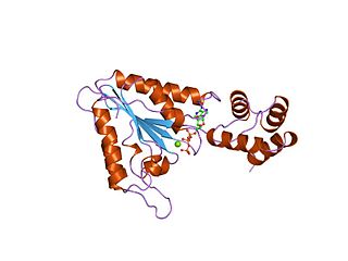
Sec14 is a cytosolic protein found in yeast which plays a role in the regulation of several cellular functions, specifically those related to intracellular transport. Encoded by the Sec14 gene, Sec14p may transport phosphatidylinositol and phosphatidylcholine produced in the endoplasmic reticulum and the Golgi body to other cellular membranes. Additionally, Sec14p potentially plays a role in the localization of lipid raft proteins. Sec14p is an essential gene in yeast, and is homologous in function to phosphatidylinositol transfer protein in mammals. A conditional mutant with non-functional Sec14p presents with Berkeley bodies and deficiencies in protein secretion.

<span class="mw-page-title-main">CDC48 N-terminal domain</span>

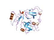
In molecular biology, the CDC48 N-terminal domain is a protein domain found in AAA ATPases including cell division protein 48 (CDC48), VCP-like ATPase and N-ethylmaleimide sensitive fusion protein. It is a substrate recognition domain which binds polypeptides, prevents protein aggregation, and catalyses refolding of permissive substrates. It is composed of two equally sized subdomains. The amino-terminal subdomain (CDC48_N) forms a double-psi beta-barrel whose pseudo-twofold symmetry is mirrored by an internal sequence repeat of 42 residues. The carboxy-terminal subdomain (CDC48_2) forms a novel six-stranded beta-clam fold. Together these subdomains form a kidney-shaped structure, in close agreement with results from electron microscopy. CDC48_N is related to numerous proteins including prokaryotic transcription factors, metabolic enzymes, the protease cofactors UFD1 and PrlF, and aspartic proteinases.

<span class="mw-page-title-main">UBXN6</span> Protein-coding gene in the species Homo sapiens

UBX domain protein 6 is a protein in humans that is encoded by the UBXN6 gene.

Raymond Joseph Deshaies is an American biochemist and cell biologist. He is senior vice president of global research at Amgen and a visiting associate at the California Institute of Technology (Caltech). Prior to that, he was a professor of biology at Caltech and an investigator of the Howard Hughes Medical Institute. He is also the co-founder of the biotechnology companies Proteolix and Cleave Biosciences. His research focuses on mechanisms and regulation of protein homeostasis in eukaryotic cells, with a particular focus on how proteins are conjugated with ubiquitin and degraded by the proteasome.

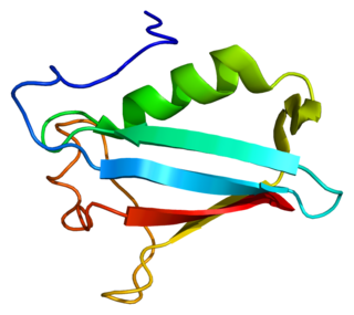
UBXD8 is a protein in the Ubiquitin regulatory X (UBX) domain-containing protein family. The UBX domain contains many eukaryotic proteins that have similarities in amino acid sequence to the tiny protein modifier ubiquitin. UBXD8 engages in a molecular interaction with p97, a protein that is essential for the degradation of membrane proteins associated with the endoplasmic reticulum (ER) through the proteasome. Ubxd8 possesses a UBA domain, alongside the UBX domain, that could interact with polyubiquitin chains. Additionally, it possesses a UAS domain of undetermined function, and this protein is used as a protein sensor that detects long chain unsaturated fatty acids (FAs), having a vital function in regulating the balance of Fatty Acids within cells to maintain cellular homeostasis.

References

  1. 1 2 3 GRCh38: Ensembl release 89: ENSG00000165280 - Ensembl, May 2017
  2. 1 2 3 GRCm38: Ensembl release 89: ENSMUSG00000028452 - Ensembl, May 2017
  3. "Human PubMed Reference:". National Center for Biotechnology Information, U.S. National Library of Medicine.
  4. "Mouse PubMed Reference:". National Center for Biotechnology Information, U.S. National Library of Medicine.
  5. Druck T, Gu Y, Prabhala G, Cannizzaro LA, Park SH, Huebner K, Keen JH (November 1995). "Chromosome localization of human genes for clathrin adaptor polypeptides AP2 beta and AP50 and the clathrin-binding protein, VCP". Genomics. 30 (1): 94–7. doi:10.1006/geno.1995.0016. PMID   8595912.
  6. 1 2 Rabouille C, Levine TP, Peters JM, Warren G (September 1995). "An NSF-like ATPase, p97, and NSF mediate cisternal regrowth from mitotic Golgi fragments". Cell. 82 (6): 905–14. doi: 10.1016/0092-8674(95)90270-8 . PMID   7553851. S2CID   2663141.
  7. "Entrez Gene: VCP valosin-containing protein".
  8. Ogura T, Wilkinson AJ (July 2001). "AAA+ superfamily ATPases: common structure--diverse function". Genes to Cells. 6 (7): 575–97. doi: 10.1046/j.1365-2443.2001.00447.x . PMID   11473577. S2CID   6740778.
  9. 1 2 Ye Y (October 2006). "Diverse functions with a common regulator: ubiquitin takes command of an AAA ATPase". Journal of Structural Biology. 156 (1): 29–40. doi:10.1016/j.jsb.2006.01.005. PMID   16529947.
  10. Moir D, Stewart SE, Osmond BC, Botstein D (April 1982). "Cold-sensitive cell-division-cycle mutants of yeast: isolation, properties, and pseudoreversion studies". Genetics. 100 (4): 547–63. doi:10.1093/genetics/100.4.547. PMC   1201831 . PMID   6749598.
  11. Koller KJ, Brownstein MJ (1987). "Use of a cDNA clone to identify a supposed precursor protein containing valosin". Nature. 325 (6104): 542–5. Bibcode:1987Natur.325..542K. doi:10.1038/325542a0. PMID   3468358. S2CID   19200775.
  12. Acharya U, Jacobs R, Peters JM, Watson N, Farquhar MG, Malhotra V (September 1995). "The formation of Golgi stacks from vesiculated Golgi membranes requires two distinct fusion events". Cell. 82 (6): 895–904. doi: 10.1016/0092-8674(95)90269-4 . PMID   7553850. S2CID   14725335.
  13. Latterich M, Fröhlich KU, Schekman R (September 1995). "Membrane fusion and the cell cycle: Cdc48p participates in the fusion of ER membranes". Cell. 82 (6): 885–93. doi: 10.1016/0092-8674(95)90268-6 . PMID   7553849. S2CID   17922017.
  14. 1 2 Xu S, Peng G, Wang Y, Fang S, Karbowski M (February 2011). "The AAA-ATPase p97 is essential for outer mitochondrial membrane protein turnover". Molecular Biology of the Cell. 22 (3): 291–300. doi:10.1091/mbc.E10-09-0748. PMC   3031461 . PMID   21118995.
  15. Ramanathan HN, Ye Y (February 2012). "The p97 ATPase associates with EEA1 to regulate the size of early endosomes". Cell Research. 22 (2): 346–59. doi:10.1038/cr.2011.80. PMC   3271578 . PMID   21556036.
  16. Madeo F, Schlauer J, Zischka H, Mecke D, Fröhlich KU (January 1998). "Tyrosine phosphorylation regulates cell cycle-dependent nuclear localization of Cdc48p". Molecular Biology of the Cell. 9 (1): 131–41. doi:10.1091/mbc.9.1.131. PMC   25228 . PMID   9436996.
  17. DeLaBarre B, Brunger AT (October 2003). "Complete structure of p97/valosin-containing protein reveals communication between nucleotide domains". Nature Structural Biology. 10 (10): 856–63. doi:10.1038/nsb972. PMID   12949490. S2CID   19281416.
  18. Davies JM, Brunger AT, Weis WI (May 2008). "Improved structures of full-length p97, an AAA ATPase: implications for mechanisms of nucleotide-dependent conformational change". Structure. 16 (5): 715–26. doi: 10.1016/j.str.2008.02.010 . PMID   18462676.
  19. Wang Q, Song C, Li CC (January 2003). "Hexamerization of p97-VCP is promoted by ATP binding to the D1 domain and required for ATPase and biological activities". Biochemical and Biophysical Research Communications. 300 (2): 253–60. doi:10.1016/s0006-291x(02)02840-1. PMID   12504076.
  20. Beuron F, Dreveny I, Yuan X, Pye VE, McKeown C, Briggs LC, Cliff MJ, Kaneko Y, Wallis R, Isaacson RL, Ladbury JE, Matthews SJ, Kondo H, Zhang X, Freemont PS (May 2006). "Conformational changes in the AAA ATPase p97-p47 adaptor complex". The EMBO Journal. 25 (9): 1967–76. doi:10.1038/sj.emboj.7601055. PMC   1456939 . PMID   16601695.
  21. Beuron F, Flynn TC, Ma J, Kondo H, Zhang X, Freemont PS (March 2003). "Motions and negative cooperativity between p97 domains revealed by cryo-electron microscopy and quantised elastic deformational model". Journal of Molecular Biology. 327 (3): 619–29. doi:10.1016/s0022-2836(03)00178-5. PMID   12634057.
  22. DeLaBarre B, Brunger AT (March 2005). "Nucleotide dependent motion and mechanism of action of p97/VCP". Journal of Molecular Biology. 347 (2): 437–52. doi:10.1016/j.jmb.2005.01.060. PMID   15740751.
  23. Rouiller I, DeLaBarre B, May AP, Weis WI, Brunger AT, Milligan RA, Wilson-Kubalek EM (December 2002). "Conformational changes of the multifunction p97 AAA ATPase during its ATPase cycle". Nature Structural Biology. 9 (12): 950–7. doi:10.1038/nsb872. PMID   12434150. S2CID   16061425.
  24. Tang WK, Li D, Li CC, Esser L, Dai R, Guo L, Xia D (July 2010). "A novel ATP-dependent conformation in p97 N-D1 fragment revealed by crystal structures of disease-related mutants". The EMBO Journal. 29 (13): 2217–29. doi:10.1038/emboj.2010.104. PMC   2905243 . PMID   20512113.
  25. Wang Q, Song C, Li CC (2004). "Molecular perspectives on p97-VCP: progress in understanding its structure and diverse biological functions". Journal of Structural Biology. 146 (1–2): 44–57. doi:10.1016/j.jsb.2003.11.014. PMID   15037236.
  26. Nishikori S, Esaki M, Yamanaka K, Sugimoto S, Ogura T (May 2011). "Positive cooperativity of the p97 AAA ATPase is critical for essential functions". The Journal of Biological Chemistry. 286 (18): 15815–20. doi: 10.1074/jbc.M110.201400 . PMC   3091191 . PMID   21454554.
  27. 1 2 Tang WK, Xia D (December 2013). "Altered intersubunit communication is the molecular basis for functional defects of pathogenic p97 mutants". The Journal of Biological Chemistry. 288 (51): 36624–35. doi: 10.1074/jbc.M113.488924 . PMC   3868774 . PMID   24196964.
  28. Ye Y, Meyer HH, Rapoport TA (July 2003). "Function of the p97-Ufd1-Npl4 complex in retrotranslocation from the ER to the cytosol: dual recognition of nonubiquitinated polypeptide segments and polyubiquitin chains". The Journal of Cell Biology. 162 (1): 71–84. doi:10.1083/jcb.200302169. PMC   2172719 . PMID   12847084.
  29. 1 2 Song C, Wang Q, Li CC (February 2003). "ATPase activity of p97-valosin-containing protein (VCP). D2 mediates the major enzyme activity, and D1 contributes to the heat-induced activity". The Journal of Biological Chemistry. 278 (6): 3648–55. doi: 10.1074/jbc.M208422200 . PMID   12446676.
  30. DeLaBarre B, Christianson JC, Kopito RR, Brunger AT (May 2006). "Central pore residues mediate the p97/VCP activity required for ERAD". Molecular Cell. 22 (4): 451–62. doi: 10.1016/j.molcel.2006.03.036 . PMID   16713576.
  31. Guedes Aguiar B, Padmanabhan PK, Dumas C, Papadopoulou B (June 2018). "Valosin-containing protein VCP/p97 is essential for the intracellular development of Leishmania and its survival under heat stress". Cellular Microbiology. 20 (10): e12867. doi: 10.1111/cmi.12867 . PMID   29895095. S2CID   48359590.
  32. Meyer HH, Kondo H, Warren G (October 1998). "The p47 co-factor regulates the ATPase activity of the membrane fusion protein, p97". FEBS Letters. 437 (3): 255–7. doi: 10.1016/s0014-5793(98)01232-0 . PMID   9824302. S2CID   33962985.
  33. Zhang X, Gui L, Zhang X, Bulfer SL, Sanghez V, Wong DE, Lee Y, Lehmann L, Lee JS, Shih PY, Lin HJ, Iacovino M, Weihl CC, Arkin MR, Wang Y, Chou TF (April 2015). "Altered cofactor regulation with disease-associated p97/VCP mutations". Proceedings of the National Academy of Sciences of the United States of America. 112 (14): E1705–14. Bibcode:2015PNAS..112E1705Z. doi: 10.1073/pnas.1418820112 . PMC   4394316 . PMID   25775548.
  34. Halawani D, LeBlanc AC, Rouiller I, Michnick SW, Servant MJ, Latterich M (August 2009). "Hereditary inclusion body myopathy-linked p97/VCP mutations in the NH2 domain and the D1 ring modulate p97/VCP ATPase activity and D2 ring conformation". Molecular and Cellular Biology. 29 (16): 4484–94. doi:10.1128/MCB.00252-09. PMC   2725746 . PMID   19506019.
  35. Weihl CC, Dalal S, Pestronk A, Hanson PI (January 2006). "Inclusion body myopathy-associated mutations in p97/VCP impair endoplasmic reticulum-associated degradation". Human Molecular Genetics. 15 (2): 189–99. doi: 10.1093/hmg/ddi426 . PMID   16321991.
  36. Ye Y, Meyer HH, Rapoport TA (December 2001). "The AAA ATPase Cdc48/p97 and its partners transport proteins from the ER into the cytosol". Nature. 414 (6864): 652–6. Bibcode:2001Natur.414..652Y. doi:10.1038/414652a. PMID   11740563. S2CID   23397533.
  37. Kondo H, Rabouille C, Newman R, Levine TP, Pappin D, Freemont P, Warren G (July 1997). "p47 is a cofactor for p97-mediated membrane fusion". Nature. 388 (6637): 75–8. Bibcode:1997Natur.388R..75K. doi: 10.1038/40411 . PMID   9214505. S2CID   32646222.
  38. Song EJ, Yim SH, Kim E, Kim NS, Lee KJ (March 2005). "Human Fas-associated factor 1, interacting with ubiquitinated proteins and valosin-containing protein, is involved in the ubiquitin-proteasome pathway". Molecular and Cellular Biology. 25 (6): 2511–24. doi:10.1128/MCB.25.6.2511-2524.2005. PMC   1061599 . PMID   15743842.
  39. Qiu L, Pashkova N, Walker JR, Winistorfer S, Allali-Hassani A, Akutsu M, Piper R, Dhe-Paganon S (January 2010). "Structure and function of the PLAA/Ufd3-p97/Cdc48 complex". The Journal of Biological Chemistry. 285 (1): 365–72. doi: 10.1074/jbc.M109.044685 . PMC   2804184 . PMID   19887378.
  40. Zhao G, Zhou X, Wang L, Li G, Schindelin H, Lennarz WJ (May 2007). "Studies on peptide:N-glycanase-p97 interaction suggest that p97 phosphorylation modulates endoplasmic reticulum-associated degradation". Proceedings of the National Academy of Sciences of the United States of America. 104 (21): 8785–90. Bibcode:2007PNAS..104.8785Z. doi: 10.1073/pnas.0702966104 . PMC   1885580 . PMID   17496150.
  41. Schaeffer V, Akutsu M, Olma MH, Gomes LC, Kawasaki M, Dikic I (May 2014). "Binding of OTULIN to the PUB domain of HOIP controls NF-κB signaling". Molecular Cell. 54 (3): 349–61. doi: 10.1016/j.molcel.2014.03.016 . PMID   24726327.
  42. Schuberth C, Buchberger A (August 2008). "UBX domain proteins: major regulators of the AAA ATPase Cdc48/p97". Cellular and Molecular Life Sciences. 65 (15): 2360–71. doi: 10.1007/s00018-008-8072-8 . PMID   18438607.
  43. Stapf C, Cartwright E, Bycroft M, Hofmann K, Buchberger A (November 2011). "The general definition of the p97/valosin-containing protein (VCP)-interacting motif (VIM) delineates a new family of p97 cofactors". The Journal of Biological Chemistry. 286 (44): 38670–8. doi: 10.1074/jbc.M111.274472 . PMC   3207395 . PMID   21896481.
  44. Ballar P, Shen Y, Yang H, Fang S (November 2006). "The role of a novel p97/valosin-containing protein-interacting motif of gp78 in endoplasmic reticulum-associated degradation". The Journal of Biological Chemistry. 281 (46): 35359–68. doi: 10.1074/jbc.M603355200 . PMID   16987818.
  45. Ballar P, Zhong Y, Nagahama M, Tagaya M, Shen Y, Fang S (November 2007). "Identification of SVIP as an endogenous inhibitor of endoplasmic reticulum-associated degradation". The Journal of Biological Chemistry. 282 (47): 33908–14. doi: 10.1074/jbc.M704446200 . PMID   17872946.
  46. Ye Y, Shibata Y, Yun C, Ron D, Rapoport TA (June 2004). "A membrane protein complex mediates retro-translocation from the ER lumen into the cytosol". Nature. 429 (6994): 841–7. Bibcode:2004Natur.429..841Y. doi:10.1038/nature02656. PMID   15215856. S2CID   4317750.
  47. Hänzelmann P, Schindelin H (November 2011). "The structural and functional basis of the p97/valosin-containing protein (VCP)-interacting motif (VIM): mutually exclusive binding of cofactors to the N-terminal domain of p97". The Journal of Biological Chemistry. 286 (44): 38679–90. doi: 10.1074/jbc.M111.274506 . PMC   3207442 . PMID   21914798.
  48. Meyer HH, Shorter JG, Seemann J, Pappin D, Warren G (May 2000). "A complex of mammalian ufd1 and npl4 links the AAA-ATPase, p97, to ubiquitin and nuclear transport pathways". The EMBO Journal. 19 (10): 2181–92. doi:10.1093/emboj/19.10.2181. PMC   384367 . PMID   10811609.
  49. Buchberger A, Schindelin H, Hänzelmann P (September 2015). "Control of p97 function by cofactor binding". FEBS Letters. 589 (19 Pt A): 2578–89. doi: 10.1016/j.febslet.2015.08.028 . PMID   26320413. S2CID   41082524.
  50. Meyer H, Bug M, Bremer S (February 2012). "Emerging functions of the VCP/p97 AAA-ATPase in the ubiquitin system". Nature Cell Biology. 14 (2): 117–23. doi:10.1038/ncb2407. PMID   22298039. S2CID   23562362.
  51. Christianson JC, Ye Y (April 2014). "Cleaning up in the endoplasmic reticulum: ubiquitin in charge". Nature Structural & Molecular Biology. 21 (4): 325–35. doi:10.1038/nsmb.2793. PMC   9397582 . PMID   24699081. S2CID   43665193.
  52. Brandman O, Stewart-Ornstein J, Wong D, Larson A, Williams CC, Li GW, Zhou S, King D, Shen PS, Weibezahn J, Dunn JG, Rouskin S, Inada T, Frost A, Weissman JS (November 2012). "A ribosome-bound quality control complex triggers degradation of nascent peptides and signals translation stress". Cell. 151 (5): 1042–54. doi:10.1016/j.cell.2012.10.044. PMC   3534965 . PMID   23178123.
  53. Defenouillère Q, Yao Y, Mouaikel J, Namane A, Galopier A, Decourty L, Doyen A, Malabat C, Saveanu C, Jacquier A, Fromont-Racine M (March 2013). "Cdc48-associated complex bound to 60S particles is required for the clearance of aberrant translation products". Proceedings of the National Academy of Sciences of the United States of America. 110 (13): 5046–51. Bibcode:2013PNAS..110.5046D. doi: 10.1073/pnas.1221724110 . PMC   3612664 . PMID   23479637.
  54. Verma R, Oania RS, Kolawa NJ, Deshaies RJ (January 2013). "Cdc48/p97 promotes degradation of aberrant nascent polypeptides bound to the ribosome". eLife. 2: e00308. doi: 10.7554/eLife.00308 . PMC   3552423 . PMID   23358411.
  55. Gallagher PS, Clowes Candadai SV, Gardner RG (May 2014). "The requirement for Cdc48/p97 in nuclear protein quality control degradation depends on the substrate and correlates with substrate insolubility". Journal of Cell Science. 127 (Pt 9): 1980–91. doi:10.1242/jcs.141838. PMC   4004975 . PMID   24569878.
  56. 1 2 Bug M, Meyer H (August 2012). "Expanding into new markets--VCP/p97 in endocytosis and autophagy". Journal of Structural Biology. 179 (2): 78–82. doi:10.1016/j.jsb.2012.03.003. PMID   22450227.
  57. Dantuma NP, Acs K, Luijsterburg MS (November 2014). "Should I stay or should I go: VCP/p97-mediated chromatin extraction in the DNA damage response". Experimental Cell Research. 329 (1): 9–17. doi:10.1016/j.yexcr.2014.08.025. PMID   25169698.
  58. Uchiyama K, Kondo H (February 2005). "p97/p47-Mediated biogenesis of Golgi and ER". Journal of Biochemistry. 137 (2): 115–9. doi:10.1093/jb/mvi028. PMID   15749824. S2CID   10459261.
  59. Jiang, Ziwen; Kuo, Yu-Hsuan; Zhong, Mengqi; Zhang, Jianchao; Zhou, Xin X.; Xing, Lijuan; Wells, James A.; Wang, Yanzhuang; Arkin, Michelle R. (2022-07-27). "Adaptor-Specific Antibody Fragment Inhibitors for the Intracellular Modulation of p97 (VCP) Protein–Protein Interactions". Journal of the American Chemical Society. 144 (29): 13218–13225. doi:10.1021/jacs.2c03665. ISSN   0002-7863. PMC   9335864 . PMID   35819848.
  60. Watts, Giles D. J.; Wymer, Jill; Kovach, Margaret J.; Mehta, Sarju G.; Mumm, Steven; Darvish, Daniel; Pestronk, Alan; Whyte, Michael P.; Kimonis, Virginia E. (2004). "Inclusion body myopathy associated with Paget disease of bone and frontotemporal dementia is caused by mutant valosin-containing protein". Nature Genetics. 36 (4): 377–381. doi: 10.1038/ng1332 . ISSN   1061-4036. PMID   15034582.
  61. Johnson, Janel O.; Mandrioli, Jessica; Benatar, Michael; Abramzon, Yevgeniya; Van Deerlin, Vivianna M.; Trojanowski, John Q.; Gibbs, J. Raphael; Brunetti, Maura; Gronka, Susan (2010-12-09). "Exome sequencing reveals VCP mutations as a cause of familial ALS". Neuron. 68 (5): 857–864. doi:10.1016/j.neuron.2010.11.036. ISSN   1097-4199. PMC   3032425 . PMID   21145000.
  62. Darwich, N.F., Phan J.M.; et al. (2020). "Autosomal dominant VCP hypomorph mutation impairs disaggregation of PHF-tau". Science. 370 (6519): eaay8826. doi:10.1126/science.aay8826. PMC   7818661 . PMID   33004675.{{cite journal}}: CS1 maint: multiple names: authors list (link)
  63. Taylor, J. Paul (2015-08-25). "Multisystem proteinopathy: intersecting genetics in muscle, bone, and brain degeneration". Neurology. 85 (8): 658–660. doi:10.1212/WNL.0000000000001862. ISSN   1526-632X. PMID   26208960. S2CID   42203997.
  64. Le Ber I, Van Bortel I, Nicolas G, Bouya-Ahmed K, Camuzat A, Wallon D, De Septenville A, Latouche M, Lattante S, Kabashi E, Jornea L, Hannequin D, Brice A (April 2014). "hnRNPA2B1 and hnRNPA1 mutations are rare in patients with "multisystem proteinopathy" and frontotemporal lobar degeneration phenotypes". Neurobiology of Aging. 35 (4): 934.e5–6. doi:10.1016/j.neurobiolaging.2013.09.016. PMID   24119545. S2CID   207160856.
  65. Fiebiger E, Hirsch C, Vyas JM, Gordon E, Ploegh HL, Tortorella D (April 2004). "Dissection of the dislocation pathway for type I membrane proteins with a new small molecule inhibitor, eeyarestatin". Molecular Biology of the Cell. 15 (4): 1635–46. doi:10.1091/mbc.E03-07-0506. PMC   379262 . PMID   14767067.
  66. Wang Q, Shinkre BA, Lee JG, Weniger MA, Liu Y, Chen W, Wiestner A, Trenkle WC, Ye Y (November 2010). "The ERAD inhibitor Eeyarestatin I is a bifunctional compound with a membrane-binding domain and a p97/VCP inhibitory group". PLOS ONE. 5 (11): e15479. Bibcode:2010PLoSO...515479W. doi: 10.1371/journal.pone.0015479 . PMC   2993181 . PMID   21124757.
  67. Wang Q, Mora-Jensen H, Weniger MA, Perez-Galan P, Wolford C, Hai T, Ron D, Chen W, Trenkle W, Wiestner A, Ye Y (February 2009). "ERAD inhibitors integrate ER stress with an epigenetic mechanism to activate BH3-only protein NOXA in cancer cells". Proceedings of the National Academy of Sciences of the United States of America. 106 (7): 2200–5. Bibcode:2009PNAS..106.2200W. doi: 10.1073/pnas.0807611106 . PMC   2629785 . PMID   19164757.
  68. Chou TF, Li K, Frankowski KJ, Schoenen FJ, Deshaies RJ (February 2013). "Structure-activity relationship study reveals ML240 and ML241 as potent and selective inhibitors of p97 ATPase". ChemMedChem. 8 (2): 297–312. doi:10.1002/cmdc.201200520. PMC   3662613 . PMID   23316025.
  69. Chou TF, Brown SJ, Minond D, Nordin BE, Li K, Jones AC, Chase P, Porubsky PR, Stoltz BM, Schoenen FJ, Patricelli MP, Hodder P, Rosen H, Deshaies RJ (March 2011). "Reversible inhibitor of p97, DBeQ, impairs both ubiquitin-dependent and autophagic protein clearance pathways". Proceedings of the National Academy of Sciences of the United States of America. 108 (12): 4834–9. Bibcode:2011PNAS..108.4834C. doi: 10.1073/pnas.1015312108 . PMC   3064330 . PMID   21383145.
  70. Magnaghi P, D'Alessio R, Valsasina B, Avanzi N, Rizzi S, Asa D, Gasparri F, Cozzi L, Cucchi U, Orrenius C, Polucci P, Ballinari D, Perrera C, Leone A, Cervi G, Casale E, Xiao Y, Wong C, Anderson DJ, Galvani A, Donati D, O'Brien T, Jackson PK, Isacchi A (September 2013). "Covalent and allosteric inhibitors of the ATPase VCP/p97 induce cancer cell death". Nature Chemical Biology. 9 (9): 548–56. doi:10.1038/nchembio.1313. PMID   23892893.
  71. Anderson DJ, Le Moigne R, Djakovic S, Kumar B, Rice J, Wong S, Wang J, Yao B, Valle E, Kiss von Soly S, Madriaga A, Soriano F, Menon MK, Wu ZY, Kampmann M, Chen Y, Weissman JS, Aftab BT, Yakes FM, Shawver L, Zhou HJ, Wustrow D, Rolfe M (November 2015). "Targeting the AAA ATPase p97 as an Approach to Treat Cancer through Disruption of Protein Homeostasis". Cancer Cell. 28 (5): 653–665. doi:10.1016/j.ccell.2015.10.002. PMC   4941640 . PMID   26555175.
  72. Zhou HJ, Wang J, Yao B, Wong S, Djakovic S, Kumar B, Rice J, Valle E, Soriano F, Menon MK, Madriaga A, Kiss von Soly S, Kumar A, Parlati F, Yakes FM, Shawver L, Le Moigne R, Anderson DJ, Rolfe M, Wustrow D (December 2015). "Discovery of a First-in-Class, Potent, Selective, and Orally Bioavailable Inhibitor of the VCP AAA ATPase (CB-5083)". Journal of Medicinal Chemistry. 58 (24): 9480–97. doi: 10.1021/acs.jmedchem.5b01346 . PMID   26565666.

Further reading