WDR12

Last updated
WDR12
Identifiers
Aliases WDR12 , YTM1, WD repeat domain 12
External IDs OMIM: 616620 MGI: 1927241 HomoloGene: 10098 GeneCards: WDR12
Orthologs
SpeciesHumanMouse
Entrez
Ensembl
UniProt
RefSeq (mRNA)

NM_018256
NM_001371664

NM_001199060
NM_001199061
NM_021312

RefSeq (protein)

NP_060726
NP_001358593

NP_001185989
NP_001185990
NP_067287

Location (UCSC) Chr 2: 202.87 – 203.01 Mb Chr 1: 60.11 – 60.14 Mb
PubMed search [3] [4]
Wikidata
View/Edit Human View/Edit Mouse

Ribosome biogenesis protein WDR12 is a protein that in humans is encoded by the WDR12 gene on chromosome 2. [5] [6] [7] It is ubiquitously expressed in many tissues and cell types. [8] WDR12 participates in ribosome biogenesis and cell proliferation as a component of the PeBoW complex. [5] This protein is associated with cardiovascular diseases such as coronary artery disease and myocardial infarction. [9] The PCSK9 gene also contains one of 27 loci associated with increased risk of coronary artery disease. [10]

Contents

Structure

Gene

The WDR12 gene resides on chromosome 2 at the band 2q33.2 and includes 13 exons. [7]

Protein

WDR12 is a member of the WD repeat WDR12/YTM1 family and contains 7 WD repeats. [11] [5] Each WD repeat typically contains a C-terminal tryptophan-aspartic acid dipeptide and an N-terminal glycine-histidine dipeptide. [12] Disruption of these 7 WD repeats tampers with the predicted propeller-like structure formed and, consequently, its nucleolar localization. [5] At the N-terminus of WDR12 lies a ubiquitin-like (UBL) domain, which contains β-grasp fold similar to that found in ubiquitin. The UBL domain binds the motor protein midasin and facilitates release of the PeBoW complex, which is composed of WDR12, Pescadillo 1 (PES1), and Block of proliferation 1 (BOP1), from pre-ribosomal particles. [12] [13]

Function

The WDR12 gene is ubiquitously expressed during embryogenesis, and high levels are found in the thymus and testis of adult mice. [5] It is a crucial factor in the mammalian ribosome biogenesis pathway that forms a stable complex named PeboW with Pes1 and Bop1. [5] [6] WDR12 is required for processing of the 32S precursor rRNA without affecting the synthesis of the 45S/47S primary transcript and it functions in the maturation of the 60S ribosomal subunit. Depletion of WDR12 severely inhibits cell proliferation. [6] It is observed that WDR12 siRNA silencing in vitro resulted in decreased phosphorylation of p38 MAPK, HSP27, and ERK1/2 in neonatal myocytes, which may partially elucidate the mechanistic role of WDR12 in the regulation of cell proliferation, differentiation, and survival. [14] [15] Given the evidence of in vitro binding of WDR12 to the cytoplasmic domain of Notch1, it is postulated that WDR12 also functions in the modulation of Notch signaling activity. [16]

Clinical significance

In humans, a large genome-wide association study (GWAS) identified several single nucleotide polymorphisms (SNPs) that were reproducible and strongly associated with a risk for coronary artery disease and myocardial infarction (i.e., heart attacks). In this large genetic study, a total of 46 genomic loci were linked to variations in susceptibility to coronary artery disease. [17] Within the 46 genome-wide SNPs, 12 indicated an association with a lipid levels and 5 showed significant association with high blood pressure. Accordingly, one of the most strongly associated variants was located on the WDR12 locus, which was also initially associated with the risk of early-onset myocardial infarction. [17] However, its exact cellular and functional role in the heart is still being identified.

Biomarker

The expression of WDR12 in the rat heart and the human heart was studied using WDR12 gene delivery to examine the direct functional and structural effects of WDR12 on cardiac maladaptive remodeling, in particular the left ventricle. This recent study revealed that overexpression of WDR12 by gene delivery could deteriorate both systolic and diastolic function of the rat heart. Likewise, subsequent analysis of a cohort of 1400 human subjects corroborated that the WDR12 variant was associated with diastolic dysfunction. [12]

Additionally, a multi-locus genetic risk score study, based on a combination of 27 loci including the WDR12 gene, identified individuals at increased risk for both incidence and recurrent coronary artery disease events, as well as an enhanced clinical benefit from statin therapy. The study was based on a community cohort study (the Malmo Diet and Cancer study) and four additional randomized controlled trials of primary prevention cohorts (JUPITER and ASCOT) and secondary prevention cohorts (CARE and PROVE IT-TIMI 22). [10]

Interactions

Interactive Pathway Map

WDR12 participates in interactions within the major pathway of rRNA processing in the nucleolus.

Related Research Articles

<span class="mw-page-title-main">Coronary artery disease</span> Disease characterized by plaque building up in the arteries of the heart

Coronary artery disease (CAD), also called coronary heart disease (CHD), ischemic heart disease (IHD), myocardial ischemia, or simply heart disease, involves the reduction of blood flow to the heart muscle due to build-up of atherosclerotic plaque in the arteries of the heart. It is the most common of the cardiovascular diseases. Types include stable angina, unstable angina, myocardial infarction, and sudden cardiac death. A common symptom is chest pain or discomfort which may travel into the shoulder, arm, back, neck, or jaw. Occasionally it may feel like heartburn. Usually symptoms occur with exercise or emotional stress, last less than a few minutes, and improve with rest. Shortness of breath may also occur and sometimes no symptoms are present. In many cases, the first sign is a heart attack. Other complications include heart failure or an abnormal heartbeat.

<span class="mw-page-title-main">Nucleolus</span> Largest structure in the nucleus of eukaryotic cells

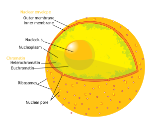
The nucleolus is the largest structure in the nucleus of eukaryotic cells. It is best known as the site of ribosome biogenesis, which is the synthesis of ribosomes. The nucleolus also participates in the formation of signal recognition particles and plays a role in the cell's response to stress. Nucleoli are made of proteins, DNA and RNA, and form around specific chromosomal regions called nucleolar organizing regions. Malfunction of nucleoli can be the cause of several human conditions called "nucleolopathies" and the nucleolus is being investigated as a target for cancer chemotherapy.

<span class="mw-page-title-main">Angina</span> Chest discomfort due to not enough blood flow to heart muscle

Angina, also known as angina pectoris, is chest pain or pressure, usually caused by insufficient blood flow to the heart muscle (myocardium). It is most commonly a symptom of coronary artery disease.

<span class="mw-page-title-main">Coronary thrombosis</span> Medical condition

Coronary thrombosis is defined as the formation of a blood clot inside a blood vessel of the heart. This blood clot may then restrict blood flow within the heart, leading to heart tissue damage, or a myocardial infarction, also known as a heart attack.

<span class="mw-page-title-main">LDL receptor</span> Mammalian protein found in Homo sapiens

The low-density lipoprotein receptor (LDL-R) is a mosaic protein of 839 amino acids that mediates the endocytosis of cholesterol-rich low-density lipoprotein (LDL). It is a cell-surface receptor that recognizes apolipoprotein B100 (ApoB100), which is embedded in the outer phospholipid layer of very low-density lipoprotein (VLDL), their remnants—i.e. intermediate-density lipoprotein (IDL), and LDL particles. The receptor also recognizes apolipoprotein E (ApoE) which is found in chylomicron remnants and IDL. In humans, the LDL receptor protein is encoded by the LDLR gene on chromosome 19. It belongs to the low density lipoprotein receptor gene family. It is most significantly expressed in bronchial epithelial cells and adrenal gland and cortex tissue.

p14ARF is an alternate reading frame protein product of the CDKN2A locus. p14ARF is induced in response to elevated mitogenic stimulation, such as aberrant growth signaling from MYC and Ras (protein). It accumulates mainly in the nucleolus where it forms stable complexes with NPM or Mdm2. These interactions allow p14ARF to act as a tumor suppressor by inhibiting ribosome biogenesis or initiating p53-dependent cell cycle arrest and apoptosis, respectively. p14ARF is an atypical protein, in terms of its transcription, its amino acid composition, and its degradation: it is transcribed in an alternate reading frame of a different protein, it is highly basic, and it is polyubiquinated at the N-terminus.

<span class="mw-page-title-main">NPM1</span> Protein-coding gene in the species Homo sapiens

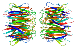
Nucleophosmin (NPM), also known as nucleolar phosphoprotein B23 or numatrin, is a protein that in humans is encoded by the NPM1 gene.

<span class="mw-page-title-main">PES1</span> Protein-coding gene in the species Homo sapiens

Pescadillo homolog is a protein that in humans is encoded by the PES1 gene.

<span class="mw-page-title-main">BOP1</span> Protein-coding gene in the species Homo sapiens

Ribosome biogenesis protein BOP1 is a protein that in humans is encoded by the BOP1 gene.

<span class="mw-page-title-main">WDR5</span> Protein-coding gene in the species Homo sapiens

WD repeat-containing protein 5 is a protein that in humans is encoded by the WDR5 gene.

<span class="mw-page-title-main">WDR36</span> Protein-coding gene in the species Homo sapiens

WD repeat-containing protein 36 is a protein that in humans is encoded by the WDR36 gene.

<span class="mw-page-title-main">GRWD1</span> Protein-coding gene in the species Homo sapiens

Glutamate-rich WD repeat-containing protein 1 is a WD40 repeat protein that in humans is encoded by the GRWD1 gene. It localizes to the nucleus and has known functions in regulating chromatin openness and loading of MCM helicase.

<span class="mw-page-title-main">Myocardial infarction</span> Interruption of blood supply to a part of the heart

A myocardial infarction (MI), commonly known as a heart attack, occurs when blood flow decreases or stops to the coronary artery of the heart, causing damage to the heart muscle. The most common symptom is chest pain or discomfort which may travel into the shoulder, arm, back, neck or jaw. Often it occurs in the center or left side of the chest and lasts for more than a few minutes. The discomfort may occasionally feel like heartburn. Other symptoms may include shortness of breath, nausea, feeling faint, a cold sweat or feeling tired. About 30% of people have atypical symptoms. Women more often present without chest pain and instead have neck pain, arm pain or feel tired. Among those over 75 years old, about 5% have had an MI with little or no history of symptoms. An MI may cause heart failure, an irregular heartbeat, cardiogenic shock or cardiac arrest.

<span class="mw-page-title-main">Myocardial scarring</span>

Myocardial scarring is the accumulation of fibrous tissue resulting after some form of trauma to the cardiac tissue. Fibrosis is the formation of excess tissue in replacement of necrotic or extensively damaged tissue. Fibrosis in the heart is often hard to detect because fibromas, scar tissue or small tumors formed in one cell line, are often formed. Because they are so small, they can be hard to detect by methods such as magnetic resonance imaging. A cell line is a path of fibrosis that follow only a line of cells.

Human HGF plasmid DNA therapy of cardiomyocytes is being examined as a potential treatment for coronary artery disease, as well as treatment for the damage that occurs to the heart after MI. After MI, the myocardium suffers from reperfusion injury which leads to death of cardiomyocytes and detrimental remodelling of the heart, consequently reducing proper cardiac function. Transfection of cardiac myocytes with human HGF reduces ischemic reperfusion injury after MI. The benefits of HGF therapy include preventing improper remodelling of the heart and ameliorating heart dysfunction post-MI.

<span class="mw-page-title-main">PHACTR1</span> Protein-coding gene in the species Homo sapiens

Phosphatase and actin regulator 1 (PHACTR1) is a protein that in humans is encoded by the PHACTR1 gene on chromosome 6. It is most significantly expressed in the globus pallidus of the brain. PHACTR1 is an actin and protein phosphatase 1 (PP1) binding protein that binds actin and regulates the reorganization of the actin cytoskeleton. This protein has been associated with coronary artery disease and migraines through genome-wide association studies. The PHACTR1 gene also contains one of 27 SNPs associated with increased risk of coronary artery disease.

<span class="mw-page-title-main">KIF6</span> Protein-coding gene in the species Homo sapiens

Kinesin family member 6 is a protein that in humans is encoded by the KIF6 gene. This gene encodes a member of the kinesin family of proteins. Members of this family are part of a multisubunit complex that functions as a microtubule motor in intracellular organelle transport.

<span class="mw-page-title-main">WD repeat domain 18</span> Protein-coding gene in the species Homo sapiens

WD repeat domain 18 is a protein that in humans is encoded by the WDR18 gene.

<span class="mw-page-title-main">BRINP3</span> Protein-coding gene in the species Homo sapiens

BMP/retinoic acid inducible neural specific 3 is a protein that in humans is encoded by the BRINP3 gene.

Cardiomyocyte proliferation refers to the ability of cardiac muscle cells to progress through the cell cycle and continue to divide. Traditionally, cardiomyocytes were believed to have little to no ability to proliferate and regenerate after birth. Although other types of cells, such as gastrointestinal epithelial cells, can proliferate and differentiate throughout life, cardiac tissue contains little intrinsic ability to proliferate, as adult human cells arrest in the cell cycle. However, a recent paradigm shift has occurred. Recent research has demonstrated that human cardiomyocytes do proliferate to a small extent for the first two decades of life. Also, cardiomyocyte proliferation and regeneration has been demonstrated to occur in various neonatal mammals in response to injury in the first week of life. Current research aims to further understand the biological mechanism underlying cardiomyocyte proliferation in hopes to turn this capability back on in adults in order to combat heart disease.

References

  1. 1 2 3 GRCh38: Ensembl release 89: ENSG00000138442 - Ensembl, May 2017
  2. 1 2 3 GRCm38: Ensembl release 89: ENSMUSG00000026019 - Ensembl, May 2017
  3. "Human PubMed Reference:". National Center for Biotechnology Information, U.S. National Library of Medicine.
  4. "Mouse PubMed Reference:". National Center for Biotechnology Information, U.S. National Library of Medicine.
  5. 1 2 3 4 5 6 Hölzel M, Rohrmoser M, Schlee M, Grimm T, Harasim T, Malamoussi A, Gruber-Eber A, Kremmer E, Hiddemann W, Bornkamm GW, Eick D (August 2005). "Mammalian WDR12 is a novel member of the Pes1-Bop1 complex and is required for ribosome biogenesis and cell proliferation". The Journal of Cell Biology. 170 (3): 367–78. doi:10.1083/jcb.200501141. PMC   2171466 . PMID   16043514.
  6. 1 2 3 Rohrmoser M, Hölzel M, Grimm T, Malamoussi A, Harasim T, Orban M, Pfisterer I, Gruber-Eber A, Kremmer E, Eick D (May 2007). "Interdependence of Pes1, Bop1, and WDR12 controls nucleolar localization and assembly of the PeBoW complex required for maturation of the 60S ribosomal subunit". Molecular and Cellular Biology. 27 (10): 3682–94. doi:10.1128/MCB.00172-07. PMC   1899993 . PMID   17353269.
  7. 1 2 "Entrez Gene: WDR12 WD repeat domain 12".
  8. "BioGPS - your Gene Portal System". biogps.org. Retrieved 2016-08-23.
  9. Myocardial Infarction Genetics Consortium (March 2009). "Genome-wide association of early-onset myocardial infarction with single nucleotide polymorphisms and copy number variants". Nature Genetics. 41 (3): 334–41. doi:10.1038/ng.327. PMC   2681011 . PMID   19198609.
  10. 1 2 Mega JL, Stitziel NO, Smith JG, Chasman DI, Caulfield MJ, Devlin JJ, Nordio F, Hyde CL, Cannon CP, Sacks FM, Poulter NR, Sever PS, Ridker PM, Braunwald E, Melander O, Kathiresan S, Sabatine MS (June 2015). "Genetic risk, coronary heart disease events, and the clinical benefit of statin therapy: an analysis of primary and secondary prevention trials". Lancet. 385 (9984): 2264–71. doi:10.1016/S0140-6736(14)61730-X. PMC   4608367 . PMID   25748612.
  11. "WDR12 - Ribosome biogenesis protein WDR12 - Homo sapiens (Human) - WDR12 gene & protein". www.uniprot.org. Retrieved 2016-08-23.
  12. 1 2 3 Moilanen AM, Rysä J, Kaikkonen L, Karvonen T, Mustonen E, Serpi R, Szabó Z, Tenhunen O, Bagyura Z, Näpänkangas J, Ohukainen P, Tavi P, Kerkelä R, Leósdóttir M, Wahlstrand B, Hedner T, Melander O, Ruskoaho H (2015-01-01). "WDR12, a Member of Nucleolar PeBoW-Complex, Is Up-Regulated in Failing Hearts and Causes Deterioration of Cardiac Function". PLOS ONE. 10 (4): e0124907. Bibcode:2015PLoSO..1024907M. doi: 10.1371/journal.pone.0124907 . PMC   4411154 . PMID   25915632.
  13. Romes EM, Sobhany M, Stanley RE (January 2016). "The Crystal Structure of the Ubiquitin-like Domain of Ribosome Assembly Factor Ytm1 and Characterization of Its Interaction with the AAA-ATPase Midasin". The Journal of Biological Chemistry. 291 (2): 882–93. doi: 10.1074/jbc.M115.693259 . PMC   4705406 . PMID   26601951.
  14. Rose BA, Force T, Wang Y (October 2010). "Mitogen-activated protein kinase signaling in the heart: angels versus demons in a heart-breaking tale". Physiological Reviews. 90 (4): 1507–46. doi:10.1152/physrev.00054.2009. PMC   3808831 . PMID   20959622.
  15. Willis MS, Patterson C (October 2010). "Hold me tight: Role of the heat shock protein family of chaperones in cardiac disease". Circulation. 122 (17): 1740–51. doi:10.1161/CIRCULATIONAHA.110.942250. PMC   2976481 . PMID   20975010.
  16. Nal B, Mohr E, Silva MI, Tagett R, Navarro C, Carroll P, Depetris D, Verthuy C, Jordan BR, Ferrier P (January 2002). "Wdr12, a mouse gene encoding a novel WD-Repeat Protein with a notchless-like amino-terminal domain". Genomics. 79 (1): 77–86. doi:10.1006/geno.2001.6682. PMID   11827460.
  17. 1 2 Kathiresan S, Voight BF, Purcell S, Musunuru K, Ardissino D, Mannucci PM, et al. (March 2009). "Genome-wide association of early-onset myocardial infarction with single nucleotide polymorphisms and copy number variants". Nature Genetics. 41 (3): 334–41. doi:10.1038/ng.327. PMC   2681011 . PMID   19198609.

Further reading