
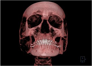
Radiography is an imaging technique using X-rays, gamma rays, or similar ionizing radiation and non-ionizing radiation to view the internal form of an object. Applications of radiography include medical and industrial radiography. Similar techniques are used in airport security,. To create an image in conventional radiography, a beam of X-rays is produced by an X-ray generator and it is projected towards the object. A certain amount of the X-rays or other radiation are absorbed by the object, dependent on the object's density and structural composition. The X-rays that pass through the object are captured behind the object by a detector. The generation of flat two-dimensional images by this technique is called projectional radiography. In computed tomography, an X-ray source and its associated detectors rotate around the subject, which itself moves through the conical X-ray beam produced. Any given point within the subject is crossed from many directions by many different beams at different times. Information regarding the attenuation of these beams is collated and subjected to computation to generate two-dimensional images on three planes which can be further processed to produce a three-dimensional image.

Radiology is the medical specialty that uses medical imaging to diagnose diseases and guide their treatment, within the bodies of humans and other animals. It began with radiography, but today it includes all imaging modalities, including those that use no ionizing electromagnetic radiation, as well as others that do, such as computed tomography (CT), fluoroscopy, and nuclear medicine including positron emission tomography (PET). Interventional radiology is the performance of usually minimally invasive medical procedures with the guidance of imaging technologies such as those mentioned above.

An upper gastrointestinal series, also called a barium swallow, barium study, or barium meal, is a series of radiographs used to examine the gastrointestinal tract for abnormalities. A contrast medium, usually a radiocontrast agent such as barium sulfate mixed with water, is ingested or instilled into the gastrointestinal tract, and X-rays are used to create radiographs of the regions of interest. The barium enhances the visibility of the relevant parts of the gastrointestinal tract by coating the inside wall of the tract and appearing white on the film. This in combination with other plain radiographs allows for the imaging of parts of the upper gastrointestinal tract such as the pharynx, larynx, esophagus, stomach, and small intestine such that the inside wall lining, size, shape, contour, and patency are visible to the examiner. With fluoroscopy, it is also possible to visualize the functional movement of examined organs such as swallowing, peristalsis, or sphincter closure. Depending on the organs to be examined, barium radiographs can be classified into "barium swallow", "barium meal", "barium follow-through", and "enteroclysis". To further enhance the quality of images, air or gas is sometimes introduced into the gastrointestinal tract in addition to barium, and this procedure is called double-contrast imaging. In this case the gas is referred to as the negative contrast medium. Traditionally the images produced with barium contrast are made with plain-film radiography, but computed tomography is also used in combination with barium contrast, in which case the procedure is called "CT enterography".

A joint dislocation, also called luxation, occurs when there is an abnormal separation in the joint, where two or more bones meet. A partial dislocation is referred to as a subluxation. Dislocations are often caused by sudden trauma on the joint like an impact or fall. A joint dislocation can cause damage to the surrounding ligaments, tendons, muscles, and nerves. Dislocations can occur in any major joint or minor joint. The most common joint dislocation is a shoulder dislocation.

A chest radiograph, chest X-ray (CXR), or chest film is a projection radiograph of the chest used to diagnose conditions affecting the chest, its contents, and nearby structures. Chest radiographs are the most common film taken in medicine.
Sir Peter James Kerley KCVO CBE (1900–1979) was an Irish radiologist famous for his role in the lung surgery of King George VI and the naming of the radiological sign in heart failure, Kerley lines.

Radiographers, also known as radiologic technologists, diagnostic radiographers and medical radiation technologists are healthcare professionals who specialise in the imaging of human anatomy for the diagnosis and treatment of pathology. Radiographers are infrequently, and almost always erroneously, known as x-ray technicians. In countries that use the title radiologic technologist they are often informally referred to as techs in the clinical environment; this phrase has emerged in popular culture such as television programmes. The term radiographer can also refer to a therapeutic radiographer, also known as a radiation therapist.

Computer-aided detection (CADe), also called computer-aided diagnosis (CADx), are systems that assist doctors in the interpretation of medical images. Imaging techniques in X-ray, MRI, Endoscopy, and ultrasound diagnostics yield a great deal of information that the radiologist or other medical professional has to analyze and evaluate comprehensively in a short time. CAD systems process digital images or videos for typical appearances and to highlight conspicuous sections, such as possible diseases, in order to offer input to support a decision taken by the professional.
Oral and maxillofacial radiology, also known as dental and maxillofacial radiology, or even more common DentoMaxilloFacial Radiology, is the specialty of dentistry concerned with performance and interpretation of diagnostic imaging used for examining the craniofacial, dental and adjacent structures.
Dr Otto Chan is a consultant radiologist, married and a father of 7 children. He is known as the whistleblower on several issues that related to patient safety, training issues in radiology. He also blew the whistle on terms of employment for flexible trainees. He is also the editor of a book, ABC of Emergency Radiology and co-editor of Ultrasound in Emergency Care.

The Biomedical Imaging and Intervention Journal is a quarterly open access peer-reviewed medical journal established in July 2005. It is financed by donations from regional and international biomedical imaging industry and the University of Malaya Research Imaging Centre. The journal also receives support from many regional associations and societies and in turn has become the official publication of them. As of 2009, it is the official publication of the ASEAN Association of Radiologists, ASEAN Society of Interventional Radiology, Asia-Oceania Federation of Organizations for Medical Physics, Asian Oceania Society of Radiology, College of Radiology, Academy of Medicine Malaysia, Southeast Asian Federation of Organisations of Medical Physics, and the South East Asian Association of Academic Radiologists.
Paediatric radiology is a subspecialty of radiology involving the imaging of fetuses, infants, children, adolescents and young adults. Many paediatric radiologists practice at children's hospitals.
Sudarshan Kumar Aggarwal is an Indian medical doctor and radiologist. He was honoured by the Government of India, in 2013, by bestowing on him the Padma Shri, the fourth highest civilian award, for his contributions to the field of medicine.

Charles Thurstan Holland was an English general practitioner in Liverpool who was best known by his pioneering research in the field of radiology. The Thurstan Holland sign is named after him.
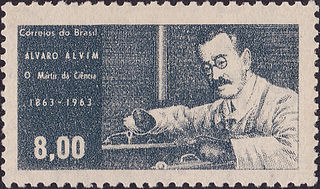
Álvaro Freire de Villalba Alvim was a Brazilian physician, pioneer in radiology and radiotherapy. He studied in France with Pierre and Marie Curie and performed the first radiograph in Brazil, which was on the xiphopagus case for Brazilian surgeon Eduardo Chapot Prévost.

Ann S. Fulcher is an American abdominal radiologist in the radiology department at Virginia Commonwealth University/Medical College of Medicine (VCU). She serves as a professor and the chair of the department of radiology at VCU.
Jeanne LaBerge is American interventional radiologist known for her work establishing the field of interventional radiology as a primary specialty in medicine. She was named a fellow of the Society of CardioVascular and Interventional Radiology in 1992.

Ali A Haydar is Lebanese physician who is an emeritus professor at the American University of Beirut and is the Chief Medical Officer at Aman Hospital, Doha, Qatar and previously the Chairman of radiology at the Clemenceau Medical Center affiliated with Johns Hopkins International since 2018. He is also a member of the Radiological Society of North America, British society of Interventional and Cardiovascular Radiology and the Cardiovascular and Interventional Radiological Society of Europe and fellow of the Pan Arab Interventional radiology society.
