Related Research Articles

The cerebral cortex, also known as the cerebral mantle, is the outer layer of neural tissue of the cerebrum of the brain in humans and other mammals. The cerebral cortex mostly consists of the six-layered neocortex, with just ten per cent consisting of allocortex. It is separated into two cortices, by the longitudinal fissure that divides the cerebrum into the left and right cerebral hemispheres. The two hemispheres are joined beneath the cortex by the corpus callosum. The cerebral cortex is the largest site of neural integration in the central nervous system. It plays a key role in attention, perception, awareness, thought, memory, language, and consciousness.

The cingulate cortex is a part of the brain situated in the medial aspect of the cerebral cortex. The cingulate cortex includes the entire cingulate gyrus, which lies immediately above the corpus callosum, and the continuation of this in the cingulate sulcus. The cingulate cortex is usually considered part of the limbic lobe.

A Brodmann area is a region of the cerebral cortex, in the human or other primate brain, defined by its cytoarchitecture, or histological structure and organization of cells.

Brodmann area 23 (BA23) is a region in the brain that lies inside the posterior cingulate cortex. It lies between Brodmann area 30 and Brodmann area 31 and is located on the medial wall of the cingulate gyrus between the callosal sulcus and the cingulate sulcus.
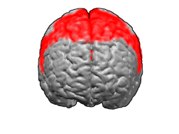
Brodmann area 6 (BA6) part of the frontal cortex in the human brain. Situated just anterior to the primary motor cortex (BA4), it is composed of the premotor cortex and, medially, the supplementary motor area (SMA). This large area of the frontal cortex is believed to play a role in planning complex, coordinated movements.

Brodmann area 10 is the anterior-most portion of the prefrontal cortex in the human brain. BA10 was originally defined broadly in terms of its cytoarchitectonic traits as they were observed in the brains of cadavers, but because modern functional imaging cannot precisely identify these boundaries, the terms anterior prefrontal cortex, rostral prefrontal cortex and frontopolar prefrontal cortex are used to refer to the area in the most anterior part of the frontal cortex that approximately covers BA10—simply to emphasize the fact that BA10 does not include all parts of the prefrontal cortex.

Brodmann area 5 is one of Brodmann's cytoarchitectural defined regions of the brain. It is involved in somatosensory processing, movement and association, and is part of the posterior parietal cortex.

Brodmann area 11 is one of Brodmann's cytologically defined regions of the brain. It is in the orbitofrontal cortex which is above the eye sockets (orbitae). It is involved in decision making and processing rewards, planning, encoding new information into long-term memory, and reasoning.

Brodmann area 4 refers to the primary motor cortex of the human brain. It is located in the posterior portion of the frontal lobe.

The insular cortex is a portion of the cerebral cortex folded deep within the lateral sulcus within each hemisphere of the mammalian brain.

The middle cerebral artery (MCA) is one of the three major paired arteries that supply blood to the cerebrum. The MCA arises from the internal carotid and continues into the lateral sulcus where it then branches and projects to many parts of the lateral cerebral cortex. It also supplies blood to the anterior temporal lobes and the insular cortices.

Brodmann area 24 is part of the anterior cingulate in the human brain.
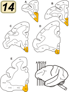
Brodmann Area 14 is one of Brodmann's subdivisions of the cerebral cortex in the brain. It was defined by Brodmann in the guenon monkey . While Brodmann, writing in 1909, argued that no equivalent structure existed in humans, later work demonstrated that area 14 has a clear homologue in the human ventromedial prefrontal cortex.

Brodmann area 13 is part of the insula, a subdivision of the cerebral cortex as defined by cytoarchitecture. The insula is covered by frontal, temporal and parietal operculum and therefore sometimes ignored as a Brodmann area.
Brodmann Area 15 is one of Brodmann's subdivisions of the cerebral cortex in the brain.

Brodmann area 26 is the name for a small part of the brain.
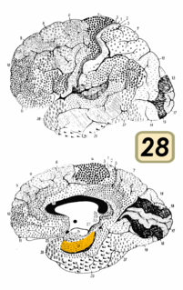
Brodmann area 28 is a subdivision of the cerebral cortex defined on the basis of cytoarchitecture. It is located on the medial aspect of the temporal lobe and is part of the entorhinal cortex (Brodmann-1909).
The primary gustatory cortex is a brain structure responsible for the perception of taste. It consists of two substructures: the anterior insula on the insular lobe and the frontal operculum on the inferior frontal gyrus of the frontal lobe. Because of its composition the primary gustatory cortex is sometimes referred to in literature as the AI/FO(Anterior Insula/Frontal Operculum). By using extracellular unit recording techniques, scientists have elucidated that neurons in the AI/FO respond to sweetness, saltiness, bitterness, and sourness, and they code the intensity of the taste stimulus.
Heterotypic cortex consists of those areas of the mature neocortex that deviate markedly from the homogeneous six-layered internal structure seen in the third trimester of human gestation. A few neocortical areas, such as Brodmann area 17 and the granular insular cortex, undergo modification to more than six layers; and in a few areas, such as Brodmann area 4, the number of layers is reduced. Heterotypic cortex is contrasted to homotypic cortex, which retains the fetal six-layered pattern into adulthood. The number of heterotypic areas is small and the specific areas differ somewhat by species.
Granular insular cortex refers to a portion of the cerebral cortex defined on the basis of internal structure in the human and macaque, the rat, and the mouse. Classified as neocortex, it is in primates distinguished from adjacent allocortex (periallocortex) by the presence of granular layers – external granular layer (II) and internal granular layer (IV) – and by differentiation of the external pyramidal layer (III) into sublayers. In primates it occupies the posterior part of the insula. In rodents it is located on the lateral surface of the cortex rostrally, dorsal to the gustatory area or, more caudally, dorsal to the agranular insula.
References
- 1 2 Mesulam M-M, Mufson EJ (1984). "5: The insula of Reil in man and monkey: Architectonics, connectivity, and function". In Peters A; Jones EG (eds.). Cerebral Cortex. pp. 179–226. OCLC 277149053.
- ↑ Carmichael ST; Price JL (1994). "Architectonic subdivision of the orbital and medial prefrontal cortex in the macaque monkey". J Comp Neurol. 346 (3): 366–402. doi:10.1002/cne.903460305. PMID 7527805. S2CID 20829291.
- ↑ Swanson LW (1998). Brain Maps: Structure of the Rat Brain (2nd Revised ed.). Amsterdam: Elsevier Science. OCLC 640898561.
- ↑ Paxinos G; Franklin KBJ (2001). The Mouse Brain in Stereotaxic Coordinates (2nd ed.). San Diego: Academic Press. OCLC 493265554.
- ↑ Swanson LW (2004). Brain Maps: Structure of the Rat Brain (3rd ed.). Oxford: Elsevier Academic Press. OCLC 225608577.