Related Research Articles

Urination is the release of urine from the urinary bladder through the urethra to the outside of the body. It is the urinary system's form of excretion. It is also known medically as micturition, voiding, uresis, or, rarely, emiction, and known colloquially by various names including peeing, weeing, pissing, and euphemistically going number one. In healthy humans and other animals, the process of urination is under voluntary control. In infants, some elderly individuals, and those with neurological injury, urination may occur as a reflex. It is normal for adult humans to urinate up to seven times during the day.

The autonomic nervous system (ANS), formerly referred to as the vegetative nervous system, is a division of the nervous system that supplies internal organs, smooth muscle and glands. The autonomic nervous system is a control system that acts largely unconsciously and regulates bodily functions, such as the heart rate, its force of contraction, digestion, respiratory rate, pupillary response, urination, and sexual arousal. This system is the primary mechanism in control of the fight-or-flight response.
The parasympathetic nervous system (PSNS) is one of the three divisions of the autonomic nervous system, the others being the sympathetic nervous system and the enteric nervous system. The enteric nervous system is sometimes considered part of the autonomic nervous system, and sometimes considered an independent system.

Tetraplegia, also known as quadriplegia, is defined as the dysfunction or loss of motor and/or sensory function in the cervical area of the spinal cord. A loss of motor function can present as either weakness or paralysis leading to partial or total loss of function in the arms, legs, trunk, and pelvis; paraplegia is similar but affects the thoracic, lumbar, and sacral segments of the spinal cord and arm function is spared. The paralysis may be flaccid or spastic. A loss of sensory function can present as an impairment or complete inability to sense light touch, pressure, heat, pinprick/pain, and proprioception. In these types of spinal cord injury, it is common to have a loss of both sensation and motor control.

A spinal nerve is a mixed nerve, which carries motor, sensory, and autonomic signals between the spinal cord and the body. In the human body there are 31 pairs of spinal nerves, one on each side of the vertebral column. These are grouped into the corresponding cervical, thoracic, lumbar, sacral and coccygeal regions of the spine. There are eight pairs of cervical nerves, twelve pairs of thoracic nerves, five pairs of lumbar nerves, five pairs of sacral nerves, and one pair of coccygeal nerves. The spinal nerves are part of the peripheral nervous system.

The somatic nervous system (SNS), or voluntary nervous system is the part of the peripheral nervous system associated with the voluntary control of body movements via skeletal muscles.

Afferent nerve fibers are axons of sensory neurons that carry sensory information from sensory receptors to the central nervous system. Many afferent projections arrive at a particular brain region.

A reflex arc is a neural pathway that controls a reflex. In vertebrates, most sensory neurons do not pass directly into the brain, but synapse in the spinal cord. This allows for faster reflex actions to occur by activating spinal motor neurons without the delay of routing signals through the brain. The brain will receive the input while the reflex is being carried out and the analysis of the signal takes place after the reflex action.

The pupillary light reflex (PLR) or photopupillary reflex is a reflex that controls the diameter of the pupil, in response to the intensity (luminance) of light that falls on the retinal ganglion cells of the retina in the back of the eye, thereby assisting in adaptation of vision to various levels of lightness/darkness. A greater intensity of light causes the pupil to constrict, whereas a lower intensity of light causes the pupil to dilate. Thus, the pupillary light reflex regulates the intensity of light entering the eye. Light shone into one eye will cause both pupils to constrict.

Onuf's nucleus is a distinct group of neurons located in the ventral part of the anterior horn of the sacral region of the human spinal cord involved in the maintenance of micturition and defecatory continence, as well as muscular contraction during orgasm. It contains motor neurons, and is the origin of the pudendal nerve. The sacral region of the spinal cord is the fourth segment of vertebrae in the spinal cord which consists of the vertebrae 26-30. While working in New York City in 1899, Bronislaw Onuf-Onufrowicz discovered this group of unique cells and originally identified it as “Group X.” “Group X” was considered distinct by Onufrowicz because the cells were different in size from the surrounding neurons in the anterolateral group, suggesting that they were independent.
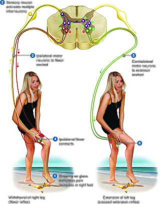
The crossed extensor reflex or crossed extensor response or crossed extension reflex is a reflex in which the contralateral limb compensates for loss of support when the ipsilateral limb withdraws from painful stimulus in a withdrawal reflex. During a withdrawal reflex, the flexors in the withdrawing limb contract and the extensors relax, while in the other limb, the opposite occurs as part of the crossed extensor reflex. Besides shifting the body weight to the other side, the reflex pathway is also associated with leg coordination when walking by flexing muscle on one side, while extending muscle on the other side. This crossed extensor response is properly part of the withdrawal reflex.

A nerve plexus is a plexus of intersecting nerves. A nerve plexus is composed of afferent and efferent fibers that arise from the merging of the anterior rami of spinal nerves and blood vessels. There are five spinal nerve plexuses, except in the thoracic region, as well as other forms of autonomic plexuses, many of which are a part of the enteric nervous system. The nerves that arise from the plexuses have both sensory and motor functions. These functions include muscle contraction, the maintenance of body coordination and control, and the reaction to sensations such as heat, cold, pain, and pressure. There are several plexuses in the body, including:

The stretch reflex, or more accurately "muscle stretch reflex", is a muscle contraction in response to stretching a muscle. The function of the reflex is generally thought be maintaining the muscle at a constant length but the response is often coordinated across multiple muscles and even joints. The term deep tendon reflex is often wrongfully used by many health workers and students to refer to this reflex. "Tendons have little to do with the response, other than being responsible for mechanically transmitting the sudden stretch from the reflex hammer to the muscle spindle. In addition, some muscles with stretch reflexes have no tendons ".
The ventral ramus is the anterior division of a spinal nerve. The ventral rami supply the antero-lateral parts of the trunk and the limbs. They are mainly larger than the dorsal rami.
Sacral nerve stimulation, also termed sacral neuromodulation, is a type of medical electrical stimulation therapy.
Pallesthesia, or vibratory sensation, is the ability to perceive vibration. This sensation, often conducted through skin and bone, is usually generated by mechanoreceptors such as Pacinian corpuscles, Merkel disk receptors, and tactile corpuscles. All of these receptors stimulate an action potential in afferent nerves found in various layers of the skin and body. The afferent neuron travels to the spinal column and then to the brain where the information is processed. Damage to the peripheral nervous system or central nervous system can result in a decline or loss of pallesthesia.
The bulbocavernosus reflex (BCR), bulbospongiosus reflex (BSR) or "Osinski reflex" is a polysynaptic reflex that is useful in testing for spinal shock and gaining information about the state of spinal cord injuries (SCI). Bulbocavernosus is an older term for bulbospongiosus, thus this reflex may also be referred to as the bulbospongiosus reflex.

The axon reflex is the response stimulated by peripheral nerves of the body that travels away from the nerve cell body and branches to stimulate target organs. Reflexes are single reactions that respond to a stimulus making up the building blocks of the overall signaling in the body's nervous system. Neurons are the excitable cells that process and transmit these reflex signals through their axons, dendrites, and cell bodies. Axons directly facilitate intercellular communication projecting from the neuronal cell body to other neurons, local muscle tissue, glands and arterioles. In the axon reflex, signaling starts in the middle of the axon at the stimulation site and transmits signals directly to the effector organ skipping both an integration center and a chemical synapse present in the spinal cord reflex. The impulse is limited to a single bifurcated axon, or a neuron whose axon branches into two divisions and does not cause a general response to surrounding tissue.

A spinal interneuron, found in the spinal cord, relays signals between (afferent) sensory neurons, and (efferent) motor neurons. Different classes of spinal interneurons are involved in the process of sensory-motor integration. Most interneurons are found in the grey column, a region of grey matter in the spinal cord.
References
- ↑ "Deep tendon reflexes" . Retrieved 5 October 2015.
- ↑ "Reflex evaluation". Archived from the original on 25 September 2020. Retrieved 5 October 2015.