The medulla oblongata is a long stem-like structure located in the brainstem. It is anterior and partially inferior to the cerebellum. It is a cone-shaped neuronal mass responsible for autonomic (involuntary) functions ranging from vomiting to sneezing. The medulla contains the cardiac, respiratory, vomiting and vasomotor centers and therefore deals with the autonomic functions of breathing, heart rate and blood pressure.

The lumbar vertebrae are, in human anatomy, the five vertebrae between the rib cage and the pelvis. They are the largest segments of the vertebral column and are characterized by the absence of the foramen transversarium within the transverse process and by the absence of facets on the sides of the body. They are designated L1 to L5, starting at the top. The lumbar vertebrae help support the weight of the body, and permit movement.

The foramen magnum is a large oval opening (foramen) in the occipital bone of the skull in humans and various other animals. It is one of the several oval or circular openings (foramina) in the base of the skull. The spinal cord, an extension of the medulla, passes through the foramen magnum as it exits the cranial cavity. Apart from the transmission of the medulla oblongata and its membranes, the foramen magnum transmits the vertebral arteries, the anterior and posterior spinal arteries, the tectorial membranes and alar ligaments. It also transmits the spinal component of the accessory nerve into the skull.

The internal carotid artery is a major paired artery, one on each side of the head and neck, in human anatomy. They arise from the common carotid arteries where these bifurcate into the internal and external carotid arteries at cervical vertebral level 3 or 4; the internal carotid artery supplies the brain, while the external carotid nourishes other portions of the head, such as face, scalp, skull, and meninges.

Dura mater is a thick membrane made of dense irregular connective tissue that surrounds the brain and spinal cord. It is the outermost of the three layers of membrane called the meninges that protect the central nervous system. The other two meningeal layers are the arachnoid mater and the pia mater. The dura surrounds the brain and the spinal cord and is responsible for keeping in the cerebrospinal fluid. It is derived from neural crest cells.

In vertebrates, cervical vertebrae are the vertebrae of the neck, immediately below the skull.
The vertebral arteries are major arteries of the neck. Typically, the vertebral arteries originate from the subclavian arteries. Each vessel courses superiorly along each side of the neck, merging within the skull to form the single, midline basilar artery. As the supplying component of the vertebrobasilar vascular system, the vertebral arteries provide supply blood to the upper spinal cord, brainstem, cerebellum, and posterior part of brain.

The rectus capitis posterior minor arises by a narrow pointed tendon from the tubercle on the posterior arch of the atlas, and, widening as it ascends, is inserted into the medial part of the inferior nuchal line of the occipital bone and the surface between it and the foramen magnum, and also takes some attachment to the spinal dura mater.
The lesser sac, also known as the omental bursa, is the cavity in the abdomen that is formed by the lesser and greater omentum. Usually found in mammals, it is connected with the greater sac via the omental foramen. In mammals, it is common for the lesser sac to contain considerable amounts of fat.
Congenital vertebral abnormalities are a collection of malformations of the spine. Most around 85% are not clinically significant, but they can cause compression of the spinal cord by deforming the vertebral canal or causing instability. This condition occurs in the womb. Congenital vertebral anomalies include alterations of the shape and number of vertebrae.

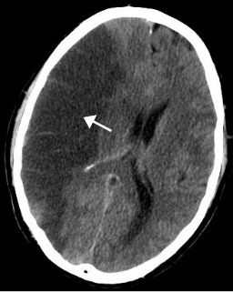
Vertebral artery dissection (VAD) is a flap-like tear of the inner lining of the vertebral artery, which is located in the neck and supplies blood to the brain. After the tear, blood enters the arterial wall and forms a blood clot, thickening the artery wall and often impeding blood flow. The symptoms of vertebral artery dissection include head and neck pain and intermittent or permanent stroke symptoms such as difficulty speaking, impaired coordination and visual loss. It is usually diagnosed with a contrast-enhanced CT or MRI scan.

The arcuate uterus is a form of a uterine anomaly or variation where the uterine cavity displays a concave contour towards the fundus. Normally the uterine cavity is straight or convex towards the fundus on anterior-posterior imaging, but in the arcuate uterus the myometrium of the fundus dips into the cavity and may form a small septation. The distinction between an arcuate uterus and a septate uterus is not standardized.
Atlanto-occipital dislocation, orthopedic decapitation, or internal decapitation describes ligamentous separation of the spinal column from the skull base. It is possible for a human to survive such an injury; however, only 30% of cases do not result in immediate death. It should not be confused with atlanto-axial dislocation, which describes ligamentous separation between the first and second cervical vertebra.
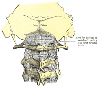
The posterior atlantooccipital membrane is a broad but thin membrane. It is connected above to the posterior margin of the foramen magnum and below to the upper border of the posterior arch of the atlas.

Spondylocostal dysostosis is a rare, heritable axial skeleton growth disorder. It is characterized by widespread and sometimes severe malformations of the vertebral column and ribs, shortened thorax, and moderate to severe scoliosis and kyphosis. Individuals with Jarcho-Levin typically appear to have a short trunk and neck, with arms appearing relatively long in comparison, and a slightly protuberant abdomen. Severely affected individuals may have life-threatening pulmonary complications due to deformities of the thorax. The syndrome was first described by Saul Jarcho and Paul M. Levin at Johns Hopkins University in 1938.
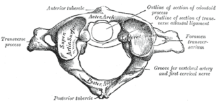
A Jefferson fracture is a bone fracture of the anterior and posterior arches of the C1 vertebra, though it may also appear as a three- or two-part fracture. The fracture may result from an axial load on the back of the head or hyperextension of the neck, causing a posterior break, and may be accompanied by a break in other parts of the cervical spine.

The spinal cord is a long, thin, tubular structure made up of nervous tissue, which extends from the medulla oblongata in the brainstem to the lumbar region of the vertebral column. It encloses the central canal of the spinal cord, which contains cerebrospinal fluid. The brain and spinal cord together make up the central nervous system (CNS). In humans, the spinal cord begins at the occipital bone, passing through the foramen magnum and entering the spinal canal at the beginning of the cervical vertebrae. The spinal cord extends down to between the first and second lumbar vertebrae, where it ends. The enclosing bony vertebral column protects the relatively shorter spinal cord. It is around 45 cm (18 in) in men and around 43 cm (17 in) long in women. The diameter of the spinal cord ranges from 13 mm in the cervical and lumbar regions to 6.4 mm in the thoracic area.

In the vertebrate spinal column, each vertebra is an irregular bone with a complex structure composed of bone and some hyaline cartilage, the proportions of which vary according to the segment of the backbone and the species of vertebrate.