Keratoconus (KC) is a disorder of the eye which results in progressive thinning of the cornea. This may result in blurry vision, double vision, nearsightedness, astigmatism, and light sensitivity. Usually both eyes are affected. In more severe cases a scarring or a circle may be seen within the cornea.

Refractive eye surgery is an eye surgery used to improve the refractive state of the eye and decrease or eliminate dependency on glasses or contact lenses. This can include various methods of surgical remodeling of the cornea (keratomileusis), lens implantation or lens replacement. The most common methods today use excimer lasers to reshape the curvature of the cornea. Successful refractive eye surgery can reduce or cure common vision disorders such as myopia, hyperopia and astigmatism, as well as degenerative disorders like keratoconus.
Fuchs' dystrophy, also referred to as Fuchs' corneal endothelial dystrophy (FCED) and Fuchs' endothelial dystrophy (FED), is a slowly progressing corneal dystrophy that usually affects both eyes and is slightly more common in women than in men. Although early signs of Fuchs' dystrophy are sometimes seen in people in their 30s and 40s, the disease rarely affects vision until people reach their 50s and 60s.

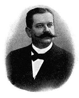
Eduard Konrad Zirm was an ophthalmologist who performed the first successful human full-thickness corneal transplant on 7 December 1905.
Recurrent corneal erosion is a disorder of the eyes characterized by the failure of the cornea's outermost layer of epithelial cells to attach to the underlying basement membrane. The condition is excruciatingly painful because the loss of these cells results in the exposure of sensitive corneal nerves. This condition can often leave patients with temporary blindness due to extreme light sensitivity photophobia.
Corneal ulcer is an inflammatory or more seriously, infective condition of the cornea involving disruption of its epithelial layer with involvement of the corneal stroma. It is a common condition in humans particularly in the tropics and the agrarian societies. In developing countries, children afflicted by Vitamin A deficiency are at high risk for corneal ulcer and may become blind in both eyes, which may persist lifelong. In ophthalmology, a corneal ulcer usually refers to having an infectious cause while the term corneal abrasion refers more to physical abrasions.

Pellucid marginal degeneration (PMD), is a degenerative corneal condition, often confused with keratoconus. It is typically characterized by a clear, bilateral thinning (ectasia) in the inferior and peripheral region of the cornea, although some cases affect only one eye. The cause of the disease remains unclear.

Meesmann corneal dystrophy is a type of corneal dystrophy and a keratin disease.

Keratoprosthesis is a surgical procedure where a diseased cornea is replaced with an artificial cornea. Traditionally, keratoprosthesis is recommended after a person has had a failure of one or more donor corneal transplants. More recently, a less invasive, non-penetrating artificial cornea has been developed which can be used in more routine cases of corneal blindness. While conventional cornea transplant uses donor tissue for transplant, an artificial cornea is used in the Keratoprosthesis procedure. The surgery is performed to restore vision in patients suffering from severely damaged cornea due to congenital birth defects, infections, injuries and burns.

Reis-Bücklers corneal dystrophy, also known as corneal dystrophy of Bowman layer, type I, is a rare, corneal dystrophy of unknown cause, in which the Bowman's layer of the cornea undergoes disintegration. The disorder is inherited in an autosomal dominant fashion, and is associated with mutations in the gene TGFB1.
Thiel–Behnke dystrophy, or Corneal dystrophy of Bowman layer, type II, is a rare form of corneal dystrophy affecting the layer that supports corneal epithelium. The dystrophy was first described in 1967 and initially suspected to denote the same entity as the earlier-described Reis-Bucklers dystrophy, but following a study in 1995 by Kuchle et al. the two look-alike dystrophies were deemed separate disorders.
Epithelial basement membrane dystrophy (EBMD), also known as map-dot-fingerprint dystrophy and Cogans's microcystic dystrophy, is a disorder of the eye that can cause pain and dryness.

Lattice corneal dystrophy type, also known as Biber-Haab-Dimmer dystrophy, is a rare form of corneal dystrophy. It has no systemic manifestations, unlike the other type of the dystrophy, Lattice corneal dystrophy type II. Lattice corneal dystrophy was first described by Swiss ophthalmologist Hugo Biber in 1890.

Granular corneal dystrophy is a slowly progressive corneal dystrophy that most often begins in early childhood.
Dua's layer, according to a 2013 paper by Harminder Singh Dua's group at the University of Nottingham, is a layer of the cornea that had not been detected previously. It is hypothetically 15 micrometres thick, the fourth caudal layer, and located between the corneal stroma and Descemet's membrane. Despite its thinness, the layer is very strong and impervious to air. It is strong enough to withstand up to 2 bars of pressure. While some scientists welcomed the announcement, other scientists cautioned that time was needed for other researchers to confirm the discovery and its significance. Others have met the claim "with incredulity". The choice of the name Dua's Layer has also been criticized.

Corneal hydrops or corneal rupture is an uncommon complication seen in people with advanced keratoconus or other corneal ectatic disorders, and is characterized by stromal edema due to leakage of aqueous humor through a tear in Descemet's membrane. Although a hydrops usually causes increased scarring of the cornea, occasionally it will benefit a patient by creating a flatter cone, aiding the fitting of contact lenses. Corneal transplantation is not usually indicated during corneal hydrops.