
Atrial septal defect (ASD) is a congenital heart defect in which blood flows between the atria of the heart. Some flow is a normal condition both pre-birth and immediately post-birth via the foramen ovale; however, when this does not naturally close after birth it is referred to as a patent (open) foramen ovale (PFO). It is common in patients with a congenital atrial septal aneurysm (ASA).

The ostium primum atrial septal defect is a defect in the atrial septum at the level of the tricuspid and mitral valves. This is sometimes known as an endocardial cushion defect because it often involves the endocardial cushion, which is the portion of the heart where the atrial septum meets the ventricular septum and the mitral valve meets the tricuspid valve.

A congenital heart defect (CHD), also known as a congenital heart anomaly, congenital cardiovascular malformation, and congenital heart disease, is a defect in the structure of the heart or great vessels that is present at birth. A congenital heart defect is classed as a cardiovascular disease. Signs and symptoms depend on the specific type of defect. Symptoms can vary from none to life-threatening. When present, symptoms are variable and may include rapid breathing, bluish skin (cyanosis), poor weight gain, and feeling tired. CHD does not cause chest pain. Most congenital heart defects are not associated with other diseases. A complication of CHD is heart failure.

Atrioventricular septal defect (AVSD) or atrioventricular canal defect (AVCD), also known as "common atrioventricular canal" or "endocardial cushion defect" (ECD), is characterized by a deficiency of the atrioventricular septum of the heart that creates connections between all four of its chambers. It is a very specific combination of 3 defects:

The interatrial septum is the wall of tissue that separates the right and left atria of the heart.
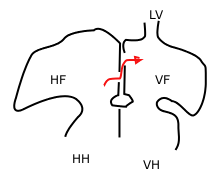
A sinus venosus atrial septal defect is a type of atrial septal defect primarily associated with the sinus venosus.

During heart development of a human embryo, the single primitive atrium becomes divided into right and left by a septum, the septum primum. The septum primum grows downward into the single atrium.

Holt–Oram syndrome is an autosomal dominant disorder that affects bones in the arms and hands and often causes heart problems. The syndrome may include an absent radial bone in the forearm, an atrial septal defect in the heart, or heart block. It affects approximately 1 in 100,000 people.
The foramen secundum, or ostium secundum is a foramen in the septum primum, a precursor to the interatrial septum of the human heart.
In the developing heart, the atria are initially open to each other, with the opening known as the primary interatrial foramen or ostium primum. The foramen lies beneath the edge of septum primum and the endocardial cushions. It progressively decreases in size as the septum grows downwards, and disappears with the formation of the atrial septum.

Atrial septostomy is a surgical procedure in which a small hole is created between the upper two chambers of the heart, the atria. This procedure is primarily used to palliate dextro-Transposition of the great arteries or d-TGA, a life-threatening cyanotic congenital heart defect seen in infants. It is performed prior to an arterial switch operation. Atrial septostomy has also seen limited use as a surgical treatment for pulmonary hypertension. The first atrial septostomy was developed by Vivien Thomas in a canine model and performed in humans by Alfred Blalock. The Rashkind balloon procedure, a common atrial septostomy technique, was developed in 1966 by American cardiologist William Rashkind at the Children's Hospital of Philadelphia.

ACTC1 encodes cardiac muscle alpha actin. This isoform differs from the alpha actin that is expressed in skeletal muscle, ACTA1. Alpha cardiac actin is the major protein of the thin filament in cardiac sarcomeres, which are responsible for muscle contraction and generation of force to support the pump function of the heart.

Homeobox protein Nkx-2.5 is a protein that in humans is encoded by the NKX2-5 gene.

Myosin heavy chain, α isoform (MHC-α) is a protein that in humans is encoded by the MYH6 gene. This isoform is distinct from the ventricular/slow myosin heavy chain isoform, MYH7, referred to as MHC-β. MHC-α isoform is expressed predominantly in human cardiac atria, exhibiting only minor expression in human cardiac ventricles. It is the major protein comprising the cardiac muscle thick filament, and functions in cardiac muscle contraction. Mutations in MYH6 have been associated with late-onset hypertrophic cardiomyopathy, atrial septal defects and sick sinus syndrome.

Protein Jumonji is a protein that in humans is encoded by the JARID2 gene. JARID2 is a member of the alpha-ketoglutarate-dependent hydroxylase superfamily.

Atrial Light Chain-2 (ALC-2) also known as Myosin regulatory light chain 2, atrial isoform (MLC2a) is a protein that in humans is encoded by the MYL7 gene. ALC-2 expression is restricted to cardiac muscle atria in healthy individuals, where it functions to modulate cardiac development and contractility. In human diseases, including hypertrophic cardiomyopathy, dilated cardiomyopathy, ischemic cardiomyopathy and others, ALC-2 expression is altered.

Miller syndrome, also known as Genée–Wiedemann syndrome, Wildervanck–Smith syndrome or postaxial acrofacial dysostosis, is an extremely rare genetic condition that manifests as craniofacial, limb and eye deformities. It is caused by a mutation in the DHODH gene. The incidence of the condition is not known, and nothing is known about its pathogenesis.
TBX20 (gene) is a member of the T-box family that encodes the transcription factor TBX20. Studies in mouse, human and fruitfly have shown that this gene is essential for early heart development, adult heart function and yolk sac vasculature remodeling and has been associated with congenital heart diseases. Tbx20 was also shown to be required for migration of hindbrain motor neurons and in facial neurons was proposed to be a positive regulator of the non-canonical Wnt signaling pathway.
The Relative Atrial Index (RAI) is a numeric parameter used to assess for cardiac shunt defects. It is calculated from the standard transthoracic Doppler echocardiogram measurements of the right atrial area divided by the left atrial area. RAI = right atrial area / left atrial area. These measurements are made from the apical four chamber view.