Axon reflex

Last updated
A flow map of the axon reflex. Stimulation of the axon can cause electric flow to all effector tissues the neuron innervates, as well as back to the soma of the neuron; this is distinct from a normal neuron firing only down the axon. Axon Reflex Drawing.png
A flow map of the axon reflex. Stimulation of the axon can cause electric flow to all effector tissues the neuron innervates, as well as back to the soma of the neuron; this is distinct from a normal neuron firing only down the axon.

The axon reflex [1] (or the flare response) [2] is the response stimulated by peripheral nerves of the body that travels away from the nerve cell body and branches to stimulate target organs. Reflexes are single reactions that respond to a stimulus making up the building blocks of the overall signaling in the body's nervous system. Neurons are the excitable cells that process and transmit these reflex signals through their axons, dendrites, and cell bodies. Axons directly facilitate intercellular communication projecting from the neuronal cell body to other neurons, local muscle tissue, glands and arterioles. In the axon reflex, signaling starts in the middle of the axon at the stimulation site and transmits signals directly to the effector organ skipping both an integration center and a chemical synapse present in the spinal cord reflex. The impulse is limited to a single bifurcated axon, [3] or a neuron whose axon branches into two divisions and does not cause a general response to surrounding tissue.

Contents

The axon reflex arc is distinct from the spinal cord reflex arc. In the spinal cord reflex pathway the afferent neuron transmits information to spinal cord interneurons. These interneurons act collectively, process and make sense of inbound stimuli, and stimulate effector neurons acting as an integration center. [4] The effector neurons leaving the integration center transmit a response to the original tissue the reflex arose resulting in a response. The axon reflex results in a localized response to only the locally innervated cells of the single neuron where the signal originated. [5] The axon reflex pathway does not include an integration center or synapse that relays communication between neurons in the spinal cord reflex. The stimulus, therefore, is diverted to the effector organ without entering the neuronal cell body and therefore indicates that the axon reflex is not a true reflex where afferent impulses pass through the central nervous system before stimulating efferent neurons.

The axon reflex was discovered and was described as "a new type of peripheral reflex" that bypasses the integration center and synapse in the central nervous system. The discovery of the axonal reflex found that the axon reflex activates local arterioles causing vasodilation and muscle contraction. This muscle contraction was observed in people with asthma where the released neuropeptides caused the smooth muscle in the airway to contract. Similarly the release of cholinergic agents at sudomotor nerve terminals evokes an axon reflex that stimulates sweat glands inducing the body to sweat in response to heat. The axon reflex is possible through the transmission of signals from the cutaneous receptors on the skin.

Research and discovery

The axon reflex was discovered by Kovalevskiy and Sokovnin, two Russian scientists in 1873. [5] They described the axon reflex as a new type of peripheral (or local) reflex where electrical signal starts in the middle of the axon and transmit immediately skipping both an integration center and a chemical synapse as typically observed in the spinal cord reflex.

In 1890 the British physiologist, John Neuport Langley, researched the hair movement on cats as they were exposed to cold temperature. Langley observed that even after stimulation, cat hair in the surrounding areas continued to rise. Langley concluded that the primary neuronal stimulation did not end after the first synapse but rather was involved in branching connections to multiple neurons causing cat hair in surrounding areas to rise. [4] Langley defined this pathway as "axon reflex."

A normal spinal cord reflex arc, whereas the axon reflex would bypass the interneuron. This distinction is important because early researchers had to differentiate between the spinal cord reflex and the axon reflex to understand the body's response to stimuli. Imgnotracat arc reflex eng.svg
A normal spinal cord reflex arc, whereas the axon reflex would bypass the interneuron. This distinction is important because early researchers had to differentiate between the spinal cord reflex and the axon reflex to understand the body's response to stimuli.

In the early 20th century, British cardiologist Sir Thomas Lewis researched mechanical abrasion to the skin. The skin demonstrated a triphasic response. First, a red spot develops and spreads outward due to the release of histamine from mast cells. Secondly, a brighter red color spread around the original spot due to arteriolar dilation. The last phase was the production of a wheal filled fluid over the original spot. Lewis believed that the skin’s response was due to the dilation of neighboring blood vessels that were triggered by the nervous system through the axon reflex. [4] This triphasic response was named the triple response of Lewis. The dilation of arterioles in the effected area is due to vasodilation. Although Lewis observed vasodilation that could be explained by axon reflex, there was not yet direct evidence explaining the branching of nerves from the center of an axon rather than a cell body or which chemical agents were responsible for the goose bump, red line, and dilated blood vessel symptoms. [4]

In the 1960s, scientists A. Janscó-Gabor and J. Szolcsányi demonstrated that when irritant chemicals and electrical stimulants are applied to the skin, cutaneous nocireceptors are stimulated. These pain sensors send signals to neighboring tissues resulting in extravasation, also known as leakage from the blood vessels. This response is similar to Lewis’s research with vasodilation as both rely on an intact sensory nerve supply that affect neighboring tissues. [5]

At the end of the 20th century more sophisticated methods for direct observation of the axon reflex arose due to more precise imaging tools and more advanced techniques. One example is laser Doppler studies which uses laser doppler imaging to observe the skin blood flow to determine vascular function. [6] These sorts of experimental collection techniques produce experimental data that suggests a mechanism to explain how the interaction of neural factors and genetic endowments make some individuals more resistant to cold. These research techniques have helped to improve medical treatment and prevention of cold-related skin damage and frostbite injuries.

Physiology

When a proximal impulse stimulates the stretch and heat receptors on one branch of a bifurcated axon, the produced signal moves backwards towards the point of axon bifurcation. The impulse then reflects down the other branch of the axon to the effector organ causing axon reflex. Axon reflexes stimulate numerous effector organs including the endocrine, vascular and circulatory systems depending on the location of the stimulation. One example is itching, a type of nociception, where the reflex often evokes a scratching desire. The compound capsaicin can be used to deplete the chemicals in the axon reflex nerve endings and reduce the symptoms of itching and pain. [5]

Physiologically, the axon reflex helps to maintain homeostasis, or regulation of the body's internal environment in response to the changing external environment, ensuring the internal environment is stable and relatively constant. The axon reflex responds to external changes in temperature, chemical concentration, and air composition. Examples of axon reflex mediated mechanisms include itching, inflammation, pain, asthma, and dermal circulation. [5]

Vasodilation

The body responds to multiple types of trauma including infection, physical injury, or toxic tissue damage through inflammation. When pain sensation increases, the axon reflex stimulates (and is responsible for) to release of many necessary chemicals that promote local tissue inflammation of the traumatized region. [5] Axon reflex regulates vasodilation, or the extra blood flow to target tissues. Axon reflex allows muscles to contract in the shortest amount of time possible by regulating the signal conduction in the neuromuscular junction.

Vasoconstriction and vasodilation, an effect that can be caused from axon reflex stimulation in certain tissues, demonstrated compared to the normal blood vessel. Vasoconstriction and Vasodilation.png
Vasoconstriction and vasodilation, an effect that can be caused from axon reflex stimulation in certain tissues, demonstrated compared to the normal blood vessel.

In dermal circulation, the axon reflex controls the temperature and circulation in the tissues through vasodilation. Small nerve fibers called thermoreceptors are sensitive to temperature and can act as sensors that initiate axon reflex mediated vasodilation. Neuromuscular diseases can be predicted early by the presence of abnormal muscle fiber reflexes and corresponding twitches. This arises because axons can generate their own action potentials when hyperexcited from the original stimulus; this is known as a fasciculation potential in the muscle fiber. [7] Fasciculations are prominent features in amyotrophic lateral sclerosis (ALS) and could be evidence of abnormal axon reflex with further research. [8]


Asthma

In asthma, the axon reflex induces the release of various neuropeptides, including substance P, neurokinin A, and calcitonin. All three of these neuropeptides cause contraction of the smooth muscle in the airway, which also happens through a similar mechanism in allergies.

This same reaction mechanism is also responsible for the loss of body heat in the extremities, demonstrated via the Hunter's Test. One clinical test for the patient that can be performed is the QSART, or the Quantitative Sudomotor Axon Reflex Testing, which stimulates the autonomic nervous system of an individual by stimulating sweat glands through the promotion of axon reflexes. [9] The skin is stimulated with electricity, causing said axon reflexes, which allows for the assessment of the type and severity of autonomic nervous disorders and peripheral neuropathies like asthma or multiple sclerosis.

Sweat response

Humans and primates use the sudomotor response to cause thermoregulation, or control of their body temperature, mainly via the sympathetic nervous system with negligible influences from the parasympathetic nervous system. [10] Heat sensitive receptors are present in the skin, viscera, and spinal cord where they receive information from the outside environment, and send it to the thermoregulatory center in the hypothalamus.

A sweat response stimulates M3 muscarinic receptors on sweat glands and a sudomotor axon reflex. In the sudomotor reflex, cholinergic agents bind to the nicotinic receptors on the sudomotor nerve terminals, evoking an impulse that travels towards the soma, or opposite of the normal impulse. At the soma of the postganglionic sympathetic sudomotor neuron, the impulse branches and travels orthodromically, or away from the soma. Finally, as this impulse reaches other sweat glands, it causes an indirect axon-reflex sweat response. Sudomotor axon reflexes can be peripherally amplified in the transmission of the action potential magnitude by acetylcholine. [10] Acetylcholine also activates sudomotor fibers and primary afferent nociceptors, triggering axon reflexes in both. However, with nerve damage (neuropathy) there is still some increase in axon reflex mediated sweating.

Mechanisms

Cutaneous receptors are sensory receptors in the skin that detect changes in temperature (thermoreceptors) and pain (nociceptors). These cutaneous receptors initiate an impulse via excitation of the main sensory axon to the spinal cord. The axon reflex is the spread of this impulse from the main axon to nearby blood vessels in the stimulated area of the skin. These impulses in the affected area release chemical agents that cause blood vessels to dilate and leak, causing the skin to sweat. Acetylcholine is released, leads to an increased extracellular calcium, which causes extracellular hyperpolarization followed by dilation of the arteriole. The redness leads to the flare response of the axon reflex. [11]

This mechanism of vasodilation is supported by research, and the effectiveness of the vasomotor response can be explained by the value of Tau (the time constant of the blood circulation over that area experiences effect from a sensor). In general, the value of Tau does not change much in temperatures of 39 °C and higher, whereas temperatures below 39 °C will exhibit a significant variance in the value of Tau. The vasodilation causing signal originates from an increase in skin temperature, approaching a threshold of around 40 °C. The cooling phase of Tau will depend on body mechanics and an individual’s ability to radiate heat from the body.

See also

Related Research Articles

Nerve Enclosed, cable-like bundle of axons in the peripheral nervous system

A nerve is an enclosed, cable-like bundle of nerve fibers called axons, in the peripheral nervous system. A nerve transmits electrical impulses and is the basic unit of the peripheral nervous system. A nerve provides a common pathway for the electrochemical nerve impulses called action potentials that are transmitted along each of the axons to peripheral organs or, in the case of sensory nerves, from the periphery back to the central nervous system. Each axon within the nerve is an extension of an individual neuron, along with other supportive cells such as some Schwann cells that coat the axons in myelin.

Nervous system Highly complex part of an animal that coordinates actions and sensory information by transmitting signals between different parts of the body

In biology, the nervous system is a highly complex part of an animal that coordinates its actions and sensory information by transmitting signals to and from different parts of its body. The nervous system detects environmental changes that impact the body, then works in tandem with the endocrine system to respond to such events. Nervous tissue first arose in wormlike organisms about 550 to 600 million years ago. In vertebrates it consists of two main parts, the central nervous system (CNS) and the peripheral nervous system (PNS). The CNS consists of the brain and spinal cord. The PNS consists mainly of nerves, which are enclosed bundles of the long fibers or axons, that connect the CNS to every other part of the body. Nerves that transmit signals from the brain are called motor or efferent nerves, while those nerves that transmit information from the body to the CNS are called sensory or afferent. Spinal nerves serve both functions and are called mixed nerves. The PNS is divided into three separate subsystems, the somatic, autonomic, and enteric nervous systems. Somatic nerves mediate voluntary movement. The autonomic nervous system is further subdivided into the sympathetic and the parasympathetic nervous systems. The sympathetic nervous system is activated in cases of emergencies to mobilize energy, while the parasympathetic nervous system is activated when organisms are in a relaxed state. The enteric nervous system functions to control the gastrointestinal system. Both autonomic and enteric nervous systems function involuntarily. Nerves that exit from the cranium are called cranial nerves while those exiting from the spinal cord are called spinal nerves.

Motor neuron Neuron whose cell body is located in the motor cortex, brainstem or the spinal cord, and whose axon projects to the spinal cord or outside of the spinal cord to directly or indirectly control effector organs, mainly muscles and glands

A motor neuron is a neuron whose cell body is located in the motor cortex, brainstem or the spinal cord, and whose axon (fiber) projects to the spinal cord or outside of the spinal cord to directly or indirectly control effector organs, mainly muscles and glands. There are two types of motor neuron – upper motor neurons and lower motor neurons. Axons from upper motor neurons synapse onto interneurons in the spinal cord and occasionally directly onto lower motor neurons. The axons from the lower motor neurons are efferent nerve fibers that carry signals from the spinal cord to the effectors. Types of lower motor neurons are alpha motor neurons, beta motor neurons, and gamma motor neurons.

Autonomic nervous system Division of the peripheral nervous system supplying smooth muscle and glands

The autonomic nervous system (ANS), formerly the vegetative nervous system, is a division of the peripheral nervous system that supplies smooth muscle and glands, and thus influences the function of internal organs. The autonomic nervous system is a control system that acts largely unconsciously and regulates bodily functions, such as the heart rate, digestion, respiratory rate, pupillary response, urination, and sexual arousal. This system is the primary mechanism in control of the fight-or-flight response.

Parasympathetic nervous system A division of the autonomic nervous system

The parasympathetic nervous system (PSNS) is one of the three divisions of the autonomic nervous system, the others being the sympathetic nervous system and the enteric nervous system.

Sympathetic nervous system Division of the autonomic nervous system

The sympathetic nervous system (SNS) is one of the three divisions of the autonomic nervous system, the others being the parasympathetic nervous system and the enteric nervous system.

Somatic nervous system Part of the peripheral nervous system

The somatic nervous system is the part of the peripheral nervous system associated with the voluntary control of body movements via skeletal muscles.

Afferent nerve fiber axonal projections that arrive at a particular brain region

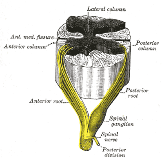
Afferent nerve fibers refer to axonal projections that arrive at a particular brain region, as opposed to efferent projections that exit the region. These terms have a slightly different meaning in the context of the peripheral nervous system (PNS) and central nervous system (CNS).

Reflex arc

A reflex arc is a neural pathway that controls a reflex. In vertebrates, most sensory neurons do not pass directly into the brain, but synapse in the spinal cord. This allows for faster reflex actions to occur by activating spinal motor neurons without the delay of routing signals through the brain. The brain will receive the sensory input while the reflex is being carried out and the analysis of the signal takes place after the reflex action.

A mechanoreceptor, also called mechanoceptor, is a sensory cell that responds to mechanical pressure or distortion. There are four main types of mechanoreceptors in glabrous, or hairless, mammalian skin: lamellar corpuscles, tactile corpuscles, Merkel nerve endings, and bulbous corpuscles. There are also mechanoreceptors in hairy skin, and the hair cells in the receptors of primates like rhesus monkeys and other mammals are similar to those of humans and also studied even in early 20th century anatomically and neurophysiologically.

The withdrawal reflex is a spinal reflex intended to protect the body from damaging stimuli. The reflex rapidly coordinates the contractions of all the flexor muscles and the relaxations of the extensors in that limb causing sudden withdrawal from the potentially damaging stimulus. Spinal reflexes are often monosynaptic and are mediated by a simple reflex arc. A withdrawal reflex is mediated by a polysynaptic reflex resulting in the stimulation of many motor neurons in order to give a quick response.

An adrenergic nerve fibre is a neuron for which the neurotransmitter is either adrenaline (epinephrine), noradrenaline or dopamine. These neurotransmitters are released at a location known as the synapse, which is a junction point between the axon of one nerve cell and the dendrite of another. The neurotransmitters are first released from the axon and then bind to the receptor site on the dendrite. Adrenergic nerve terminals are found in the secondary neurons of the sympathetic nervous system, one of two divisions of the autonomic nervous system which is responsible for the fight-or-flight response. This system increases heart rate, slows digestion, dilates pupils, and also controls the secretion of apocrine sweat glands in the dermal layer of skin, in addition to other responses.

Lateral grey column

The lateral grey column is one of the three grey columns of the spinal cord ; the others being the anterior and posterior grey columns. The lateral grey column is primarily involved with activity in the sympathetic division of the autonomic motor system. It projects to the side as a triangular field in the thoracic and upper lumbar regions of the postero-lateral part of the anterior grey column.

Alpha motor neuron

Alpha (α) motor neurons (also called alpha motoneurons), are large, multipolar lower motor neurons of the brainstem and spinal cord. They innervate extrafusal muscle fibers of skeletal muscle and are directly responsible for initiating their contraction. Alpha motor neurons are distinct from gamma motor neurons, which innervate intrafusal muscle fibers of muscle spindles.

The triceps reflex, a deep tendon reflex, is a reflex as it elicits involuntary contraction of the triceps brachii muscle. It is initiated by the Cervical spinal nerve 7 nerve root. The reflex is tested as part of the neurological examination to assess the sensory and motor pathways within the C7 and C8 spinal nerves.

Cutaneous innervation refers to the area of the skin which is supplied by a specific cutaneous nerve.

Pallesthesia, or vibratory sensation, is the ability to perceive vibration. This sensation, often conducted through skin and bone, is usually generated by mechanoreceptors such as Pacinian corpuscles, Merkel disk receptors, and tactile corpuscles. All of these receptors stimulate an action potential in afferent nerves found in various layers of the skin and body. The afferent neuron travels to the spinal column and then to the brain where the information is processed. Damage to the peripheral nervous system or central nervous system can result in a decline or loss of pallesthesia.

The Golgi tendon reflex (also called inverse stretch reflex, autogenic inhibition, tendon reflex) is an inhibitory effect on the muscle resulting from the muscle tension stimulating Golgi tendon organs (GTO) of the muscle, and hence it is self-induced. The reflex arc is a negative feedback mechanism preventing too much tension on the muscle and tendon. When the tension is extreme, the inhibition can be so great it overcomes the excitatory effects on the muscle's alpha motoneurons causing the muscle to suddenly relax. This reflex is also called the inverse myotatic reflex, because it is the inverse of the stretch reflex.

Cutaneous reflex in human locomotion

Cutaneous, or skin reflexes, are activated by skin receptors and play a valuable role in locomotion, providing quick responses to unexpected environmental challenges. They have been shown to be important in responses to obstacles or stumbling, in preparing for visually challenging terrain, and for assistance in making adjustments when instability is introduced. In addition to the role in normal locomotion, cutaneous reflexes are being studied for their potential in enhancing rehabilitation therapy (physiotherapy) for people with gait abnormalities.

Golgi tendon organ Proprioceptive sensory receptor organ that senses changes in muscle tension

The Golgi tendon organ (GTO) is a proprioceptive sensory receptor organ that senses changes in muscle tension. It lies at the origins and insertion of skeletal muscle fibers into the tendons of skeletal muscle. It provides the sensory component of the Golgi tendon reflex.

References

  1. Langley, J. N. (1900-08-29). "On axon-reflexes in the pre-ganglionic fibres of the sympathetic system". The Journal of Physiology. 25 (5): 364–398. doi:10.1113/jphysiol.1900.sp000803. ISSN   1469-7793. PMC   1516700 . PMID   16992541.
  2. "Applications. Peripheral Autonomic Neuropathy and Axon Reflex. Moor Instruments". Moor Instruments. Retrieved 2014-05-07.
  3. Farlex Partner Medical Dictionary. "Axon Reflex". The Free Dictionary by Farlex. Retrieved 2016-03-31.
  4. 1 2 3 4 Lisney, S. J. W.; Bharali, L. a. M. (1989-04-01). "The Axon Reflex: An Outdated Idea or a Valid Hypothesis?". Physiology. 4 (2): 45–48. doi:10.1152/physiologyonline.1989.4.2.45. ISSN   1548-9213.
  5. 1 2 3 4 5 6 Yaprak, Mevlut (2008). "The axon reflex" (PDF). Neuroanatomy. 7: 17–19. ISSN   1303-1775.
  6. Wårdell, K.; Naver, H. K.; Nilsson, G. E.; Wallin, B. G. (1993). "The cutaneous vascular axon reflex in humans characterized by laser Doppler perfusion imaging". The Journal of Physiology. 460 (2): 185–199. doi:10.1113/jphysiol.1993.sp019466. PMC   1175208 . PMID   8487191.
  7. Kudina, Lydia P.; Andreeva, Regina E. (2015-08-04). "Motor unit firing pattern: evidence for motoneuronal or axonal discharge origin?". Neurological Sciences. 37 (1): 37–43. doi:10.1007/s10072-015-2354-3. ISSN   1590-1874. PMID   26238963.
  8. Kuwabara, Satoshi; Shibuya, Kazumoto; Misawa, Sonoko (2014). "Fasciculations, axonal hyperecitability, and motoneuronal death in amyotrophic lateral sclerosis". Clinical Neurophysiology. 125 (5): 872–873. doi:10.1016/j.clinph.2013.11.014. ISSN   1388-2457. PMID   24345315.
  9. Crnošija, Luka; Adamec, Ivan; Lovrić, Mila; Junaković, Anamari; Skorić, Magdalena Krbot; Lušić, Ivo; Habek, Mario (2016-01-01). "Autonomic dysfunction in clinically isolated syndrome suggestive of multiple sclerosis" (PDF). Clinical Neurophysiology. 127 (1): 864–869. doi:10.1016/j.clinph.2015.06.010. ISSN   1388-2457. PMID   26138149.
  10. 1 2 Illigens, Ben M.W.; Gibbons, Christopher H. (2009-04-01). "Sweat testing to evaluate autonomic function". Clinical Autonomic Research. 19 (2): 79–87. doi:10.1007/s10286-008-0506-8. ISSN   0959-9851. PMC   3046462 . PMID   18989618.
  11. Tuma, Ronald. Microcirculation. Academic Press, 2011, p. 297.