BioBrick

Last updated
Synthetic Biology Open Language (SBOL) standard visual symbols for use with BioBricks Standard Synthetic Biology Open Language (SBOL) standard visual symbols.svg
Synthetic Biology Open Language (SBOL) standard visual symbols for use with BioBricks Standard

BioBrick parts are DNA sequences which conform to a restriction-enzyme assembly standard. [1] [2] These building blocks are used to design and assemble larger synthetic biological circuits from individual parts and combinations of parts with defined functions, which would then be incorporated into living cells such as Escherichia coli cells to construct new biological systems. [3] Examples of BioBrick parts include promoters, ribosomal binding sites (RBS), coding sequences and terminators.

Contents

Overview

Abstraction hierarchy allows the breakdown of complexity. Abstraction hierarchy.jpg
Abstraction hierarchy allows the breakdown of complexity.

The BioBrick parts are used by applying engineering principles of abstraction and modularization. BioBrick parts form the base of the hierarchical system on which synthetic biology is based. There are three levels to the hierarchy:

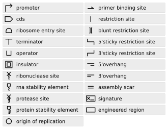
  1. Parts: Pieces of DNA that form a functional unit (for example promoter, RBS, etc.)
  2. Device: Collection set of parts with defined function. In simple terms, a set of complementary BioBrick parts put together forms a device.
  3. System: Combination of a set of devices that performs high-level tasks.

The development of standardized biological parts allows for the rapid assembly of sequences. The ability to test individual parts and devices to be independently tested and characterized also improves the reliability of higher-order systems. [2]

History

The first attempt to create a list of standard biological parts was in 1996, by Rebatchouk et al. This team introduced a cloning strategy for the assembly of short DNA fragments. However, this early attempt was not widely recognised by the scientific research community at the time. [2] [4] In 1999, Arkin and Endy realized that the heterogeneous elements that made up a genetic circuit were lacking standards, so they proposed a list of standard biological parts. [5] BioBricks were described and introduced by Tom Knight at MIT in 2003. [1] Since then, various research groups have utilized the BioBrick standard parts to engineer novel biological devices and systems.

BioBricks Foundation

The BioBricks Foundation was formed in 2006 by engineers and scientists alike as a not-for-profit organization to standardize biological parts across the field. [6] The Foundation focuses on improving in areas of Technology, Law, Education and the Global Community as they apply to synthetic biology. BioBricks Foundation's activities include hosting SBx.0 Conferences, technical and educational programs. The SBx.0 conferences are international conferences on synthetic biology hosted across the world. Technical programs are aimed at the production of a series of standard biological parts, and their education expansion is creating acts which help create open, standardized sources of biological parts. [7]

BioBricks Public Agreement

As an alternative to traditional biotechnology patent systems and in an effort to allow BioBricks to be utilized as an open-source community standard, the BioBricks Foundation created the BioBrick Public Agreement, which consists of a Contributor Agreement and a User Agreement. Those who want to give a part to the community sign the Contributor Agreement, agreeing not to assert against Users Contributor-held intellectual property rights that might limit the use of the contributed materials. Signers of the User Agreement may freely use the whole collection of parts given by contributors. There is no requirement for users to contribute to the community in order to use the parts, and users may assert intellectual property rights to inventions developed by using the parts. [8] The User Agreement allows users to establish invention of uses of parts, to disclose patents on parts combinations, and to freely build on the contributions of other users. [9] [10]

BioBrick Assembly standard

The BioBrick assembly standard was introduced to overcome the lack of standardization posed by traditional molecular cloning methods. The BioBrick assembly standard is a more reliable approach for combining parts to form larger composites. The assembly standard enables two groups of synthetic biologists in different parts of the world to re-use a BioBrick part without going through the whole cycle of design and manipulation. [2] This means the newly designed part can be used by other teams of researchers more easily. Besides that, when compared to the old-fashioned ad hoc cloning method, the assembly standard process is faster and promotes automation. [11] The BioBrick assembly standard 10 was the first assembly standard to be introduced. Over the years, several other assembly standards, such as the Biofusion standard and Freiburg standard have been developed.

BioBrick assembly standard 10

Standard assembly of two BioBrick parts (promoter and coding sequence) by digestion and ligation which forms a "scar" site(M). Standard assembly 10.jpg
Standard assembly of two BioBrick parts (promoter and coding sequence) by digestion and ligation which forms a "scar" site(M).

Assembly standard 10 was developed by Tom Knight, and is the most widely used assembly standard. It involves the use of restriction enzymes. Every BioBrick part is a DNA sequence which is carried by a circular plasmid, which acts as a vector. [12] The vector acts as a transport system to carry the BioBrick parts. The first approach towards a BioBrick standard was the introduction of standard sequences, the prefix and suffix sequences, which flank the 5 and 3 ends of the DNA part respectively. [13] These standard sequences encode specific restriction enzyme sites. The prefix sequence encodes EcoRI (E) and Xbal (X) sites, while the suffix sequence encodes SpeI (S) and PstI (P) sites. The prefix and the suffix are not considered part of the BioBrick part. [3] To facilitate the assembly process, the BioBrick part itself must not contain any of these restriction sites. During the assembly of two different parts, one of the plasmids is digested with EcoRI and SpeI. The plasmid carrying the other BioBrick part is digested with EcoRI and Xbal. This leaves both plasmids with 4 base pair (bp) overhangs at the 5 and 3 ends. The EcoRI sites will ligate since they are complementary to each other. The Xbal and SpeI sites will also ligate as the digestion produces compatible ends. Now, both the DNA parts are in one plasmid. The ligation produces an 8 base pair "scar" site between the two BioBrick parts. Since the scar site is a hybrid of the Xbal and SpeI sites, it is not recognized by either restriction enzyme. [13] The prefix and suffix sequences remain unchanged by this digestion and ligation process, which allows for subsequent assembly steps with more BioBrick parts.

This assembly is an idempotent process: multiple applications do not change the end product, and maintain the prefix and suffix. Although the BioBrick standard assembly allows for the formation of functional modules, there is a limitation to this standard 10 approach. The 8 bp scar site does not allow the creation of a fusion protein. [12] The scar site causes a frame shift which prevents the continuous reading of codons, which is required for the formation of fusion protein.

Tom Knight later developed the BB-2 assembly standard in 2008 to address problems with joining the scars of protein domains and that the scars consist of eight bases, which will yield an altered reading frame when joining protein domains. The enzymes used for digestion of the initial parts are almost the same, but with modified prefixes and suffixes. [14]

BglBricks assembly standard

The BglBrick assembly standard was proposed by J. Christopher Anderson, John E. Dueber, Mariana Leguia, Gabriel C. Wu, Jonathan C. Goler, Adam P. Arkin, and Jay D. Keasling in September 2009 as a standard very similar in concept to BioBrick, but enabling the generation of fusion proteins without altering the reading frame or introducing stop codons and while creating a relatively neutral amino acid linker scar (GlySer). A BglBrick part is as a DNA sequence flanked by 5 EcoRI and BglII sites (GAATTCaaaAGATCT) and 3 BamHI and XhoI sites (GGATCCaaaCTCGAG), and lacking in these same restriction sites internally. The upstream part in the pairwise assembly is purified from an EcoRI/BamHI digest, and the downstream part+vector is purified from an EcoRI/BglII digest. Ligation of these two fragments creates a composite part reforming the original flanking sites required in the part definition and leaving a GGATCT scar sequence at the junction of the parts, a scar that encodes the amino acids glycine and serine when fusing CDS parts together in-frame, convenient due to the GlySer dipeptide being a popular linker of protein domains. [2]

Silver (Biofusion) standard

Biofusion assembly of two BioBrick parts.The schematic diagram shows the 6 base pair scar site made due to the deletion and insertion of nucleotide in the XbaI and SpeI sites. Biofusion standard.jpg
Biofusion assembly of two BioBrick parts.The schematic diagram shows the 6 base pair scar site made due to the deletion and insertion of nucleotide in the XbaI and SpeI sites.

Pam Silver's lab created the Silver assembly standard to overcome the issue surrounding the formation of fusion protein. This assembly standard is also known as Biofusion standard, and is an improvement of the BioBrick assembly standard 10. Silver's standard involves deletion of one nucleotide from the Xbal and SpeI site, which shortens the scar site by 2 nucleotides, which now forms a 6 bp scar sequence. The 6 bp sequence allows the reading frame to be maintained. The scar sequence codes for the amino acid threonine (ACT) and arginine (AGA). [15] This minor improvement allows for the formation of in-frame fusion protein. However, arginine's being a large, charged amino acid is a disadvantage to the Biofusion assembly technique: these properties of arginine result in the destabilisation of the protein by the N-end rule.

Freiburg standard

The 2007 Freiburg iGEM team [16] introduced a new assembly standard to overcome the disadvantages of the existing Biofusion standard technique. The Freiburg team created a new set of prefix and suffix sequences by introducing additional restriction enzyme sites, AgeI and NgoMIV to the existing prefix and suffix respectively. These newly introduced restriction enzyme sites are BioBrick standard compatible. The Freiburg standard still forms a 6 bp scar site, but the scar sequence (ACCGGC) now codes for threonine and glycine respectively. This scar sequence results in a much more stable protein [17] as the glycine forms a stable N-terminal, unlike the arginine, which signals for N-terminal degradation. The assembly technique proposed by the Freiburg team diminishes the limitations of the Biofusion standard.

Assembly method

Different methods are used when it comes to assembling BioBricks. This is because some standards require different materials and methods (use of different restriction enzymes), while others are due to preferences in protocol because some methods of assembly have higher efficiency and is user-friendly.

3 Antibiotic (3A) Assembly

The 3A assembly method is the most commonly used, as its compatible with assembly Standard 10, Silver standard as well as the Freiburg standard. This assembly method involves two BioBrick parts and a destination plasmid. The destination plasmid contains a toxic (lethal) gene, to ease the selection of a correctly assembled plasmid. The destination plasmids also have different antibiotic resistance genes than the plasmids carrying the BioBrick parts. All three plasmids are digested with an appropriate restriction enzyme and then allowed to ligate. Only the correctly assembled part will produce a viable composite part contained in the destination plasmid. This allows a good selection as only the correctly assembled BioBrick parts survive.

Amplified Insert Assembly

The amplified insert assembly method does not depend on prefix and suffix sequences, allowing to be used in combination with a majority of assembly standards. It also has a higher transformation rate than 3A assembly, and it does not require the involved plasmids to have different antibiotic resistance genes. This method reduces noise from uncut plasmids by amplifying a desired insert using PCR prior to digestion and treating the mixture with the restriction enzyme DpnI, which digests methylated DNA like plasmids. Eliminating the template plasmids with DpnI leaves only the insert to be amplified by PCR. To decrease the possibility of creating plasmids with unwanted combinations of insert and backbone, the backbone can be treated with phosphatase to prevent its religation. [14]

Gibson Scarless Assembly

The Gibson scarless assembly method allows the joining of multiple BioBricks simultaneously. This method requires the desired sequences to have an overlap of 20 to 150 bps. Because BioBricks do not have this overlap, this method requires PCR primers to create overhangs between adjacent BioBricks. T5 exonuclease attacks the 5 ends of sequences, creating single-stranded DNA in the ends of all sequences where the different components are designed to anneal. DNA polymerase then adds DNA parts to gaps in the anneal components, and a Taq ligase can seal the final strands. [14]

Methylase-assisted (4R/2M) Assembly

The 4R/2M assembly method was designed to combine parts (BioBrick Assembly Standard 10 or Silver Standard) within existing plasmids (i.e. without PCR or subcloning). The plasmids are reacted in vivo with sequence-specific DNA methyltransferases, so that each is modified and protected from one of two restriction endonucleases that are later used to linearize undesired circular ligation products. [18]

Parts Registry

The MIT group led by Tom Knight that developed BioBricks and International Genetically Engineered Machines (iGEM) competition are also the pioneers of The Registry of Standard Biological Parts (Registry). [19] Registry being one of the foundations of synthetic biology, provides web-based information and data on over 20,000 BioBrick parts. The Registry contains:

Every BioBrick part has its unique identification code which makes the search for the desired BioBrick part easier (for example, BBa_J23100, a constitutive promoter). [2] The registry is open access, whereby anyone can submit a BioBrick part. Most of the BioBrick submission is from students participating in the annual iGEM competition hosted every summer. [20] The Registry allows exchange of data and materials online which allows rapid re-use and modifications of parts by the participating community.

Professional parts registries have also been developed. Since most of the BioBrick parts are submitted by undergraduates as part of the iGEM competition, the parts may lack important characterisation data and metadata which would be essential when it comes to designing and modelling the functional components. [19] One example of a professional parts registry is the USA-based publicly funded facility, The International Open Facility Advancing Biotechnology (BIOFAB), which contains detailed descriptions of each biological part. It is also an open-source registry, and is available commercially. BIOFAB aims to catalogue high-quality BioBrick parts to accommodate the needs of professional synthetic biology community.

The BioBrick Foundation (BBF) is a public-benefit organization established to promote the use of standardized BioBrick parts on a scale beyond the iGEM competition. The BBF is currently working on the derivation of standard framework to promote the production high quality BioBrick parts which would be freely available to everyone. [21]

See also

Related Research Articles

A restriction enzyme, restriction endonuclease, REase, ENase orrestrictase is an enzyme that cleaves DNA into fragments at or near specific recognition sites within molecules known as restriction sites. Restriction enzymes are one class of the broader endonuclease group of enzymes. Restriction enzymes are commonly classified into five types, which differ in their structure and whether they cut their DNA substrate at their recognition site, or if the recognition and cleavage sites are separate from one another. To cut DNA, all restriction enzymes make two incisions, once through each sugar-phosphate backbone of the DNA double helix.

Protein engineering is the process of developing useful or valuable proteins through the design and production of unnatural polypeptides, often by altering amino acid sequences found in nature. It is a young discipline, with much research taking place into the understanding of protein folding and recognition for protein design principles. It has been used to improve the function of many enzymes for industrial catalysis. It is also a product and services market, with an estimated value of $168 billion by 2017.

<span class="mw-page-title-main">Cloning vector</span> Small piece of maintainable DNA

A cloning vector is a small piece of DNA that can be stably maintained in an organism, and into which a foreign DNA fragment can be inserted for cloning purposes. The cloning vector may be DNA taken from a virus, the cell of a higher organism, or it may be the plasmid of a bacterium. The vector contains features that allow for the convenient insertion of a DNA fragment into the vector or its removal from the vector, for example through the presence of restriction sites. The vector and the foreign DNA may be treated with a restriction enzyme that cuts the DNA, and DNA fragments thus generated contain either blunt ends or overhangs known as sticky ends, and vector DNA and foreign DNA with compatible ends can then be joined by molecular ligation. After a DNA fragment has been cloned into a cloning vector, it may be further subcloned into another vector designed for more specific use.

Site-directed mutagenesis is a molecular biology method that is used to make specific and intentional mutating changes to the DNA sequence of a gene and any gene products. Also called site-specific mutagenesis or oligonucleotide-directed mutagenesis, it is used for investigating the structure and biological activity of DNA, RNA, and protein molecules, and for protein engineering.

<span class="mw-page-title-main">Synthetic biology</span> Interdisciplinary branch of biology and engineering

Synthetic biology (SynBio) is a multidisciplinary field of science that focuses on living systems and organisms, and it applies engineering principles to develop new biological parts, devices, and systems or to redesign existing systems found in nature.

A DNA construct is an artificially-designed segment of DNA borne on a vector that can be used to incorporate genetic material into a target tissue or cell. A DNA construct contains a DNA insert, called a transgene, delivered via a transformation vector which allows the insert sequence to be replicated and/or expressed in the target cell. This gene can be cloned from a naturally occurring gene, or synthetically constructed. The vector can be delivered using physical, chemical or viral methods. Typically, the vectors used in DNA constructs contain an origin of replication, a multiple cloning site, and a selectable marker. Certain vectors can carry additional regulatory elements based on the expression system involved.

A restriction digest is a procedure used in molecular biology to prepare DNA for analysis or other processing. It is sometimes termed DNA fragmentation. Hartl and Jones describe it this way:

This enzymatic technique can be used for cleaving DNA molecules at specific sites, ensuring that all DNA fragments that contain a particular sequence at a particular location have the same size; furthermore, each fragment that contains the desired sequence has the sequence located at exactly the same position within the fragment. The cleavage method makes use of an important class of DNA-cleaving enzymes isolated primarily from bacteria. These enzymes are called restriction endonucleases or restriction enzymes, and they are able to cleave DNA molecules at the positions at which particular short sequences of bases are present.

<span class="mw-page-title-main">Multiple cloning site</span>

A multiple cloning site (MCS), also called a polylinker, is a short segment of DNA which contains many restriction sites - a standard feature of engineered plasmids. Restriction sites within an MCS are typically unique, occurring only once within a given plasmid. The purpose of an MCS in a plasmid is to allow a piece of DNA to be inserted into that region.

Synthetic genomics is a nascent field of synthetic biology that uses aspects of genetic modification on pre-existing life forms, or artificial gene synthesis to create new DNA or entire lifeforms.

Biomolecular engineering is the application of engineering principles and practices to the purposeful manipulation of molecules of biological origin. Biomolecular engineers integrate knowledge of biological processes with the core knowledge of chemical engineering in order to focus on molecular level solutions to issues and problems in the life sciences related to the environment, agriculture, energy, industry, food production, biotechnology and medicine.

<span class="mw-page-title-main">Artificial gene synthesis</span> Group of methods in synthetic biology

Artificial gene synthesis, or simply gene synthesis, refers to a group of methods that are used in synthetic biology to construct and assemble genes from nucleotides de novo. Unlike DNA synthesis in living cells, artificial gene synthesis does not require template DNA, allowing virtually any DNA sequence to be synthesized in the laboratory. It comprises two main steps, the first of which is solid-phase DNA synthesis, sometimes known as DNA printing. This produces oligonucleotide fragments that are generally under 200 base pairs. The second step then involves connecting these oligonucleotide fragments using various DNA assembly methods. Because artificial gene synthesis does not require template DNA, it is theoretically possible to make a completely synthetic DNA molecule with no limits on the nucleotide sequence or size.

The Gateway cloning method, invented and commercialized by Invitrogen since the late 1990s, is the cloning method of the integration and excision recombination reactions that take place when bacteriophage lambda infects bacteria. This technology provides a fast and highly efficient way to transport DNA sequences into multi-vector systems for functional analysis and protein expression using Gateway att sites, and two proprietary enzyme mixes called BP Clonase and LR Clonase. In vivo, these recombination reactions are facilitated by the recombination of attachment sites from the lambda/phage chromosome (attP) and the bacteria (attB). As a result of recombination between the attP and attB sites, the phage integrates into the bacterial genome flanked by two new recombination sites. The removal of the phage from the bacterial chromosome and the regeneration of attP and attB sites can both result from the attL and attR sites recombining under specific circumstances.

The Registry of Standard Biological Parts is a collection of genetic parts that are used in the assembly of systems and devices in synthetic biology. The registry was founded in 2003 at the Massachusetts Institute of Technology. The registry, as of 2018, contains over 20,000 parts. Recipients of the genetic parts include academic labs, established scientists, and student teams participating in the iGEM Foundation's annual synthetic biology competition.

Gibson assembly is a molecular cloning method that allows for the joining of multiple DNA fragments in a single, isothermal reaction. It is named after its creator, Daniel G. Gibson, who is the chief technology officer and co-founder of the synthetic biology company, Telesis Bio.

<span class="mw-page-title-main">Molecular cloning</span> Set of methods in molecular biology

Molecular cloning is a set of experimental methods in molecular biology that are used to assemble recombinant DNA molecules and to direct their replication within host organisms. The use of the word cloning refers to the fact that the method involves the replication of one molecule to produce a population of cells with identical DNA molecules. Molecular cloning generally uses DNA sequences from two different organisms: the species that is the source of the DNA to be cloned, and the species that will serve as the living host for replication of the recombinant DNA. Molecular cloning methods are central to many contemporary areas of modern biology and medicine.

<span class="mw-page-title-main">Genetic engineering techniques</span> Methods used to change the DNA of organisms

Genetic engineering techniques allow the modification of animal and plant genomes. Techniques have been devised to insert, delete, and modify DNA at multiple levels, ranging from a specific base pair in a specific gene to entire genes. There are a number of steps that are followed before a genetically modified organism (GMO) is created. Genetic engineers must first choose what gene they wish to insert, modify, or delete. The gene must then be isolated and incorporated, along with other genetic elements, into a suitable vector. This vector is then used to insert the gene into the host genome, creating a transgenic or edited organism.

<span class="mw-page-title-main">Golden Gate Cloning</span>

Golden Gate Cloning or Golden Gate assembly is a molecular cloning method that allows a researcher to simultaneously and directionally assemble multiple DNA fragments into a single piece using Type IIS restriction enzymes and T4 DNA ligase. This assembly is performed in vitro. Most commonly used Type IIS enzymes include BsaI, BsmBI, and BbsI.

<span class="mw-page-title-main">Cassette mutagenesis</span>

Cassette mutagenesis is a type of site-directed mutagenesis that uses a short, double-stranded oligonucleotide sequence to replace a fragment of target DNA. It uses complementary restriction enzyme digest ends on the target DNA and gene cassette to achieve specificity. It is different from methods that use single oligonucleotide in that a single gene cassette can contain multiple mutations. Unlike many site directed mutagenesis methods, cassette mutagenesis also does not involve primer extension by DNA polymerase.

ATUM is an American biotechnology company which provides tools for the life sciences, from design and synthesis of optimized DNA to protein production and GMP cell line development.

No-SCAR genome editing is an editing method that is able to manipulate the Escherichia coli genome. The system relies on recombineering whereby DNA sequences are combined and manipulated through homologous recombination. No-SCAR is able to manipulate the E. coli genome without the use of the chromosomal markers detailed in previous recombineering methods. Instead, the λ-Red recombination system facilitates donor DNA integration while Cas9 cleaves double-stranded DNA to counter-select against wild-type cells. Although λ-Red and Cas9 genome editing are widely used technologies, the no-SCAR method is novel in combining the two functions; this technique is able to establish point mutations, gene deletions, and short sequence insertions in several genomic loci with increased efficiency and time sensitivity.

References

  1. 1 2 Knight, Thomas (2003). "Idempotent Vector Design for Standard Assembly of Biobricks". hdl:1721.1/21168.
  2. 1 2 3 4 5 6 Knight, Thomas F; Reshma P Shetty; Drew Endy (14 April 2008). "Engineering BioBrick vectors from BioBrick parts". Journal of Biological Engineering. 2 (5): 5. doi:10.1186/1754-1611-2-5. ISSN   1754-1611. PMC   2373286 . PMID   18410688.
  3. 1 2 "SynBio Standards -BioBrick" (PDF). Archived from the original (PDF) on 27 March 2014. Retrieved 27 March 2014.
  4. Rebatchouk, Dmitri; Daraselia, N.; Narita, J. O. (1 October 1996). "NOMAD: a versatile strategy for in vitro DNA manipulation applied to promoter analysis and vector design". Proceedings of the National Academy of Sciences. 93 (20): 10891–10896. Bibcode:1996PNAS...9310891R. doi: 10.1073/pnas.93.20.10891 . PMC   38253 . PMID   8855278.
  5. Arkin, Adam. "A Standard Parts List for Biological Circuitry" (PDF). Retrieved 27 March 2014.
  6. "About - BioBricks Foundation". BioBricks Foundation. Archived from the original on 2015-11-13. Retrieved 2015-11-04.
  7. "Programs - BioBricks Foundation". BioBricks Foundation. Archived from the original on 2015-09-17. Retrieved 2015-11-04.
  8. "Frequently Asked Questions | BioBricks Foundation" . Retrieved 2021-10-19.
  9. Mark, Fischer; Lee, Crews; Jennifer, Lynch; Jason, Schultz; David, Grewal; Drew, Endy (2009-10-18). "The BioBrick Public Agreement v1 (draft)". hdl:1721.1/49434.{{cite journal}}: Cite journal requires |journal= (help)
  10. Smolke, Christina D (2009). "Building outside of the box: iGEM and the BioBricks Foundation". Nature Biotechnology. 27 (12): 1099–1102. doi:10.1038/nbt1209-1099. PMID   20010584. S2CID   5486814.
  11. "The BioBrick approach". j5.jbei.org.
  12. 1 2 Sleight, S. C.; Bartley, B. A.; Lieviant, J. A.; Sauro, H. M. (12 April 2010). "In-Fusion BioBrick assembly and re-engineering". Nucleic Acids Research. 38 (8): 2624–2636. doi:10.1093/nar/gkq179. PMC   2860134 . PMID   20385581.
  13. 1 2 Shetty, R.; Lizarazo, M.; Rettberg, R.; Knight, T. F. (2011). Assembly of BioBrick standard biological parts using three antibiotic assembly. Methods in Enzymology. Vol. 498. pp. 311–26. doi:10.1016/B978-0-12-385120-8.00013-9. hdl:1721.1/65066. ISBN   9780123851208. PMID   21601683.
  14. 1 2 3 Røkke, G.; Korvald, E.; Pahr, J.; Oyås, O.; Lale, R. (2014-01-01). Valla, Svein; Lale, Rahmi (eds.). BioBrick Assembly Standards and Techniques and Associated Software Tools. Methods in Molecular Biology. Vol. 1116. Humana Press. pp. 1–24. doi:10.1007/978-1-62703-764-8_1. ISBN   978-1-62703-763-1. PMID   24395353.
  15. Silver, Pamela A.; Ira E. Phillips (April 18, 2006). "A New Biobrick Assembly Strategy Designed for Facile Protein Engineering" (PDF). Harvard Medical School: 1–6.
  16. "Freiburg07/report fusion parts - 2007.igem.org". August 29, 2021. Archived from the original on 2021-08-29.
  17. Muller, Kristian M. "dspace.mit.edu/bitstream/handle/1721.1/45140/BBF_RFC%2025.pdf?sequence=1" (PDF). MIT. Retrieved 27 March 2014.
  18. Matsumura I. 2020. Methylase-assisted subcloning for high throughput BioBrick assembly. PeerJ 8:e9841 https://doi.org/10.7717/peerj.9841
  19. 1 2 Baldwin, Geoff (2012). Synthetic Biology A Primer. London: Imperial College Pr. ISBN   978-1848168633.
  20. "Main Page - ung.igem.org". igem.org. Retrieved 2015-11-10.
  21. "About BioBricks Foundation". Archived from the original on 13 November 2015. Retrieved 27 March 2014.