Related Research Articles

A cataract is a cloudy area in the lens of the eye that leads to a decrease in vision of the eye. Cataracts often develop slowly and can affect one or both eyes. Symptoms may include faded colours, blurry or double vision, halos around light, trouble with bright lights, and difficulty seeing at night. This may result in trouble driving, reading, or recognizing faces. Poor vision caused by cataracts may also result in an increased risk of falling and depression. Cataracts cause 51% of all cases of blindness and 33% of visual impairment worldwide.

Cataract surgery, also called lens replacement surgery, is the removal of the natural lens of the eye that has developed a cataract, an opaque or cloudy area. The eye's natural lens is usually replaced with an artificial intraocular lens (IOL) implant.

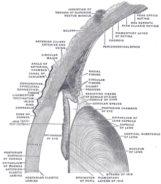
The lens capsule is a component of the globe of the eye. It is a clear elastic basement membrane similar in composition to other basement membranes in the body. The capsule is a very thick basement membrane and the thickness varies in different areas on the lens surface and with the age of the animal. It is composed of various types of fibers such as collagen IV, laminin, etc. and these help it stay under constant tension. The capsule is attached to the surrounding eye by numerous suspensory ligaments and in turn suspends the rest of the lens in an appropriate position. As the lens grows throughout life so must the capsule. Due to the shape of the capsule, the lens naturally tends towards a rounder or more globular configuration, a shape it must assume for the eye to focus at a near distance. Tension on the capsule is varied to allow the lens to subtly change shape to allow the eye to focus in a process called accommodation.

Larsen syndrome (LS) is a congenital disorder discovered in 1950 by Larsen and associates when they observed dislocation of the large joints and face anomalies in six of their patients. Patients with Larsen syndrome normally present with a variety of symptoms, including congenital anterior dislocation of the knees, dislocation of the hips and elbows, flattened facial appearance, prominent foreheads, and depressed nasal bridges. Larsen syndrome can also cause a variety of cardiovascular and orthopedic abnormalities. This rare disorder is caused by a genetic defect in the gene encoding filamin B, a cytoplasmic protein that is important in regulating the structure and activity of the cytoskeleton. The gene that influences the emergence of Larsen syndrome is found in chromosome region, 3p21.1-14.1, a region containing human type VII collagen gene. Larsen syndrome has recently been described as a mesenchyme disorder that affects the connective tissue of an individual. Autosomal dominant and recessive forms of the disorder have been reported, although most cases are autosomal dominant. Reports have found that in Western societies, Larsen syndrome can be found in one in every 100,000 births, but this is most likely an underestimate because the disorder is frequently unrecognized or misdiagnosed.

The posterior chamber is a narrow space behind the peripheral part of the iris, and in front of the suspensory ligament of the lens and the ciliary processes. The posterior chamber consists of small space directly posterior to the iris but anterior to the lens. The posterior chamber is part of the anterior segment and should not be confused with the vitreous chamber.

Gamma-crystallin D is a protein that in humans is encoded by the CRYGD gene.

Eyes absent homolog 1 is a protein that in humans is encoded by the EYA1 gene.

Disintegrin and metalloproteinase domain-containing protein 9 is an enzyme that in humans is encoded by the ADAM9 gene.

Crystallin, gamma C, also known as CRYGC, is a protein which in humans is encoded by the CRYGC gene.

Transcription factor Maf, also known as proto-oncogene c-Maf or V-maf musculoaponeurotic fibrosarcoma oncogene homolog, is a transcription factor that in humans is encoded by the MAF gene.

Gap junction alpha-3 protein is a protein that in humans is encoded by the GJA3 gene.

Beta-crystallin A3 is a protein that in humans is encoded by the CRYBA1 gene.

Gap junction alpha-8 protein is a protein that in humans is encoded by the GJA8 gene. It is also known as connexin 50.

Heat shock factor protein 4 is a protein that in humans is encoded by the HSF4 gene.

Pituitary homeobox 3 is a protein that in humans is encoded by the PITX3 gene.

Persistent fetal vasculature(PFV), also known as persistent fetal vasculature syndrome (PFVS), and until 1997 known primarily as persistent hyperplastic primary vitreous (PHPV), is a rare congenital anomaly which occurs when blood vessels within the developing eye, known as the embryonic hyaloid vasculature network, fail to regress as they normally would in-utero after the eye is fully developed. Defects which arise from this lack of vascular regression are diverse; as a result, the presentation, symptoms, and prognosis of affected patients vary widely, ranging from clinical insignificance to irreversible blindness. The underlying structural causes of PFV are considered to be relatively common, and the vast majority of cases do not warrant additional intervention. When symptoms do manifest, however, they are often significant, causing detrimental and irreversible visual impairment. Persistent fetal vasculature heightens the lifelong risk of glaucoma, cataracts, intraocular hemorrhages, and Retinal detachments, accounting for the visual loss of nearly 5% of the blind community in the developed world. In diagnosed cases of PFV, approximately 90% of patients with a unilateral disease have associated poor vision in the affected eye.

Forkhead box protein E3 (FOXE3) also known as forkhead-related transcription factor 8 (FREAC-8) is a protein that in humans is encoded by the FOXE3 gene located on the short arm of chromosome 1.

Congenital cataracts are a lens opacity that is present at birth. Congenital cataracts occur in a broad range of severity. Some lens opacities do not progress and are visually insignificant, others can produce profound visual impairment.

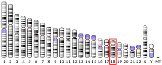
Retinal homeobox protein Rx also known as retina and anterior neural fold homeobox is a protein that in humans is encoded by the RAX gene. The RAX gene is located on chromosome 18 in humans, mice, and rats.

Cataract-microcornea syndrome is a rare genetic syndrome characterized by congenital cataracts and microcornea in the absence of any other systemic anomaly or dysmorphism. Clinical findings include a reduction in corneal diameter in both meridians in an otherwise normal eye, as well as an inherited cataract, that is primarily bilateral posterior polar with opacification within the lens periphery which advances to form a total cataract after visual maturity is achieved. Other ocular manifestations, such as myopia, iris coloboma, sclerocornea, and Peters anomaly, may be observed.
References
- ↑ "Human PubMed Reference:". National Center for Biotechnology Information, U.S. National Library of Medicine.
- ↑ "Entrez Gene: Cataract, anterior polar 2" . Retrieved 2018-10-22.