
The carpal bones are the eight small bones that make up the wrist (carpus) that connects the hand to the forearm. The term "carpus" and "carpal" is derived from the Latin carpus and the Greek καρπός (karpós), meaning "wrist". In human anatomy, the main role of the carpal bones is to articulate with the radial and ulnar heads to form a highly mobile condyloid joint, to provide attachments for thenar and hypothenar muscles, and to form part of the rigid carpal tunnel which allows the median nerve and tendons of the anterior forearm muscles to be transmitted to the hand and fingers.

In human anatomy, the wrist is variously defined as (1) the carpus or carpal bones, the complex of eight bones forming the proximal skeletal segment of the hand; (2) the wrist joint or radiocarpal joint, the joint between the radius and the carpus and; (3) the anatomical region surrounding the carpus including the distal parts of the bones of the forearm and the proximal parts of the metacarpus or five metacarpal bones and the series of joints between these bones, thus referred to as wrist joints. This region also includes the carpal tunnel, the anatomical snuff box, bracelet lines, the flexor retinaculum, and the extensor retinaculum.

In human anatomy, the metacarpal bones or metacarpus, also known as the "palm bones", are the appendicular bones that form the intermediate part of the hand between the phalanges (fingers) and the carpal bones, which articulate with the forearm. The metacarpal bones are homologous to the metatarsal bones in the foot.

The capitate bone is a bone in the human wrist found in the center of the carpal bone region, located at the distal end of the radius and ulna bones. It articulates with the third metacarpal bone and forms the third carpometacarpal joint. The capitate bone is the largest of the carpal bones in the human hand. It presents, above, a rounded portion or head, which is received into the concavity formed by the scaphoid and lunate bones; a constricted portion or neck; and below this, the body. The bone is also found in many other mammals, and is homologous with the "third distal carpal" of reptiles and amphibians.

The hamate bone, or unciform bone, Latin os hamatum and occasionally abbreviated as just hamatum, is a bone in the human wrist readily distinguishable by its wedge shape and a hook-like process ("hamulus") projecting from its palmar surface.

The upper limbs or upper extremities are the forelimbs of an upright-postured tetrapod vertebrate, extending from the scapulae and clavicles down to and including the digits, including all the musculatures and ligaments involved with the shoulder, elbow, wrist and knuckle joints. In humans, each upper limb is divided into the arm, forearm and hand, and is primarily used for climbing, lifting and manipulating objects.

Bone age is the degree of a person's skeletal development. In children, bone age serves as a measure of physiological maturity and aids in the diagnosis of growth abnormalities, endocrine disorders, and other medical conditions. As a person grows from fetal life through childhood, puberty, and finishes growth as a young adult, the bones of the skeleton change in size and shape. These changes can be seen by x-ray and other imaging techniques. A comparison between the appearance of a patient's bones to a standard set of bone images known to be representative of the average bone shape and size for a given age can be used to assign a "bone age" to the patient.
In human anatomy, the extensor pollicis longus muscle (EPL) is a skeletal muscle located dorsally on the forearm. It is much larger than the extensor pollicis brevis, the origin of which it partly covers and acts to stretch the thumb together with this muscle.

The opponens digiti minimi is a muscle in the hand. It is of a triangular form, and placed immediately beneath the palmaris brevis, abductor digiti minimi and flexor digiti minimi brevis. It is one of the three hypothenar muscles that control the little finger.

The carpometacarpal (CMC) joints are five joints in the wrist that articulate the distal row of carpal bones and the proximal bases of the five metacarpal bones.
The intermetacarpal joints are in the hand formed between the metacarpal bones. The bases of the second, third, fourth and fifth metacarpal bones articulate with one another by small surfaces covered with cartilage. The metacarpal bones are connected together by dorsal, palmar, and interosseous ligaments.

The intercarpal joints can be subdivided into three sets of joints : Those of the proximal row of carpal bones, those of the distal row of carpal bones, and those of the two rows with each other.
The midcarpal joint is formed by the scaphoid, lunate, and triquetral bones in the proximal row, and the trapezium, trapezoid, capitate, and hamate bones in the distal row. The distal pole of the scaphoid articulates with two trapezial bones as a gliding type of joint. The proximal end of the scaphoid combines with the lunate and triquetrum to form a deep concavity that articulates with the convexity of the combined capitate and hamate in a form of diarthrodial, almost condyloid joint.
The Palmar carpometacarpal ligaments are a series of bands on the palmar surface of the carpometacarpal joints that connect the carpal bones to the second through fifth metacarpal bones. The second metacarpal is connected to the trapezium. The third metacarpal is connected to the trapezium, to the capitate, and to the hamate. The fourth and fifth metacarpals are connected to the hamate.

The carpometacarpus is a bone found in the hands of birds. It results from the fusion of the carpal and metacarpal bone, and is essentially a single fused bone between the wrist and the knuckles. It is a smallish bone in most birds, generally flattened and with a large hole in the middle. In flightless birds, however, its shape may be slightly different, or it might be absent entirely.

A hand is a prehensile, multi-fingered appendage located at the end of the forearm or forelimb of primates such as humans, chimpanzees, monkeys, and lemurs. A few other vertebrates such as the koala are often described as having "hands" instead of paws on their front limbs. The raccoon is usually described as having "hands" though opposable thumbs are lacking.

The joints in the hand are joints found at the distal end of the upper limb.
The hand is a very complex organ with multiple joints, different types of ligament, tendons and nerves. Hand disease injuries are common in society and can result from excessive use, degenerative disorders or trauma.

The extrinsic extensor muscles of the hand are located in the back of the forearm and have long tendons connecting them to bones in the hand, where they exert their action. Extrinsic denotes their location outside the hand. Extensor denotes their action which is to extend, or open flat, joints in the hand. They include the extensor carpi radialis longus (ECRL), extensor carpi radialis brevis (ECRB), extensor digitorum (ED), extensor digiti minimi (EDM), extensor carpi ulnaris (ECU), abductor pollicis longus (APL), extensor pollicis brevis (EPB), extensor pollicis longus (EPL), and extensor indicis (EI).

Trapeziometacarpal osteoarthritis (TMC OA) is, also known as osteoarthritis at the base of the thumb, thumb carpometacarpal osteoarthritis, basilar (or basal) joint arthritis, or as rhizarthrosis. This joint is formed by the trapezium bone of the wrist and the metacarpal bone of the thumb. This is one of the joints where most humans develop osteoarthritis with age. Osteoarthritis is age-related loss of the smooth surface of the bone where it moves against another bone (cartilage of the joint). In reaction to the loss of cartilage, the bones thicken at the joint surface, resulting in subchondral sclerosis. Also, bony outgrowths, called osteophytes (also known as “bone spurs”), are formed at the joint margins.
 Micro-radiography of 8 weeks human embryo hand
 Second metacarpal bone
 Third metacarpal bone
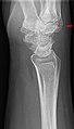 Carpal boss in plain X-Ray.
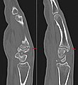 Carpal boss in CT.


