Cellular anastasis

Last updated

Anastasis is a cellular phenomenon characterized by recovery of cells threatened by cell death; it essentially reverses the process of programmed cell death, or apoptosis. Contrary to the prior assumption that apoptosis is irreversible, some cells have been discovered to resist the stimuli that trigger apoptosis. Some of these cells can survive even following the activation of executioner caspases, forming the basis of anastasis. The initial phase of recovery begins when transcription is initiated and the cell recovers from previous stressors. Finally, the cell's cytoskeleton undergoes reorganization, reinforcing its structure and encouraging migration. The relatively recent discovery of anastasis is a key factor in the survival of cancer cells exposed to chemotherapy and changed the way scientists approach the topic of cell death. [1] Anastasis is possible even during advanced stages of cell death, leading researchers to believe that further research on the topic can have therapeutic and pathological implications. Further exploration of the phenomenon could potentially bring forth information including treatment for neurodegenerative diseases and anti-aging therapy.

Contents

History

Apoptosis

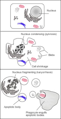
Labelled diagram of a cell undergoing apoptosis. Apoptosis.png
Labelled diagram of a cell undergoing apoptosis.

Apoptosis, or programmed cell death, was discovered in 1842 by Carl Vogt and was initially believed to be irreversible. [2] Once a cell exhibited signs of apoptosis, the cell was doomed. Apoptosis is triggered by external or internal signals, such as developmental cues or cellular damage, which activate cellular pathways leading to apoptosis. Cells at risk of cell death display shrinkage and membrane blebbing. [3] Once the pathway to cellular death is initiated, an enzyme known as caspase, a proteolytic cysteine, is activated. Caspase breaks down proteins and DNA in the cells, preparing the cell for removal by phagocytes. [4] Should apoptosis be restricted or prevented, uncontrolled cell division and tumor growth may occur. [5] Apoptosis has a significant role in maintaining tissue homeostasis by eliminating damaged cells and preventing tumor formation. Cells that are no longer needed or damaged are targeted for apoptosis, aiding in the regulation of normal conditions and body functioning.

Anastasis

Apoptosis was once considered irreversible and unavoidable before the recent discovery of the process of anastasis. It is a rapid process with many initiating factors that were once believed to be permanent. [6] Anastasis, meaning rising to life, was a term coined by siblings Ho Man Tang and Ho Lam Tang following their discovery at the University of Hong Kong in 2007. [7] [8] The Tang siblings executed their experiment by exposing breast cancer cells to various toxic chemicals and waiting for signs of apoptosis. After the cells displayed these characteristics, they then washed the cells with fresh medium and allowed them to incubate. The cancer cells were induced into apoptosis by treating them with ethanol, and the results showed that survival of these cells was possible. [9] Many cells in the original study survived apoptosis and appeared normal once again following the washing of the cells by fresh medium. Tang's results were not initially well received due to the popular opinion at the time that apoptosis was irreversible. However, their research eventually became more accepted and challenged the traditional understanding of apoptosis as an irreversible process. The discovery of anastasis suggested that cells have the potential to reverse the process of cell death under certain circumstances.

Etymology

Alek Rapoport, Anastasis I, 1996 Alek Rapoport - Anastasis-1 - 1996.jpg
Alek Rapoport, Anastasis I, 1996

The word Anastasis comes from the Greek word for resurrection, ανάσταση. [10] The prefix ana- means "upward" or "again", and the root sta- means "to stand", forming a combined meaning of "standing again" or "resurrection". In Christianity, the term anastasis refers to the resurrection of Jesus Christ. The term is used to describe the notion of rising or standing again after a period of death or dormancy. The use of the word anastasis began increasing steadily following the Tang siblings' discovery in 2007. [11]

Process

Anastasis begins in response to stimuli such as DNA damage, chemical stress, and other indicators of approaching cellular death. During early stages of apoptosis, mechanisms allow the cell to evade destruction and halt the process of cell death. Once the apoptotic process is halted, the cell undergoes recovery and repair. The process of transcription resumes, allowing the cell to synthesize vital proteins. Normal cellular morphology and function are restored, and the cell is no longer in danger of cell death. The recovery of cells can limit the damage done to tissue by injury or infection. [12]

Clinical applications

Cancer treatment

HeLa cervical cancer cells HeLa cells stained with antibody to actin (green) , vimentin (red) and DNA (blue).jpg
HeLa cervical cancer cells

Some cancer cells can undergo the process of anastasis after they are exposed to chemotherapy. Anastasis can help cancer cells by enhancing their migration, metastasis, and resistance to chemotherapy. [13] [14] The process of anastasis can be one explanation for the survival of cancer cells after they are treated with cytotoxic drugs; apoptotic cells are able to recover via anastasis following the elimination of such compounds. [15] [16] Similarly to the University of Hong Kong study on breast cancer cells, a study of HeLa cancer cells showed that the cells were able to recover from the presence of caspase and ethanol after being washed with fresh medium. [17] Another study suggested that anastasis in normal cells can even induce carcinomatous results. [18] Due to these effects, tumors can progress and grow in size thanks to anastasis. By understanding this phenomenon, cancer treatments may be improved.

Related Research Articles

<span class="mw-page-title-main">Apoptosis</span> Type of programmed cell death in multicellular organisms

Apoptosis is a form of programmed cell death that occurs in multicellular organisms and in some eukaryotic, single-celled microorganisms such as yeast. Biochemical events lead to characteristic cell changes (morphology) and death. These changes include blebbing, cell shrinkage, nuclear fragmentation, chromatin condensation, DNA fragmentation, and mRNA decay. The average adult human loses 50 to 70 billion cells each day due to apoptosis. For the average human child between 8 and 14 years old, each day the approximate loss is 20 to 30 billion cells.

<span class="mw-page-title-main">Caspase</span> Family of cysteine proteases

Caspases are a family of protease enzymes playing essential roles in programmed cell death. They are named caspases due to their specific cysteine protease activity – a cysteine in its active site nucleophilically attacks and cleaves a target protein only after an aspartic acid residue. As of 2009, there are 12 confirmed caspases in humans and 10 in mice, carrying out a variety of cellular functions.

Programmed cell death is the death of a cell as a result of events inside of a cell, such as apoptosis or autophagy. PCD is carried out in a biological process, which usually confers advantage during an organism's lifecycle. For example, the differentiation of fingers and toes in a developing human embryo occurs because cells between the fingers apoptose; the result is that the digits are separate. PCD serves fundamental functions during both plant and animal tissue development.

<span class="mw-page-title-main">Reactive oxygen species</span> Highly reactive molecules formed from diatomic oxygen (O₂)

In chemistry and biology, reactive oxygen species (ROS) are highly reactive chemicals formed from diatomic oxygen (O2), water, and hydrogen peroxide. Some prominent ROS are hydroperoxide (O2H), superoxide (O2-), hydroxyl radical (OH.), and singlet oxygen. ROS are pervasive because they are readily produced from O2, which is abundant. ROS are important in many ways, both beneficial and otherwise. ROS function as signals, that turn on and off biological functions. They are intermediates in the redox behavior of O2, which is central to fuel cells. ROS are central to the photodegradation of organic pollutants in the atmosphere. Most often however, ROS are discussed in a biological context, ranging from their effects on aging and their role in causing dangerous genetic mutations.

<span class="mw-page-title-main">Cell death</span> Biological cell ceasing to carry out its functions

Cell death is the event of a biological cell ceasing to carry out its functions. This may be the result of the natural process of old cells dying and being replaced by new ones, as in programmed cell death, or may result from factors such as diseases, localized injury, or the death of the organism of which the cells are part. Apoptosis or Type I cell-death, and autophagy or Type II cell-death are both forms of programmed cell death, while necrosis is a non-physiological process that occurs as a result of infection or injury.

<span class="mw-page-title-main">Death effector domain</span> InterPro Domain

The death-effector domain (DED) is a protein interaction domain found only in eukaryotes that regulates a variety of cellular signalling pathways. The DED domain is found in inactive procaspases and proteins that regulate caspase activation in the apoptosis cascade such as FAS-associating death domain-containing protein (FADD). FADD recruits procaspase 8 and procaspase 10 into a death induced signaling complex (DISC). This recruitment is mediated by a homotypic interaction between the procaspase DED and a second DED that is death effector domain in an adaptor protein that is directly associated with activated TNF receptors. Complex formation allows proteolytic activation of procaspase into the active caspase form which results in the initiation of apoptosis. Structurally the DED domain are a subclass of protein motif known as the death fold and contains 6 alpha helices, that closely resemble the structure of the Death domain (DD).

<span class="mw-page-title-main">Death-inducing signaling complex</span>

The death-inducing signaling complex or DISC is a multi-protein complex formed by members of the death receptor family of apoptosis-inducing cellular receptors. A typical example is FasR, which forms the DISC upon trimerization as a result of its ligand (FasL) binding. The DISC is composed of the death receptor, FADD, and caspase 8. It transduces a downstream signal cascade resulting in apoptosis.

<span class="mw-page-title-main">FADD</span> Human protein and coding gene

FAS-associated death domain protein, also called MORT1, is encoded by the FADD gene on the 11q13.3 region of chromosome 11 in humans.

<span class="mw-page-title-main">BH3 interacting-domain death agonist</span> Protein-coding gene in the species Homo sapiens

The BH3 interacting-domain death agonist, or BID, gene is a pro-apoptotic member of the Bcl-2 protein family. Bcl-2 family members share one or more of the four characteristic domains of homology entitled the Bcl-2 homology (BH) domains, and can form hetero- or homodimers. Bcl-2 proteins act as anti- or pro-apoptotic regulators that are involved in a wide variety of cellular activities.

<span class="mw-page-title-main">Caspase-9</span> Enzyme found in humans

Caspase-9 is an enzyme that in humans is encoded by the CASP9 gene. It is an initiator caspase, critical to the apoptotic pathway found in many tissues. Caspase-9 homologs have been identified in all mammals for which they are known to exist, such as Mus musculus and Pan troglodytes.

Pyroptosis is a highly inflammatory form of lytic programmed cell death that occurs most frequently upon infection with intracellular pathogens and is likely to form part of the antimicrobial response. This process promotes the rapid clearance of various bacterial, viral, fungal and protozoan infections by removing intracellular replication niches and enhancing the host's defensive responses. Pyroptosis can take place in immune cells and is also reported to occur in keratinocytes and some epithelial cells.

Inhibitors of apoptosis are a group of proteins that mainly act on the intrinsic pathway that block programmed cell death, which can frequently lead to cancer or other effects for the cell if mutated or improperly regulated. Many of these inhibitors act to block caspases, a family of cysteine proteases that play an integral role in apoptosis. Some of these inhibitors include the Bcl-2 family, viral inhibitor crmA, and IAP's.

<span class="mw-page-title-main">XIAP</span> Protein-coding gene in the species Homo sapiens

X-linked inhibitor of apoptosis protein (XIAP), also known as inhibitor of apoptosis protein 3 (IAP3) and baculoviral IAP repeat-containing protein 4 (BIRC4), is a protein that stops apoptotic cell death. In humans, this protein (XIAP) is produced by a gene named XIAP gene located on the X chromosome.

<span class="mw-page-title-main">Caspase 10</span> Enzyme found in humans

Caspase-10 is an enzyme that, in humans, is encoded by the CASP10 gene.

<span class="mw-page-title-main">ENDOG</span> Protein-coding gene in the species Homo sapiens

Endonuclease G, mitochondrial is an enzyme that in humans is encoded by the ENDOG gene. This protein primarily participates in caspase-independent apoptosis via DNA degradation when translocating from the mitochondrion to nucleus under oxidative stress. As a result, EndoG has been implicated in cancer, aging, and neurodegenerative diseases such as Parkinson's disease (PD). Regulation of its expression levels thus holds potential to treat or ameliorate those conditions.

HAMLET is a complex between alpha-lactalbumin and oleic acid that has been shown in cell culture experiments to induce cell death in tumor cells, but not in healthy cells.

<span class="mw-page-title-main">Necroptosis</span> Programmed form of necrosis, or inflammatory cell death

Necroptosis is a programmed form of necrosis, or inflammatory cell death. Conventionally, necrosis is associated with unprogrammed cell death resulting from cellular damage or infiltration by pathogens, in contrast to orderly, programmed cell death via apoptosis. The discovery of necroptosis showed that cells can execute necrosis in a programmed fashion and that apoptosis is not always the preferred form of cell death. Furthermore, the immunogenic nature of necroptosis favors its participation in certain circumstances, such as aiding in defence against pathogens by the immune system. Necroptosis is well defined as a viral defense mechanism, allowing the cell to undergo "cellular suicide" in a caspase-independent fashion in the presence of viral caspase inhibitors to restrict virus replication. In addition to being a response to disease, necroptosis has also been characterized as a component of inflammatory diseases such as Crohn's disease, pancreatitis, and myocardial infarction.

In cellular biology, dependence receptors are proteins that mediate programmed cell death by monitoring the absence of certain trophic factors that otherwise serve as ligands (interactors) for the dependence receptors. A trophic ligand is a molecule whose protein binding stimulates cell growth, differentiation, and/or survival. Cells depend for their survival on stimulation that is mediated by various receptors and sensors, and integrated via signaling within the cell and between cells. The withdrawal of such trophic support leads to a form of cellular suicide.

cIAP1 is the abbreviation for a human protein, cellular inhibitor of apoptosis protein-1. It belongs to the IAP family of proteins and therefore contains at least one BIR domain. cIAP1 is a multi-functional protein which can be found in the cytoplasm of cells and in the nucleus of tumor cells. Its function in this particular case is yet to be understood. However, it is well known that this protein has a big influence in the growth of diverse cancers. cIAP1 is involved in the development process of osteosarcoma and gastric cancer among others.

<span class="mw-page-title-main">Denise Montell</span> American biochemist and researcher

Denise Johnson Montell is an American biologist who is the Duggan Professor of Molecular, Cellular, and Developmental Biology at the University of California, Santa Barbara. Her research considers the oogenesis process in Drosophila and border cell migration. She has served as president of the Genetics Society of America and was elected to the National Academy of Sciences in 2021.

References

  1. Sun, Gongping; Montell, Denise J. (2017-10-24). "Q&A: Cellular near death experiences—what is anastasis?". BMC Biology. 15 (1): 92. doi: 10.1186/s12915-017-0441-z . ISSN   1741-7007. PMC   5655817 . PMID   29065871.
  2. Peter, Marcus E.; Heufelder, Armin E.; Hengartner, Michael O. (1997-11-25). "Advances in apoptosis research". Proceedings of the National Academy of Sciences. 94 (24): 12736–12737. Bibcode:1997PNAS...9412736P. doi: 10.1073/pnas.94.24.12736 . ISSN   0027-8424. PMC   34166 . PMID   9398063.
  3. Sun, Gongping; Montell, Denise J. (2017-10-24). "Q&A: Cellular near death experiences—what is anastasis?". BMC Biology. 15 (1): 92. doi: 10.1186/s12915-017-0441-z . ISSN   1741-7007. PMC   5655817 . PMID   29065871.
  4. Kroemer, G.; Galluzzi, L.; Vandenabeele, P.; Abrams, J.; Alnemri, E. S.; Baehrecke, E. H.; Blagosklonny, M. V.; El-Deiry, W. S.; Golstein, P.; Green, D. R.; Hengartner, M.; Knight, R. A.; Kumar, S.; Lipton, S. A.; Malorni, W. (January 2009). "Classification of cell death: recommendations of the Nomenclature Committee on Cell Death 2009". Cell Death & Differentiation. 16 (1): 3–11. doi:10.1038/cdd.2008.150. ISSN   1476-5403. PMC   2744427 . PMID   18846107.
  5. "Apoptosis". www.genome.gov. Retrieved 2024-04-12.
  6. Tang, Ho Man; Tang, Ho Lam (October 2018). "Correction to: 'Anastasis: recovery from the brink of cell death'". Royal Society Open Science. 5 (10): 181629. Bibcode:2018RSOS....581629T. doi:10.1098/rsos.181629. ISSN   2054-5703. PMC   6227989 . PMID   30475350.
  7. "Cell Death Processes Are Reversible". The Scientist Magazine®. Retrieved 2024-03-08.
  8. Sun, Gongping; Montell, Denise J. (2017-10-24). "Q&A: Cellular near death experiences—what is anastasis?". BMC Biology. 15 (1): 92. doi: 10.1186/s12915-017-0441-z . ISSN   1741-7007. PMC   5655817 . PMID   29065871.
  9. Mohammed, Rebar N.; Khosravi, Mohsen; Rahman, Heshu Sulaiman; Adili, Ali; Kamali, Navid; Soloshenkov, Pavel Petrovich; Thangavelu, Lakshmi; Saeedi, Hossein; Shomali, Navid; Tamjidifar, Rozita; Isazadeh, Alireza; Aslaminabad, Ramin; Akbari, Morteza (2022-06-03). "Anastasis: cell recovery mechanisms and potential role in cancer". Cell Communication and Signaling. 20 (1): 81. doi: 10.1186/s12964-022-00880-w . ISSN   1478-811X. PMC   9166643 . PMID   35659306.
  10. "Google Translate". translate.google.com. Retrieved 2024-04-02.
  11. "Anastasia | Etymology of the name Anastasia by etymonline". www.etymonline.com. Retrieved 2024-04-02.
  12. Sun, Gongping; Guzman, Elmer; Balasanyan, Varuzhan; Conner, Christopher M.; Wong, Kirsten; Zhou, Hongjun Robin; Kosik, Kenneth S.; Montell, Denise J. (2017-10-02). "A molecular signature for anastasis, recovery from the brink of apoptotic cell death". Journal of Cell Biology. 216 (10): 3355–3368. doi:10.1083/jcb.201706134. ISSN   0021-9525. PMC   5626555 . PMID   28768686.
  13. Wang, Ru; Wang, Yuxing; Liu, Xiaohe; Liu, Menghao; Sun, Lili; Pan, Xiaohua; Hu, Huili; Jiang, Baichun; Zou, Yongxin; Liu, Qiao; Gong, Yaoqin; Wang, Molin; Sun, Gongping (2023-06-30). "Anastasis enhances metastasis and chemoresistance of colorectal cancer cells through upregulating cIAP2/NFκB signaling". Cell Death & Disease. 14 (6): 388. doi:10.1038/s41419-023-05916-8. ISSN   2041-4889. PMC   10313691 . PMID   37391410.
  14. Mohammed, Rebar N.; Khosravi, Mohsen; Rahman, Heshu Sulaiman; Adili, Ali; Kamali, Navid; Soloshenkov, Pavel Petrovich; Thangavelu, Lakshmi; Saeedi, Hossein; Shomali, Navid; Tamjidifar, Rozita; Isazadeh, Alireza; Aslaminabad, Ramin; Akbari, Morteza (December 2022). "Anastasis: cell recovery mechanisms and potential role in cancer". Cell Communication and Signaling. 20 (1): 81. doi: 10.1186/s12964-022-00880-w . ISSN   1478-811X. PMC   9166643 . PMID   35659306.
  15. Zaitceva, Victoria; Kopeina, Gelina S.; Zhivotovsky, Boris (January 2021) [22 July 2021]. "Anastasis: Return Journey from Cell Death". Cancers. 13 (15): 3671. doi: 10.3390/cancers13153671 . ISSN   2072-6694. PMC   8345212 . PMID   34359573.
  16. Mohammed, Rebar N.; Khosravi, Mohsen; Rahman, Heshu Sulaiman; Adili, Ali; Kamali, Navid; Soloshenkov, Pavel Petrovich; Thangavelu, Lakshmi; Saeedi, Hossein; Shomali, Navid; Tamjidifar, Rozita; Isazadeh, Alireza; Aslaminabad, Ramin; Akbari, Morteza (2022-06-16). "Correction: Anastasis: cell recovery mechanisms and potential role in cancer". Cell Communication and Signaling. 20 (1): 91. doi: 10.1186/s12964-022-00914-3 . ISSN   1478-811X. PMC   9202085 . PMID   35710488.
  17. Tang, Ho Man; Tang, Ho Lam (October 2018). "Correction to: 'Anastasis: recovery from the brink of cell death'". Royal Society Open Science. 5 (10): 181629. Bibcode:2018RSOS....581629T. doi:10.1098/rsos.181629. ISSN   2054-5703. PMC   6227989 . PMID   30475350.
  18. Sun, Gongping; Montell, Denise J. (December 2017). "Q&A: Cellular near death experiences—what is anastasis?". BMC Biology. 15 (1): 92. doi: 10.1186/s12915-017-0441-z . ISSN   1741-7007. PMC   5655817 . PMID   29065871.