Related Research Articles

The skull is a bone protective cavity for the brain. The skull is composed of four types of bone i.e., cranial bones, facial bones, ear ossicles and hyoid bone. However two parts are more prominent: the cranium and the mandible. In humans, these two parts are the neurocranium and the viscerocranium that includes the mandible as its largest bone. The skull forms the anterior-most portion of the skeleton and is a product of cephalisation—housing the brain, and several sensory structures such as the eyes, ears, nose, and mouth. In humans these sensory structures are part of the facial skeleton.

Snoring is the vibration of respiratory structures and the resulting sound due to obstructed air movement during breathing while sleeping. The sound may be soft or loud and unpleasant. Snoring during sleep may be a sign, or first alarm, of obstructive sleep apnea (OSA). Research suggests that snoring is one of the factors of sleep deprivation.

A dental implant is a prosthesis that interfaces with the bone of the jaw or skull to support a dental prosthesis such as a crown, bridge, denture, or facial prosthesis or to act as an orthodontic anchor. The basis for modern dental implants is a biological process called osseointegration, in which materials such as titanium or zirconia form an intimate bond to the bone. The implant fixture is first placed so that it is likely to osseointegrate, then a dental prosthetic is added. A variable amount of healing time is required for osseointegration before either the dental prosthetic is attached to the implant or an abutment is placed which will hold a dental prosthetic/crown.

Treacher Collins syndrome (TCS) is a genetic disorder characterized by deformities of the ears, eyes, cheekbones, and chin. The degree to which a person is affected, however, may vary from mild to severe. Complications may include breathing problems, problems seeing, cleft palate, and hearing loss. Those affected generally have normal intelligence.

Macroglossia is the medical term for an unusually large tongue. Severe enlargement of the tongue can cause cosmetic and functional difficulties in speaking, eating, swallowing and sleeping. Macroglossia is uncommon, and usually occurs in children. There are many causes. Treatment depends upon the exact cause.

Prognathism, also called Habsburg jaw or Habsburgs' jaw primarily in the context of its prevalence amongst members of the House of Habsburg, is a positional relationship of the mandible or maxilla to the skeletal base where either of the jaws protrudes beyond a predetermined imaginary line in the coronal plane of the skull. In general dentistry, oral and maxillofacial surgery, and orthodontics, this is assessed clinically or radiographically (cephalometrics). The word prognathism derives from Greek πρό and γνάθος. One or more types of prognathism can result in the common condition of malocclusion, in which an individual's top teeth and lower teeth do not align properly.

In orthodontics, a malocclusion is a misalignment or incorrect relation between the teeth of the upper and lower dental arches when they approach each other as the jaws close. The English-language term dates from 1864; Edward Angle (1855-1930), the "father of modern orthodontics", popularised it. The word "malocclusion" derives from occlusion, and refers to the manner in which opposing teeth meet.

A bone-anchored hearing aid (BAHA) is a type of hearing aid based on bone conduction. It is primarily suited for people who have conductive hearing losses, unilateral hearing loss, single-sided deafness and people with mixed hearing losses who cannot otherwise wear 'in the ear' or 'behind the ear' hearing aids. They are more expensive than conventional hearing aids, and their placement involves invasive surgery which carries a risk of complications, although when complications do occur, they are usually minor.
Oral myology is the field of study that involves the evaluation and treatment of the oral and facial musculature, including the muscles of the tongue, lips, cheeks, and jaw.

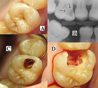
Pericoronitis is inflammation of the soft tissues surrounding the crown of a partially erupted tooth, including the gingiva (gums) and the dental follicle. The soft tissue covering a partially erupted tooth is known as an operculum, an area which can be difficult to access with normal oral hygiene methods. The hyponym operculitis technically refers to inflammation of the operculum alone.

Gorham's disease, also known as Gorham vanishing bone disease and phantom bone disease, is a very rare skeletal condition of unknown cause, characterized by the uncontrolled proliferation of distended, thin-walled vascular or lymphatic channels within bone, which leads to resorption and replacement of bone with angiomas and/or fibrosis.
Dental radiographs, commonly known as X-rays, are radiographs used to diagnose hidden dental structures, malignant or benign masses, bone loss, and cavities.
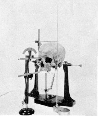
Cephalometry is the study and measurement of the head, usually the human head, especially by medical imaging such as radiography. Craniometry, the measurement of the cranium (skull), is a large subset of cephalometry. Cephalometry also has a history in phrenology, which is the study of personality and character as well as physiognomy, which is the study of facial features. Cephalometry as applied in a comparative anatomy context informs biological anthropology. In clinical contexts such as dentistry and oral and maxillofacial surgery, cephalometric analysis helps in treatment and research; cephalometric landmarks guide surgeons in planning and operating.

Facial trauma, also called maxillofacial trauma, is any physical trauma to the face. Facial trauma can involve soft tissue injuries such as burns, lacerations and bruises, or fractures of the facial bones such as nasal fractures and fractures of the jaw, as well as trauma such as eye injuries. Symptoms are specific to the type of injury; for example, fractures may involve pain, swelling, loss of function, or changes in the shape of facial structures.
Cephalometric analysis is the clinical application of cephalometry. It is analysis of the dental and skeletal relationships of a human skull. It is frequently used by dentists, orthodontists, and oral and maxillofacial surgeons as a treatment planning tool. Two of the more popular methods of analysis used in orthodontology are the Steiner analysis and the Downs analysis. There are other methods as well which are listed below.
A jaw abnormality is a disorder in the formation, shape and/or size of the jaw. In general abnormalities arise within the jaw when there is a disturbance or fault in the fusion of the mandibular processes. The mandible in particular has the most differential typical growth anomalies than any other bone in the human skeleton. This is due to variants in the complex symmetrical growth pattern which formulates the mandible.
Orofacial Myofunctional Disorders (OMD) are muscle disorders of the face, mouth, lips, or jaw due to chronic mouth breathing.
A cyst is a pathological epithelial lined cavity that fills with fluid or soft material and usually grows from internal pressure generated by fluid being drawn into the cavity from osmosis. The bones of the jaws, the mandible and maxilla, are the bones with the highest prevalence of cysts in the human body. This is due to the abundant amount of epithelial remnants that can be left in the bones of the jaws. The enamel of teeth is formed from ectoderm, and so remnants of epithelium can be left in the bone during odontogenesis. The bones of the jaws develop from embryologic processes which fuse, and ectodermal tissue may be trapped along the lines of this fusion. This "resting" epithelium is usually dormant or undergoes atrophy, but, when stimulated, may form a cyst. The reasons why resting epithelium may proliferate and undergo cystic transformation are generally unknown, but inflammation is thought to be a major factor. The high prevalence of tooth impactions and dental infections that occur in the bones of the jaws is also significant to explain why cysts are more common at these sites.

Most bony fishes have two sets of jaws made mainly of bone. The primary oral jaws open and close the mouth, and a second set of pharyngeal jaws are positioned at the back of the throat. The oral jaws are used to capture and manipulate prey by biting and crushing. The pharyngeal jaws, so-called because they are positioned within the pharynx, are used to further process the food and move it from the mouth to the stomach.
Human jaw shrinkage is the phenomenon of continued size reduction of the human mandible and maxilla over the past 12,000 to 15,000 years. Modern human lifestyles and diets are vastly different now from what they were for most of human evolutionary history. Human jaws, as well as oral cavities, have been shrinking ever since the Neolithic agricultural revolution. This has been confirmed by bone remains dated to this time period. Researchers are able to infer the basic lifestyle practices of past cultures, enabling them to link jaw size with lifestyle practice/behaviors. Bones from burial sites of past hunter-gatherer societies are associated with larger jaws and mouths, while bones retrieved from former farming cultures have decreased jaw size. Bones from farming societies also indicate the presence of dental malocclusions, commonly known as non-straight teeth. Within recent centuries, as food has become more processed and soft in form, a rapid increase in non-straight teeth, smaller jaws, and mouths; a lack of space for wisdom teeth; and associated health conditions have been observed. Such conditions include sleep apnea, constricted airways, and decreased respiratory fitness. Medical professionals have been making similar observations and documenting them for hundreds of years. Changes in diet, lifestyle, and breathing patterns have led to maladaptive phenotypic expression in terms of morphological craniofacial development that starts in childhood but persists throughout the lifespan.
References
- ↑ Swennen, G.R.J.; Schutyser, F.A.C.; Hausamen, J.E. (2005). Three-Dimensional Cephalometry: A Color Atlas and Manual. Springer. p. 10. ISBN 978-3-540-25440-9.