
In biology, histones are highly basic proteins abundant in lysine and arginine residues that are found in eukaryotic cell nuclei. They act as spools around which DNA winds to create structural units called nucleosomes. Nucleosomes in turn are wrapped into 30-nanometer fibers that form tightly packed chromatin. Histones prevent DNA from becoming tangled and protect it from DNA damage. In addition, histones play important roles in gene regulation and DNA replication. Without histones, unwound DNA in chromosomes would be very long. For example, each human cell has about 1.8 meters of DNA if completely stretched out, however when wound about histones, this length is reduced to about 90 micrometers (0.09 mm) of 30 nm diameter chromatin fibers.

Histone acetyltransferases (HATs) are enzymes that acetylate conserved lysine amino acids on histone proteins by transferring an acetyl group from acetyl-CoA to form ε-N-acetyllysine. DNA is wrapped around histones, and, by transferring an acetyl group to the histones, genes can be turned on and off. In general, histone acetylation increases gene expression.
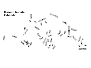
Constitutive heterochromatin domains are regions of DNA found throughout the chromosomes of eukaryotes. The majority of constitutive heterochromatin is found at the pericentromeric regions of chromosomes, but is also found at the telomeres and throughout the chromosomes. In humans there is significantly more constitutive heterochromatin found on chromosomes 1, 9, 16, 19 and Y. Constitutive heterochromatin is composed mainly of high copy number tandem repeats known as satellite repeats, minisatellite and microsatellite repeats, and transposon repeats. In humans these regions account for about 200Mb or 6.5% of the total human genome, but their repeat composition makes them difficult to sequence, so only small regions have been sequenced.

A chromomere, also known as an idiomere, is one of the serially aligned beads or granules of a eukaryotic chromosome, resulting from local coiling of a continuous DNA thread. Chromeres are regions of chromatin that have been compacted through localized contraction. In areas of chromatin with the absence of transcription, condensing of DNA and protein complexes will result in the formation of chromomeres. It is visible on a chromosome during the prophase of meiosis and mitosis. Giant banded (Polytene) chromosomes resulting from the replication of the chromosomes and the synapsis of homologs without cell division is a process called endomitosis. These chromosomes consist of more than 1000 copies of the same chromatid that are aligned and produce alternating dark and light bands when stained. The dark bands are the chromomere.
The family of heterochromatin protein 1 (HP1) consists of highly conserved proteins, which have important functions in the cell nucleus. These functions include gene repression by heterochromatin formation, transcriptional activation, regulation of binding of cohesion complexes to centromeres, sequestration of genes to nuclear periphery, transcriptional arrest, maintenance of heterochromatin integrity, gene repression at the single nucleosome level, gene repression by heterochromatization of euchromatin and DNA repair. HP1 proteins are fundamental units of heterochromatin packaging that are enriched at the centromeres and telomeres of nearly all Eukaryotic chromosomes with the notable exception of budding yeast, in which a yeast-specific silencing complex of SIR proteins serve a similar function. Members of the HP1 family are characterized by an N-terminal chromodomain and a C-terminal chromoshadow domain, separated by a Hinge region. HP1 is also found at euchromatic sites, where its binding correlates with gene repression. HP1 was originally discovered by Tharappel C James and Sarah Elgin in 1986 as a factor in the phenomenon known as position effect variegation in Drosophila melanogaster.

A chromodomain is a protein structural domain of about 40–50 amino acid residues commonly found in proteins associated with the remodeling and manipulation of chromatin. The domain is highly conserved among both plants and animals, and is represented in a large number of different proteins in many genomes, such as that of the mouse. Some chromodomain-containing genes have multiple alternative splicing isoforms that omit the chromodomain entirely. In mammals, chromodomain-containing proteins are responsible for aspects of gene regulation related to chromatin remodeling and formation of heterochromatin regions. Chromodomain-containing proteins also bind methylated histones and appear in the RNA-induced transcriptional silencing complex. In histone modifications, chromodomains are very conserved. They function by identifying and binding to methylated lysine residues that exist on the surface of chromatin proteins and thereby regulate gene transcription.
RNA-induced transcriptional silencing (RITS) is a form of RNA interference by which short RNA molecules – such as small interfering RNA (siRNA) – trigger the downregulation of transcription of a particular gene or genomic region. This is usually accomplished by posttranslational modification of histone tails which target the genomic region for heterochromatin formation. The protein complex that binds to siRNAs and interacts with the methylated lysine 9 residue of histones H3 is the RITS complex.

Chromobox protein homolog 1 is a protein that in humans is encoded by the CBX1 gene.

Chromobox protein homolog 3 is a protein that is encoded by the CBX3 gene in humans.

High-mobility group protein HMG-I/HMG-Y is a protein that in humans is encoded by the HMGA1 gene.

Tripartite motif-containing 28 (TRIM28), also known as transcriptional intermediary factor 1β (TIF1β) and KAP1, is a protein that in humans is encoded by the TRIM28 gene.

Chromodomain-helicase-DNA-binding protein 3 is an enzyme that in humans is encoded by the CHD3 gene.

Tripartite motif-containing 24 (TRIM24) also known as transcriptional intermediary factor 1α (TIF1α) is a protein that, in humans, is encoded by the TRIM24 gene.

The Chromodomain-Helicase DNA-binding 1 is a protein that, in humans, is encoded by the CHD1 gene. CHD1 is a chromatin remodeling protein that is widely conserved across many eukaryotic organisms, from yeast to humans. CHD1 is named for three of its protein domains: two tandem chromodomains, its ATPase catalytic domain, and its DNA-binding domain.

Chromobox protein homolog 5 is a protein that in humans is encoded by the CBX5 gene. It is a highly conserved, non-histone protein part of the heterochromatin family. The protein itself is more commonly called HP1α. Heterochromatin protein-1 (HP1) has an N-terminal domain that acts on methylated lysines residues leading to epigenetic repression. The C-terminal of this protein has a chromo shadow-domain (CSD) that is responsible for homodimerizing, as well as interacting with a variety of chromatin-associated, non-histone proteins.
In molecular biology, the BEN domain is a protein domain which is found in diverse proteins including:

Nucleic acidquaternary structure refers to the interactions between separate nucleic acid molecules, or between nucleic acid molecules and proteins. The concept is analogous to protein quaternary structure, but as the analogy is not perfect, the term is used to refer to a number of different concepts in nucleic acids and is less commonly encountered. Similarly other biomolecules such as proteins, nucleic acids have four levels of structural arrangement: primary, secondary, tertiary, and quaternary structure. Primary structure is the linear sequence of nucleotides, secondary structure involves small local folding motifs, and tertiary structure is the 3D folded shape of nucleic acid molecule. In general, quaternary structure refers to 3D interactions between multiple subunits. In the case of nucleic acids, quaternary structure refers to interactions between multiple nucleic acid molecules or between nucleic acids and proteins. Nucleic acid quaternary structure is important for understanding DNA, RNA, and gene expression because quaternary structure can impact function. For example, when DNA is packed into chromatin, therefore exhibiting a type of quaternary structure, gene transcription will be inhibited.
M33 is a gene. It is a mammalian homologue of Drosophila Polycomb. It localises to euchromatin within interphase nuclei, but it is enriched within the centromeric heterochromatin of metaphase chromosomes. In mice, the official symbol of M33 gene styled Cbx2 and the official name chromobox 2 are maintained by the MGI. Also known as pc; MOD2. In human ortholog CBX2, synonyms CDCA6, M33,SRXY5 from orthology source HGNC. M33 was isolated by means of the structural similarity of its chromodomain. It contains a region of homology shared by Xenopus and Drosophila in the fifth exon. Polycomb genes in Drosophila mediate changes in higher-order chromatin structure to maintain the repressed state of developmentally regulated genes. M33 deficiency interferes with steps upstream of the Y-chromosome-specific SRY gene may cause sex reversal. It may also involved in the campomelic syndrome and neoplastic disorders linked to allele loss in this region. Disruption of the murine M33 gene, displayed posterior transformation of the sternal ribs and vertebral columns.
Protein methylation is a type of post-translational modification featuring the addition of methyl groups to proteins. It can occur on the nitrogen-containing side-chains of arginine and lysine, but also at the amino- and carboxy-termini of a number of different proteins. In biology, methyltransferases catalyze the methylation process, activated primarily by S-adenosylmethionine. Protein methylation has been most studied in histones, where the transfer of methyl groups from S-adenosyl methionine is catalyzed by histone methyltransferases. Histones that are methylated on certain residues can act epigenetically to repress or activate gene expression.

Thomas Jenuwein is a German scientist working in the fields of epigenetics, chromatin biology, gene regulation and genome function.
