D, or d, is the fourth letter of the Latin alphabet, used in the modern English alphabet, the alphabets of other western European languages and others worldwide. Its name in English is dee, plural dees.

In humans, the kidneys are two reddish-brown bean-shaped blood-filtering organs that are a multilobar, multipapillary form of mammalian kidneys, usually without signs of external lobulation. They are located on the left and right in the retroperitoneal space, and in adult humans are about 12 centimetres in length. They receive blood from the paired renal arteries; blood exits into the paired renal veins. Each kidney is attached to a ureter, a tube that carries excreted urine to the bladder.

Kidney stone disease, also known as renal calculus disease, nephrolithiasis or urolithiasis, is a crystallopathy where a solid piece of material develops in the urinary tract. Renal calculi typically form in the kidney and leave the body in the urine stream. A small calculus may pass without causing symptoms. If a stone grows to more than 5 millimeters, it can cause blockage of the ureter, resulting in sharp and severe pain in the lower back that often radiates downward to the groin. A calculus may also result in blood in the urine, vomiting, or painful urination. About half of people who have had a renal calculus are likely to have another within ten years.

In vertebrate anatomy, ribs are the long curved bones which form the rib cage, part of the axial skeleton. In most tetrapods, ribs surround the thoracic cavity, enabling the lungs to expand and thus facilitate breathing by expanding the thoracic cavity. They serve to protect the lungs, heart, and other vital organs of the thorax. In some animals, especially snakes, ribs may provide support and protection for the entire body.

The rib cage or thoracic cage is an endoskeletal enclosure in the thorax of most vertebrates that comprises the ribs, vertebral column and sternum, which protect the vital organs of the thoracic cavity, such as the heart, lungs and great vessels and support the shoulder girdle to form the core part of the axial skeleton.

Hematuria or haematuria is defined as the presence of blood or red blood cells in the urine. "Gross hematuria" occurs when urine appears red, brown, or tea-colored due to the presence of blood. Hematuria may also be subtle and only detectable with a microscope or laboratory test. Blood that enters and mixes with the urine can come from any location within the urinary system, including the kidney, ureter, urinary bladder, urethra, and in men, the prostate. Common causes of hematuria include urinary tract infection (UTI), kidney stones, viral illness, trauma, bladder cancer, and exercise. These causes are grouped into glomerular and non-glomerular causes, depending on the involvement of the glomerulus of the kidney. But not all red urine is hematuria. Other substances such as certain medications and foods can cause urine to appear red. Menstruation in women may also cause the appearance of hematuria and may result in a positive urine dipstick test for hematuria. A urine dipstick test may also give an incorrect positive result for hematuria if there are other substances in the urine such as myoglobin, a protein excreted into urine during rhabdomyolysis. A positive urine dipstick test should be confirmed with microscopy, where hematuria is defined by three or more red blood cells per high power field. When hematuria is detected, a thorough history and physical examination with appropriate further evaluation can help determine the underlying cause.

Rheumatism or rheumatic disorders are conditions causing chronic, often intermittent pain affecting the joints or connective tissue. Rheumatism does not designate any specific disorder, but covers at least 200 different conditions, including arthritis and "non-articular rheumatism", also known as "regional pain syndrome" or "soft tissue rheumatism". There is a close overlap between the term soft tissue disorder and rheumatism. Sometimes the term "soft tissue rheumatic disorders" is used to describe these conditions.

Abdominal pain, also known as a stomach ache, is a symptom associated with both non-serious and serious medical issues. Since the abdomen contains most of the body's vital organs, it can be an indicator of a wide variety of diseases. Given that, approaching the examination of a person and planning of a differential diagnosis is extremely important.

Phenylbutazone, often referred to as "bute", is a nonsteroidal anti-inflammatory drug (NSAID) for the short-term treatment of pain and fever in animals.

Cystinuria is an inherited autosomal recessive disease characterized by high concentrations of the amino acid cystine in the urine, leading to the formation of cystine stones in the kidneys, ureters, and bladder. It is a type of aminoaciduria. "Cystine", not "cysteine," is implicated in this disease; the former is a dimer of the latter.

Pyelonephritis is inflammation of the kidney, typically due to a bacterial infection. Symptoms most often include fever and flank tenderness. Other symptoms may include nausea, burning with urination, and frequent urination. Complications may include pus around the kidney, sepsis, or kidney failure.
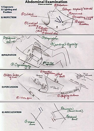
An abdominal examination is a portion of the physical examination which a physician or nurse uses to clinically observe the abdomen of a patient for signs of disease. The abdominal examination is conventionally split into four different stages: first, inspection of the patient and the visible characteristics of their abdomen. Auscultation (listening) of the abdomen with a stethoscope. Palpation of the patient's abdomen. Finally, percussion (tapping) of the patient's abdomen and abdominal organs. Depending on the need to test for specific diseases such as ascites, special tests may be performed as a part of the physical examination. An abdominal examination may be performed because the physician suspects a disease of the organs inside the abdominal cavity (including the liver, spleen, large or small intestines), or simply as a part of a complete physical examination for other conditions. In a complete physical examination, the abdominal exam classically follows the respiratory examination and cardiovascular examination.

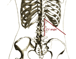
Costovertebral angle (CVA) tenderness is pain that results from touching the region inside of the costovertebral angle. The CVA is formed by the 12th rib and the spine. Assessing for CVA tenderness is part of the abdominal exam, and CVA tenderness often indicates kidney pathology.

Nail–patella syndrome is a genetic disorder that results in small, poorly developed nails and kneecaps, but can also affect many other areas of the body, such as the elbows, chest, and hips. The name "nail–patella" can be very misleading because the syndrome often affects many other areas of the body, including even the production of certain proteins. The severity of these effects varies depending on the individual. It is also referred to as iliac horn syndrome, hereditary onychoosteodysplasia, Fong disease or Turner–Kieser syndrome.
Costovertebral may refer to:

Abdominal ultrasonography is a form of medical ultrasonography to visualise abdominal anatomical structures. It uses transmission and reflection of ultrasound waves to visualise internal organs through the abdominal wall. For this reason, the procedure is also called a transabdominal ultrasound, in contrast to endoscopic ultrasound, the latter combining ultrasound with endoscopy through visualize internal structures from within hollow organs.

Loin pain hematuria syndrome (LPHS) is the combination of debilitating unilateral or bilateral flank pain and microscopic or macroscopic amounts of blood in the urine that is otherwise unexplained.
Lloyd's sign indicates the presence of renal calculus or pyelonephritis when pain is elicited by deep percussion in the back between the 12th rib and the spine. It is closely related to costovertebral angle tenderness, as the area of percussion is the same. However, Lloyd's sign is specifically defined as positive costovertebral angle tenderness along with the absence of tenderness when normal pressure is applied.

The human abdomen is divided into quadrants and regions by anatomists and physicians for the purposes of study, diagnosis, and treatment. The division into four quadrants allows the localisation of pain and tenderness, scars, lumps, and other items of interest, narrowing in on which organs and tissues may be involved. The quadrants are referred to as the left lower quadrant, left upper quadrant, right upper quadrant and right lower quadrant. These terms are not used in comparative anatomy, since most other animals do not stand erect.
Mathe's sign is an ultrasonographic sign that suggests the existence of an abscess in the proximity of a kidney. Normally during inspiration when standing, the kidneys move distally to some extent. When one of them does not move at all or moves downwards scarcely, either in erect position or deep inspiration, the test is called positive. Along with fever, urinary symptoms, and costovertebral angle tenderness, this will almost always indicate a perinephric abscess.