Related Research Articles

Color or colour is the visual perception based on the electromagnetic spectrum. Though color is not an inherent property of matter, color perception is related to an object's light absorption, reflection, emission spectra and interference. For most humans, colors are perceived in the visible light spectrum with three types of cone cells (trichromacy). Other animals may have a different number of cone cell types or have eyes sensitive to different wavelength, such as bees that can distinguish ultraviolet, and thus have a different color sensitivity range. Animal perception of color originates from different light wavelength or spectral sensitivity in cone cell types, which is then processed by the brain.

Eyes are organs of the visual system. They provide living organisms with vision, the ability to receive and process visual detail, as well as enabling several photo response functions that are independent of vision. Eyes detect light and convert it into electro-chemical impulses in neurons (neurones). In higher organisms, the eye is a complex optical system which collects light from the surrounding environment, regulates its intensity through a diaphragm, focuses it through an adjustable assembly of lenses to form an image, converts this image into a set of electrical signals, and transmits these signals to the brain through complex neural pathways that connect the eye via the optic nerve to the visual cortex and other areas of the brain. Eyes with resolving power have come in ten fundamentally different forms, and 96% of animal species possess a complex optical system. Image-resolving eyes are present in molluscs, chordates and arthropods.

Night vision is the ability to see in low-light conditions, either naturally with scotopic vision or through a night-vision device. Night vision requires both sufficient spectral range and sufficient intensity range. Humans have poor night vision compared to many animals such as cats, foxes and rabbits, in part because the human eye lacks a tapetum lucidum, tissue behind the retina that reflects light back through the retina thus increasing the light available to the photoreceptors.

Color vision, a feature of visual perception, is an ability to perceive differences between light composed of different frequencies independently of light intensity. Color perception is a part of the larger visual system and is mediated by a complex process between neurons that begins with differential stimulation of different types of photoreceptors by light entering the eye. Those photoreceptors then emit outputs that are propagated through many layers of neurons and then ultimately to the brain. Color vision is found in many animals and is mediated by similar underlying mechanisms with common types of biological molecules and a complex history of evolution in different animal taxa. In primates, color vision may have evolved under selective pressure for a variety of visual tasks including the foraging for nutritious young leaves, ripe fruit, and flowers, as well as detecting predator camouflage and emotional states in other primates.
Chromatic adaptation is the human visual system’s ability to adjust to changes in illumination in order to preserve the appearance of object colors. It is responsible for the stable appearance of object colors despite the wide variation of light which might be reflected from an object and observed by our eyes. A chromatic adaptation transform (CAT) function emulates this important aspect of color perception in color appearance models.
The flicker fusion threshold, also known as critical flicker frequency or flicker fusion rate, is the frequency at which a flickering light appears steady to the average human observer. It is concept studied in vision science, more specifically in the psychophysics of visual perception. A traditional term for "flicker fusion" is "persistence of vision", but this has also been used to describe positive afterimages or motion blur. Although flicker can be detected for many waveforms representing time-variant fluctuations of intensity, it is conventionally, and most easily, studied in terms of sinusoidal modulation of intensity.

An afterimage is an image that continues to appear in the eyes after a period of exposure to the original image. An afterimage may be a normal phenomenon or may be pathological (palinopsia). Illusory palinopsia may be a pathological exaggeration of physiological afterimages. Afterimages occur because photochemical activity in the retina continues even when the eyes are no longer experiencing the original stimulus.
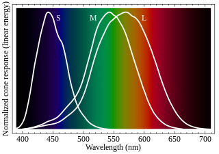
Cone cells, or cones, are photoreceptor cells in the retinas of vertebrates' eyes, including the human eye. They respond differently to light of different wavelengths, and the combination of their responses is responsible for color vision. Cones function best in relatively bright light, called the photopic region, as opposed to rod cells, which work better in dim light, or the scotopic region. Cone cells are densely packed in the fovea centralis, a 0.3 mm diameter rod-free area with very thin, densely packed cones which quickly reduce in number towards the periphery of the retina. Conversely, they are absent from the optic disc, contributing to the blind spot. There are about six to seven million cones in a human eye, with the highest concentration being towards the macula.
In visual physiology, adaptation is the ability of the retina of the eye to adjust to various levels of light. Natural night vision, or scotopic vision, is the ability to see under low-light conditions. In humans, rod cells are exclusively responsible for night vision as cone cells are only able to function at higher illumination levels. Night vision is of lower quality than day vision because it is limited in resolution and colors cannot be discerned; only shades of gray are seen. In order for humans to transition from day to night vision they must undergo a dark adaptation period of up to two hours in which each eye adjusts from a high to a low luminescence "setting", increasing sensitivity hugely, by many orders of magnitude. This adaptation period is different between rod and cone cells and results from the regeneration of photopigments to increase retinal sensitivity. Light adaptation, in contrast, works very quickly, within seconds.

Visual acuity (VA) commonly refers to the clarity of vision, but technically rates a person's ability to recognize small details with precision. Visual acuity depends on optical and neural factors. Optical factors of the eye influence the sharpness of an image on its retina. Neural factors include the health and functioning of the retina, of the neural pathways to the brain, and of the interpretative faculty of the brain.

The fovea centralis is a small, central pit composed of closely packed cones in the eye. It is located in the center of the macula lutea of the retina.

Nyctalopia, also called night-blindness, is a condition making it difficult or impossible to see in relatively low light. It is a symptom of several eye diseases. Night blindness may exist from birth, or be caused by injury or malnutrition. It can be described as insufficient adaptation to darkness.

The Purkinje effect or Purkinje phenomenon is the tendency for the peak luminance sensitivity of the eye to shift toward the blue end of the color spectrum at low illumination levels as part of dark adaptation. In consequence, reds will appear darker relative to other colors as light levels decrease. The effect is named after the Czech anatomist Jan Evangelista Purkyně. While the effect is often described from the perspective of the human eye, it is well established in a number of animals under the same name to describe the general shifting of spectral sensitivity due to pooling of rod and cone output signals as a part of dark/light adaptation.
In neuroscience and psychophysics, an absolute threshold was originally defined as the lowest level of a stimulus – light, sound, touch, etc. – that an organism could detect. Under the influence of signal detection theory, absolute threshold has been redefined as the level at which a stimulus will be detected a specified percentage of the time. The absolute threshold can be influenced by several different factors, such as the subject's motivations and expectations, cognitive processes, and whether the subject is adapted to the stimulus.
The absolute threshold can be compared to the difference threshold, which is the measure of how different two stimuli must be for the subject to notice that they are not the same.
In the study of human visual perception, scotopic vision is the vision of the eye under low-light conditions. The term comes from Greek skotos, meaning "darkness", and -opia, meaning "a condition of sight". In the human eye, cone cells are nonfunctional in low visible light. Scotopic vision is produced exclusively through rod cells, which are most sensitive to wavelengths of around 498 nm (blue-green) and are insensitive to wavelengths longer than about 640 nm (red-orange). This condition is called the Purkinje effect.

Dark adaptor goggles, also called red adaptation goggles, are used in the field of meteorology and astronomy for adapting the eyes to the dark prior to an observation at night. They also aid with the identification of clouds during bright sunshine or glare from snow. The goggles are made with red-tinted plastic lenses. Such goggles or glasses are often used by pilots and weather observers to preserve their natural night vision.
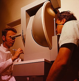
A visual field test is an eye examination that can detect dysfunction in central and peripheral vision which may be caused by various medical conditions such as glaucoma, stroke, pituitary disease, brain tumours or other neurological deficits. Visual field testing can be performed clinically by keeping the subject's gaze fixed while presenting objects at various places within their visual field. Simple manual equipment can be used such as in the tangent screen test or the Amsler grid. When dedicated machinery is used it is called a perimeter.
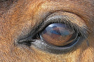
The equine eye is one of the largest of any land mammal. Its visual abilities are directly related to the animal's behavior; for example, it is active during both day and night, and it is a prey animal. Both the strengths and weaknesses of the horse's visual abilities should be taken into consideration when training the animal, as an understanding of the horse's eye can help to discover why the animal behaves the way it does in various situations.
Mammals normally have a pair of eyes. Although mammalian vision is not so excellent as bird vision, it is at least dichromatic for most of mammalian species, with certain families possessing a trichromatic color perception.

Vision is an important sensory system for most species of fish. Fish eyes are similar to the eyes of terrestrial vertebrates like birds and mammals, but have a more spherical lens. Birds and mammals normally adjust focus by changing the shape of their lens, but fish normally adjust focus by moving the lens closer to or further from the retina. Fish retinas generally have both rod cells and cone cells, and most species have colour vision. Some fish can see ultraviolet and some are sensitive to polarised light.
References
- ↑ Ferri, Fred F. (2022-06-21). Ferri's Clinical Advisor 2023. Elsevier Health Sciences. p. 1615. ISBN 978-0-323-75574-0.
- ↑ Saunders, David (2021-01-12). Museum Lighting: A Guide for Conservators and Curators. Getty Publications. p. 168. ISBN 978-1-60606-728-4.