Dental explorer

Last updated
A No. 23 explorer, also known as a 'sickle probe' Dentalexplorer01.jpg
A No. 23 explorer, also known as a 'sickle probe'

Dental explorers, also known as sickle probes, are tools found in the dental arsenal that are frequently utilised. The explorer is designed with a sharp tip at the end to improve tactile perception.

Contents

In the past it was usual for dentists to use the explorer to probe teeth for the presence of cavities. Some dental professionals have questioned this practice in twenty-first century. [1] [2] The use of a sharp explorer to diagnose caries in pit and fissure sites is no longer recommended, and clinicians instead should rely on "sharp eyes and a blunt explorer or probe." Penetration by a sharp explorer causes cavitation in areas that are remineralizing or could be remineralized. Dental lesions initially develop a subsurface lesion. Early lesions may be reversed with meticulous patient self-care and application of fluoride, as long as the thin surface layer remains intact. The use of a dental explorer with firm pressure to probe suspicious areas may result in the rupture of the surface layer covering early lesions. [3] Instead, they argue that fluoride and oral hygiene should be used to remineralize the enamel and prevent it from decaying further. This continues because sometimes decay can be difficult to diagnose without tactile verification. Additionally, radiographs and other products designed to identify decay (such as measuring fluorescence from a laser) help the dental professional make a final diagnosis of tooth decay.

There are various types of explorers, though the most common one is the No. 23 explorer, which is also known as a "shepherd's hook". Other types include the 3CH (also known as "cowhorn" or "pigtail") and No. 17 explorers, which are useful for the interproximal areas between teeth.

The 11/12 Explorer

An 11/12 type explorer is a double-ended dental assessment instrument with mirror image working-ends. [4] The 11/12 dental explorer should be considered a dental hygienists most important instrument for the detection of subgingival and supragingival calculus. This instrument is used with the modified pen grasp and a feather light grip allowing for detection of both grainy and bindable deposits. Learning its adaptability is critical for the successful tactile sensations needed for the confirmation of calculus removal.

11/12 Dental Explorer Dental Explorer 11-12.jpg
11/12 Dental Explorer

Design characteristics

90 degree angle Dental Explorer 11-12; 90 degree angle.jpg
90 degree angle

Use: Assessment of root surfaces on anterior and posterior teeth. [6]

The assessment stroke

Also known as the exploratory stroke, is used to detect calculus deposits and root surface irregularities. These kinds of strokes do require a high degree of precision for the accurate detection of unseen anomalies. Tactile sensitivity is key with this stroke, it is used to detect tooth irregularities by feeling vibration transferred from the explorer tip to the handle. [7]

The table below outlines the assessment stroke with an explorer: [4]

Assessment Stroke with an Explorer
Grasp: Relaxed grasp; middle finger rests lightly on shank [4]
Adaptation: 1–2 mm of the side of the tip is adapted; often referred to as "the tip 1/3" [4]
Lateral Pressure: Feather light pressure with working-end against tooth [4]
Activation: Wrist activation is usually recommended, however digital activation is acceptable with an explorer because physical strength is NOT required for assessment strokes. [4]
Stroke Characteristics: Fluid, sweeping strokes [4]
Stroke Number: Many close, overlapping multidirectional strokes are used to cover all of the root surface [4]

Common errors with the use of the 11/12 explorer is the use of a tight, tense "death grip" on the handle blanching the fingers at the joints. This disables the tactile sense needed to feel for the irregularities on the root surfaces. It is important to avoid pressing the middle finger against the instrument shank, thus reducing the tactile information to the finger. [5]

Adaptation of the 11/12 Dental Explorer at the MB line angle Adaptation of assessment dental explorer F.jpg
Adaptation of the 11/12 Dental Explorer at the MB line angle

Adaptability

Adaptation of the 11/12 Dental Explorer at the DB line angle Adaptation of assessment dental explorer C.jpg
Adaptation of the 11/12 Dental Explorer at the DB line angle

It is crucial to keep the terminal shank parallel to the long axis of the tooth and to use the tip third of the instrument at all times. If adapted correctly the instrument "elbow" will be angled towards the distal.[ citation needed ] [8]

For posterior placement, the instrument is inserted subgingival at the distal line angle of the tooth and using small muti-directional angles moving the tip third in contact with the tooth into the direction of the interproximal space. The instrument can be removed, reinserted at the distal line angle, now with the tip third in the direction toward the mesial of the tooth. Using the same multi-directional movements with the feather light grasp, follow the contour of the buccal or lingual aspects of the tooth around to the mesial line angle, here roll the instrument with the index finger the thumb into the interproximal space. [8]

Subgingival exploring

Without the use of our 11/12 explorer we are only assuming we have removed all subgingival and supragingival calculus. It is imperative to use many different overlapping stokes for subgingival assessment of residual deposits.[ citation needed ] [9]

Steps for subgingival assessment

Source: [4]

  1. Insertion: Adapt the explorers tip 1/3 against the tooth surface above the gingival margin and carefully slide the tip beneath the gingival margin.
  2. Reach the base of the sulcus: Always keep the tip in contact with the root surface to avoid laceration of the gingiva. You will know you are at the base of the sulcus when the tissue feels soft and elastic.
  3. Begin assessment stroke in coronal direction: Direct the tip 1/3 of the instrument forward slightly and use a oblique or vertical stroke along the root surface.
  4. Control the length of the stroke: Do not remove the explorer from the sulcus.
  5. Mentally section the tooth into apical, middle, and cervical sections: Allow 2-3mm length assessment strokes.
    1. For a healthy sulcus of less than 3mm; strokes will extend from the base of the sulcus to just beneath the gingival margin. Be carful not to come out of the sulcus.
    2. For a deeper sulcus depth begin exploration of the root at the base of the pocket. Then advance the tip 1/3 to the middle section on the root, followed by the coronal portion.
  6. Proximal surfaces: For assessing the mesial and distal proximal surfaces lead with the tip of the explorer. These strokes should reach under the contact area, so that the proximal surface is explored half from the facial/buccal aspect and the other half from the lingual aspect.

Anterior teeth technique

Anterior teeth are assessed using our clock positioning as fundamentals to proper adaptation of the 11/12 explorer.[ citation needed ] [10]

Clock positions for the right-handed clinicians: [10]

Clock positions for the left-handed clinicians: [10]

Related Research Articles

<span class="mw-page-title-main">Calculus (dental)</span> Form of hardened dental plaque

In dentistry, calculus or tartar is a form of hardened dental plaque. It is caused by precipitation of minerals from saliva and gingival crevicular fluid (GCF) in plaque on the teeth. This process of precipitation kills the bacterial cells within dental plaque, but the rough and hardened surface that is formed provides an ideal surface for further plaque formation. This leads to calculus buildup, which compromises the health of the gingiva (gums). Calculus can form both along the gumline, where it is referred to as supragingival, and within the narrow sulcus that exists between the teeth and the gingiva, where it is referred to as subgingival.

<span class="mw-page-title-main">Gums</span> Soft tissue surrounding the roots of the teeth

The gums or gingiva consist of the mucosal tissue that lies over the mandible and maxilla inside the mouth. Gum health and disease can have an effect on general health.

<span class="mw-page-title-main">Bridge (dentistry)</span> Dental restoration for missing teeth

A bridge is a fixed dental restoration used to replace one or more missing teeth by joining an artificial tooth definitively to adjacent teeth or dental implants.

<span class="mw-page-title-main">Cementoenamel junction</span>

Cementoenamel junction (CEJ) is defined as the area of the union of cementum and enamel at the cervical region of the tooth. It is a slightly visible anatomical border identified on a tooth. It is the location where the enamel, which covers the anatomical crown of a tooth, and the cementum, which covers the anatomical root of a tooth, meet. Informally it is known as the neck of the tooth. The border created by these two dental tissues has much significance as it is usually the location where the gingiva attaches to a healthy tooth by fibers called the gingival fibers.

Periodontology or periodontics is the specialty of dentistry that studies supporting structures of teeth, as well as diseases and conditions that affect them. The supporting tissues are known as the periodontium, which includes the gingiva (gums), alveolar bone, cementum, and the periodontal ligament. A periodontist is a dentist that specializes in the prevention, diagnosis and treatment of periodontal disease and in the placement of dental implants.

<span class="mw-page-title-main">Periodontal probe</span>

A periodontal probe is an instrument in dentistry commonly used in the dental armamentarium. It is usually long, thin, and blunted at the end. Its main function is to evaluate the depth of the pockets surrounding a tooth in order to determine the periodontium's overall health. For accuracy and readability, the instrument's head has markings written on it.

<span class="mw-page-title-main">Maxillary central incisor</span> Tooth

The maxillary central incisor is a human tooth in the front upper jaw, or maxilla, and is usually the most visible of all teeth in the mouth. It is located mesial to the maxillary lateral incisor. As with all incisors, their function is for shearing or cutting food during mastication (chewing). There is typically a single cusp on each tooth, called an incisal ridge or incisal edge. Formation of these teeth begins at 14 weeks in utero for the deciduous (baby) set and 3–4 months of age for the permanent set.

<span class="mw-page-title-main">Veterinary dentistry</span> Branch of veterinary medicine

Veterinary dentistry is the field of dentistry applied to the care of animals. It is the art and science of prevention, diagnosis, and treatment of conditions, diseases, and disorders of the oral cavity, the maxillofacial region, and its associated structures as it relates to animals.

Dentin hypersensitivity is dental pain which is sharp in character and of short duration, arising from exposed dentin surfaces in response to stimuli, typically thermal, evaporative, tactile, osmotic, chemical or electrical; and which cannot be ascribed to any other dental disease.

<span class="mw-page-title-main">Gingival sulcus</span> Space between tooth and gums

The gingival sulcus is an area of potential space between a tooth and the surrounding gingival tissue and is lined by sulcular epithelium. The depth of the sulcus is bounded by two entities: apically by the gingival fibers of the connective tissue attachment and coronally by the free gingival margin. A healthy sulcular depth is three millimeters or less, which is readily self-cleansable with a properly used toothbrush or the supplemental use of other oral hygiene aids.

This is a list of definitions of commonly used terms of location and direction in dentistry. This set of terms provides orientation within the oral cavity, much as anatomical terms of location provide orientation throughout the body.

<span class="mw-page-title-main">Scaling and root planing</span> Dental procedure

Scaling and root planing, also known as conventional periodontal therapy, non-surgical periodontal therapy or deep cleaning, is a procedure involving removal of dental plaque and calculus and then smoothing, or planing, of the (exposed) surfaces of the roots, removing cementum or dentine that is impregnated with calculus, toxins, or microorganisms, the agents that cause inflammation. It is a part of non-surgical periodontal therapy. This helps to establish a periodontium that is in remission of periodontal disease. Periodontal scalers and periodontal curettes are some of the tools involved.

The periodontal curette is a type of hand-activated instrument used in dentistry and dental hygiene for the purpose of scaling and root planing. The periodontal curette is considered a treatment instrument and is classified into two main categories: universal curettes and Gracey curettes. Periodontal curettes have one face, one or two cutting edges and a rounded back and rounded toe. They are typically the instrument of choice for subgingival calculus removal.

<span class="mw-page-title-main">Periodontal scaler</span> Dental tool

Periodontal scalers are dental instruments used in the prophylactic and periodontal care of teeth, including scaling and root planing. The working ends come in a variety of shapes and sizes, but they are always narrow at the tip, so as to allow for access to narrow embrasure spaces between teeth. They differ from periodontal curettes, which possess a blunt tip.

<span class="mw-page-title-main">Tooth polishing</span>

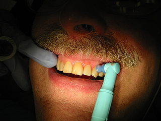
Tooth polishing procedures are done to smooth the surfaces of teeth and restorations. The purpose of polishing is to remove extrinsic stains, remove dental plaque accumulation, increase aesthetics and to reduce corrosion of metallic restorations. Tooth polishing has little therapeutic value and is usually done as a cosmetic procedure after debridement and before fluoride application. Common practice is to use a prophy cup—a small motorized rubber cup—along with an abrasive polishing compound.

<span class="mw-page-title-main">Debridement (dental)</span> Removal of plaque and calculus from teeth

In dentistry, debridement refers to the removal by dental cleaning of accumulations of plaque and calculus (tartar) in order to maintain dental health. Debridement may be performed using ultrasonic instruments, which fracture the calculus, thereby facilitating its removal, as well as hand tools, including periodontal scaler and curettes, or through the use of chemicals such as hydrogen peroxide.

<span class="mw-page-title-main">Furcation defect</span>

In dentistry, a furcation defect is bone loss, usually a result of periodontal disease, affecting the base of the root trunk of a tooth where two or more roots meet. The extent and configuration of the defect are factors in both diagnosis and treatment planning.

Height of curvature in the tooth can be defined as the line encircling a tooth at its greatest bulge to a selected path of insertion. The height of curvature is the same as the height of contour.

<span class="mw-page-title-main">Periodontal abscess</span> Medical condition

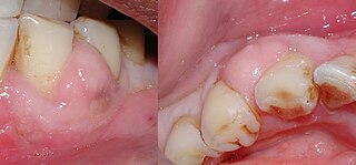
A periodontal abscess, is a localized collection of pus within the tissues of the periodontium. It is a type of dental abscess. A periodontal abscess occurs alongside a tooth, and is different from the more common periapical abscess, which represents the spread of infection from a dead tooth. To reflect this, sometimes the term "lateral (periodontal) abscess" is used. In contrast to a periapical abscess, periodontal abscesses are usually associated with a vital (living) tooth. Abscesses of the periodontium are acute bacterial infections classified primarily by location.

Chronic periodontitis is one of the seven categories of periodontitis as defined by the American Academy of Periodontology 1999 classification system. Chronic periodontitis is a common disease of the oral cavity consisting of chronic inflammation of the periodontal tissues that is caused by the accumulation of profuse amounts of dental plaque. Periodontitis initially begins as gingivitis and can progress onto chronic and subsequent aggressive periodontitis according to the 1999 classification.

References

  1. George Stookey "USE OF AN EXPLORER CAN LEAD TO MISDIAGNOSIS AND DISRUPT REMINERALIZATION", The Journal of the American Dental Association, November 2005, accessed November 17, 2011.
  2. George Stookey "The Evolution of Caries Detection", 2003, Dimensions of Dental Hygiene, accessed November 17, 2011.
  3. Gehrig, Jill (2016). Fundamentals of Periodontal Instrumentation & Advanced Root Instrumentation (8th ed.). Philadelphia: Wolters Kluwer. p. 315. ISBN   978-1-4963-2020-9.
  4. 1 2 3 4 5 6 7 8 9 10 11 Gehrig, Jill S. (2019). Fundamentals of periodontal instrumentation & advanced root instrumentation. Rebecca Sroda, Darlene Saccuzzo (Eighth edition, revised reprint ed.). Philadelphia. p. 291. ISBN   978-1-9751-1750-4. OCLC   1038036350.{{cite book}}: CS1 maint: location missing publisher (link)
  5. 1 2 Darby, Michele (2012). Mosby's comprehensive review of dental hygiene. Michele Leonardi Darby (7th ed.). St. Louis, Mo.: Elsevier/Mosby. p. 642. ISBN   978-0-323-07963-1. OCLC   712765588.
  6. Fundamentals of Periodontal Instrumentation and Advanced Root Instrumentation (8 ed.). Wolters Kluwer. 2019. p. 288. ISBN   978-1-975117-50-4.
  7. Linda D. Boyd, RDH, RD, EdD; Lisa F. Mallonee, MPH, RDH, RD, LD; Charlotte J. Wyche, BSDH, MS; Jane F. Halaris, ASDH, MA (2021). Wilkin's Clinical Practice of the Dental Hygienist (13th ed.). Jones and Bartlett. ISBN   9781496396273.{{cite book}}: CS1 maint: multiple names: authors list (link)
  8. 1 2 Fundamentals of Periodontal Instrumentation and Advanced Root Instrumentation (8 ed.). Wolters and Kluwer. 2019. pp. 300–306. ISBN   978-1-9751-1750-4.
  9. Fundamentals of Periodontal Instrumentation and Advanced Root Instrumentation (8 ed.). Wolter Kluwer. 2019. p. 292. ISBN   978-1-9751-1750-4.
  10. 1 2 3 Fundamentals of Periodontal Instrumentation and Advanced Root Instrumentation (8 ed.). Wolters Kluwer. 2019. p. 293. ISBN   978-1-9751-1750-4.