Related Research Articles

In anatomy, flexor carpi radialis is a muscle of the human forearm that acts to flex and (radially) abduct the hand. The Latin carpus means wrist; hence flexor carpi is a flexor of the wrist.

Froment's sign is a special test of the wrist for palsy of the ulnar nerve, specifically, the action of adductor pollicis. Froment's maneuver can also refer to the cogwheel effect from contralateral arm movements seen in Parkinson's disease.

The ulnar collateral ligament (UCL) or internal lateral ligament is a thick triangular ligament at the medial aspect of the elbow uniting the distal aspect of the humerus to the proximal aspect of the ulna.

Wheeless is an unincorporated community in Cimarron County, Oklahoma, United States. The post office was established February 12, 1907, and discontinued September 27, 1963. Nearby are the ruins of Camp Nichols, a military encampment on the Santa Fe Trail, which is a National Historic Landmark.

The ulnar canal or ulnar tunnel (also known as Guyon's canal or tunnel) is a semi-rigid longitudinal canal in the wrist that allows passage of the ulnar artery and ulnar nerve into the hand. The roof of the canal is made up of the superficial palmar carpal ligament, while the deeper flexor retinaculum and hypothenar muscles comprise the floor. The space is medially bounded by the pisiform and pisohamate ligament more proximally, and laterally bounded by the hook of the hamate more distally. It is approximately 4 cm long, beginning proximally at the transverse carpal ligament and ending at the aponeurotic arch of the hypothenar muscles.

A Salter–Harris fracture is a fracture that involves the epiphyseal plate or growth plate of a bone, specifically the zone of provisional calcification. It is thus a form of child bone fracture. It is a common injury found in children, occurring in 15% of childhood long bone fractures. This type of fracture and its classification system is named for Robert B. Salter and William H. Harris who created and published this classification system in the Journal of Bone and Joint Surgery in 1963.

The anatomical neck of the humerus is obliquely directed, forming an obtuse angle with the body of the humerus. It represents the fused epiphyseal plate.

Gamekeeper's thumb is a type of injury to the ulnar collateral ligament (UCL) of the thumb. The UCL may be merely stretched, or it may be torn from its insertion site into the proximal phalanx of the thumb; in approximately 90% of cases part of the bone is actually avulsed from the joint. This condition is commonly observed among gamekeepers and Scottish fowl hunters, as well as athletes. It also occurs among people who sustain a fall onto an outstretched hand while holding a rod, frequently skiers grasping ski poles.

A Barton's fracture is an intra-articular fracture of the distal radius with dislocation of the radiocarpal joint.

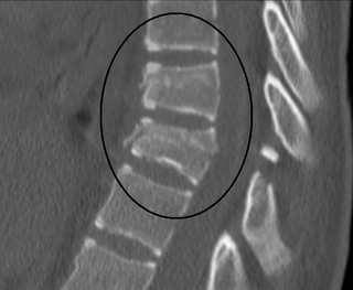
A Chance fracture is a type of vertebral fracture that results from excessive flexion of the spine. Symptoms may include abdominal bruising, or less commonly paralysis of the legs. In around half of cases there is an associated abdominal injury such as a splenic rupture, small bowel injury, pancreatic injury, or mesenteric tear. Injury to the bowel may not be apparent in the first day.

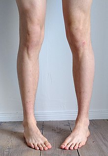
Pigeon toe, also known as in-toeing, is a condition which causes the toes to point inward when walking. It is most common in infants and children under two years of age and, when not the result of simple muscle weakness, normally arises from underlying conditions, such as a twisted shin bone or an excessive anteversion resulting in the twisting of the thigh bone when the front part of a person's foot is turned in.
Kanavel's sign is a clinical sign found in patients with infection of a flexor tendon sheath in the hand, a serious condition which can cause rapid loss of function of the affected finger.
Triple arthrodesis is a surgical procedure whose purpose is to relieve pain in the rear part of the foot, improve stability of the foot, and in some cases correct deformity of the foot, by fusing of the three main joints of the hindfoot: the subtalar joint, calcaneocuboid joint and the talonavicular joint. It is commonly carried out on patients with joint degeneration resulting from arthritis or a severe flat foot deformity.
A Bankart repair is an operation for habitual anterior shoulder dislocation. The joint capsule is sewed to the detached glenoid labrum, without duplication of the subscapularis tendon.
An enterocele is a protrusion of the small intestines and peritoneum into the vaginal canal. It may be treated transvaginally or by laparoscopy.

Freiberg disease, also known as a Freiberg infraction, is a form of avascular necrosis in the metatarsal bone of the foot. It generally develops in the second metatarsal, but can occur in any metatarsal. Physical stress causes multiple tiny fractures where the middle of the metatarsal meets the growth plate. These fractures impair blood flow to the end of the metatarsal resulting in the death of bone cells (osteonecrosis). It is an uncommon condition, occurring most often in young women, athletes, and those with abnormally long metatarsals. Approximately 80% of those diagnosed are women.

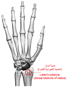
Lister's tubercle or dorsal tubercle of radius is a bony prominence located at the distal end of the radius. It is palpable on the dorsum of the wrist.
Frykman classification is a system of categorizing Colles' fractures. In the Frykman classification system there are four types of fractures.
The anterior colliculus is the anterior portion of the medial malleolus of the distal tibia, forming part of the ankle mortise. It has an attachment of the anterior tibiotalar ligament.
The posterior colliculus is the posterior portion of the medial malleolus of the distal tibia which is smaller in size comparing to the anterior colliculus. It has an attachment of the posterior tibiotalar ligament which is a part of deltoid ligament on the medial side of the ankle.
References
- ↑ "Wheeless' Textbook of Orthopaedics". Wheeless Online. 22 July 2020.